Review
. 2020 Dec;68(12):2819-2830.
doi: 10.4103/ijo.IJO_2399_20.

Advanced epithelial mapping for refractive surgery

Affiliations
Review

Advanced epithelial mapping for refractive surgery

Pooja Khamar et al. Indian J Ophthalmol. 2020 Dec.

Abstract

One of the leading challenges in refractive surgery today is the presence of underlying subclinical early-stage keratoconus (KC), which can lead to iatrogenic post laser in situ keratomileusis ectasia. Timely detection of this condition could aid the refractive surgeons in better decision-making. This includes being able to defer refractive surgery in subclinical cases as well as providing treatment for the same in the form of appropriate corneal collagen crosslinking treatments. Corneal topography is considered the gold standard for the diagnosis of corneal ectatic disorders. However, there is a likelihood that topographers are overlooking certain subclinical cases. The corneal epithelium is known to remodel, which may mask underlying stromal irregularities. Imaging and analyzing corneal epithelium and stroma independently will undoubtedly open newer avenues to supplement our understanding of postrefractive surgery outcomes and KC. This review encapsulates the various Optical coherence tomography-based epithelial mapping devices particularly RTVue (Optovue, Fremont, USA) and MS-39 (Costruzione Strumenti Oftalmici, Florence, Italy) in terms of their utility in these conditions. It will help guide the clinician on how including an epithelial mapping in clinical practice can aid in diagnosis, management, and interpretation of outcomes both for refractive surgery as well as KC.

Keywords: Epithelial mapping; keratoconus; preferred practices; refractive surgery.


Conflict of interest statement

None

Figures

Figure 1
Figure 1
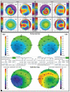
Pachymetric map of RTVue® (Optovue Inc, Fremont, CA) showing (red box).1. Minimum: The thinnest corneal thickness. 2. Minimum–Maximum: The thinnest corneal thickness minus the thickest corneal thickness. 3. SN-IT: Average thickness of Supero-Nasal (SN) octant minus average thickness of Infero-Temporal (IT) octant. 4.S-I: Average thickness of the superior (S) octant minus average thickness of the inferior (I) octant
Figure 2
Figure 2
Case 1: A patient with normal cornea topography on Pentacam HR refrative 4 maps (a), normal BAD-D (b); D 1.56 (c) and ET of 121μm (d)
Figure 3
Figure 3
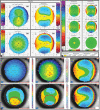
(a) Case 2: A case of epithelial hyperplasia masquerading as Regression post refractive surgery. (b) epithelial map of a patient with both eye contact lens warpage and the corresponding axial/saggital map on the Pentacam (c). (d) Case 3: A patient who underwent LASIK, presenting with blurring of vision and glares postoperatively, found to have irregularity of epithelium as the underlying cause. (e) Epithelial map showing donut-shaped rim of thickened peripheral epithelium in a case of keratoconus
Figure 4
Figure 4
(a) Case 4: Pentacam refractive four maps showing grade 2 KC in both the eyes. (b) Corneal pachymetry map of Keratoconic patient characterized by the inferotemporal thinning of the cornea, along with the corresponding thinning of the overlying epithelium in cone area
Figure 5
Figure 5
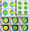
OD Refractive 4 map showing an asymmetric bowtie pattern with inferior steepening (a), Normal BAD-D, (b) Suspicious Df 1.76 (c), and (d) OD MS-39 6 map – Epithelial map showing a an increased epithelial thickness with no obvious stromal elevation and posterior elevation in the area corresponding to the inferior steepening (red circles)
Figure 6
Figure 6
OS Refractive 4 map on Pentacam HR showing inferior steepening on the axial/sagittal map (a) with an normal BAD-D display ((b and c). (d) OS MS-39 6 map scan showing an area of increased epithelial thickness inferiorly with no stromal elevation (red circles)
Figure 7
Figure 7
OD axial/sagittal map showing asymmetric bowtie pattern with inferior steepening (a). Normal BAD-D examination (b). (c) OD MS-39 6 map scan showing epithelial irregularity corresponding to the area of steepening on sagittal map. However, the area of posterior and stromal elevation on MS-39 does not coincide with the area of steepening (red circles)
Figure 8
Figure 8
(a) OD comparative map on the Pentacam showing progression in right eye (three points of steepening in the cone area > 1D). (b) OD MS 39 comparative map shows an area of compensatory hypertrophy around the area of thinning (donut sign) on the epithelial maps and an increase in the stomal elevation map, documented two visits apart
Figure 9
Figure 9
(a) OD comparative map on the Pentacam showing progression in the left eye (three points of steepening in the cone area >1D). (b) OD MS 39 Comparative epithelial map shows an obvious epithelial remodeling (b) with no evidence of progression on the stromal or posterior elevation comparative map (b)
Figure 10
Figure 10
(a) OS Refractive 4 map on Pentacam HR showing subclinical keratoconus. (b) OS Pachymetric and epithelial map showing epithelial hyperplasia. (c) OS MS-39 epithelial map showing hyperplasia with no evidence of elevation on the stromal elevation map

References

    1. Li Y, Chamberlain W, Tan O, Brass R, Weiss JL, Huang D. Subclinical keratoconus detection by pattern analysis of corneal and epithelial thickness maps with optical coherence tomography. J Cataract Refract Surg. 2016;42:284–95. - PMC - PubMed
    1. Gaster RN, Caiado Canedo AL, Rabinowitz YS. Corneal collagen cross-linking for keratoconus and post-LASIK ectasia. Int Ophthalmol Clin. 2013;53:79–90. - PMC - PubMed
    1. Amri AM. Prevalence of keratoconus in a refractive surgery population. J Ophthalmol. 2018;2018:5983530. doi: 10.1155/2018/5983530. - PMC - PubMed
    1. Goebels S, Eppig T, Wagenpfeil S, Cayless A, Seitz B, Langenbucher A. Complementary keratoconus indices based on topographical interpretation of biomechanical waveform parameters: A supplement to established keratoconus indices? Comput Math Methods Med. 2017;2017:5293573. doi: 10.1155/2017/5293573. - PMC - PubMed
    1. Ambrósio R, Jr, Dawson DG, Salomão M, Guerra FP, Caiado AL, Belin MW. Corneal ectasia after LASIK despite low preoperative risk: Tomographic and biomechanical findings in the unoperated, stable, fellow eye. J Refract Surg. 2010;26:906–11. - PubMed