

Study on change in corneal biomechanics and effect of percent tissue altered in myopic laser-assisted in situ keratomileusis
- PMID: 33229679
- PMCID: PMC7856945
- DOI: 10.4103/ijo.IJO_1453_20
Study on change in corneal biomechanics and effect of percent tissue altered in myopic laser-assisted in situ keratomileusis
Abstract
Purpose: To evaluate corneal biomechanical changes and their correlation with the percentage of tissue altered (PTA) in myopic femtosecond (FS)-flap LASIK.
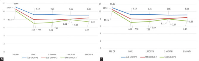
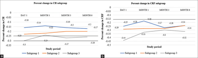
Methods: Prospective longitudinal observational study of 80 eyes of FS LASIK. Demographic details, LASIK parameters, preoperative and postoperative (day 1, month 1, 3, and 6), UCVA, BCVA, refraction, corneal topography, corneal hysteresis (CH), and a corneal resistance factor (CRF) were noted. Change in CH and CRF and its correlation with PTA were analyzed. Data were analyzed in three subgroups [subgroup 1: PTA 23 to <27%; subgroup 2: 27 to <33%; subgroup 3: 33 to <40%].
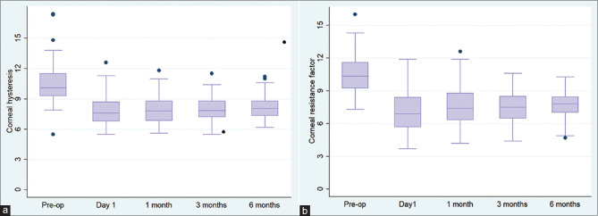
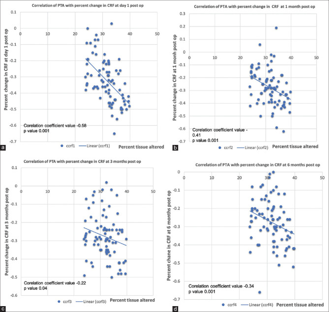
Results: FS LASIK for MRSE -3.5D ± 1.6D with mean PTA of 31.6 ± 4.4% (range 23.8-39.8%), showed statistically significant decrease in CH and CRF. Mean CH decreased from a preoperative value of 10.4 ± 1.9 to 8.1 ± 1.1; mean CRF from 10.5 ± 1.6 to 7.5 ± 1.3 at 6-months postoperative period, respectively. Mean preoperative CH decreased by 25%, 24%, 23%, and 21% and mean preoperative CRF decreased by 34%, 28%, 28%, and 28% at postoperative day 1, month 1, 3, and 6 follow-ups. Mean CH and CRF showed a significant negative correlation with PTA (CH: r = - 0.33 [P = <0.0001], CRF: r = -0.34 [P = <0.001]. Subgroup analysis noted greater decrease in CRF and CH in eyes with higher PTA (subgroup 3).
Conclusion: Myopic FS LASIK causes a decrease in corneal biomechanics with a significant negative correlation with PTA indicating a greater decrease in corneal biomechanics with higher PTA.
Keywords: Ablation depth; CH; CRF; LASIK; PTA; biomechanics; cornea; cornea resistance factor; correlation; flap thickness; hysteresis; laser ablation; myopia; resistance.
Conflict of interest statement
None
Figures


Similar articles
-
Comparison Between Q-Adjusted LASIK and Small-Incision Lenticule Extraction for Correction of Myopia and Myopic Astigmatism.Eye Contact Lens. 2018 Nov;44 Suppl 2:S426-S432. doi: 10.1097/ICL.0000000000000532. Eye Contact Lens. 2018. PMID: 30024453
-
Corneal biomechanical measurements before and after laser in situ keratomileusis.J Cataract Refract Surg. 2008 Nov;34(11):1886-91. doi: 10.1016/j.jcrs.2008.06.035. J Cataract Refract Surg. 2008. PMID: 19006734
-
Predictors affecting myopic regression in - 6.0D to - 10.0D myopia after laser-assisted subepithelial keratomileusis and laser in situ keratomileusis flap creation with femtosecond laser-assisted or mechanical microkeratome-assisted.Int Ophthalmol. 2020 Jan;40(1):213-225. doi: 10.1007/s10792-019-01179-5. Epub 2019 Sep 30. Int Ophthalmol. 2020. PMID: 31571091
-
Comparison of corneal biomechanical changes after refractive surgery by noncontact tonometry: small-incision lenticule extraction versus flap-based refractive surgery - a systematic review.Acta Ophthalmol. 2019 Mar;97(2):127-136. doi: 10.1111/aos.13906. Epub 2018 Sep 10. Acta Ophthalmol. 2019. PMID: 30203530
-
Corneal biomechanical properties after SMILE versus FLEX, LASIK, LASEK, or PRK: a systematic review and meta-analysis.BMC Ophthalmol. 2019 Aug 1;19(1):167. doi: 10.1186/s12886-019-1165-3. BMC Ophthalmol. 2019. PMID: 31370817 Free PMC article.
Cited by
-
Clinical Evaluation of Corneal Biomechanics following Laser Refractive Surgery in Myopic Eyes: A Review of the Literature.J Clin Med. 2022 Dec 28;12(1):243. doi: 10.3390/jcm12010243. J Clin Med. 2022. PMID: 36615041 Free PMC article. Review.
References
-
- Schmack I, Dawson DG, McCarey BE, Waring GO, 3rd, Grossniklaus HE, Edelhauser HF. Cohesive tensile strength of human LASIK wounds with histologic, ultrastructural, and clinical correlations. J Refract Surg. 2005;21:433–45. - PubMed
-
- Sonigo B, Iordanidou V, Chong-Sit D, Auclin F, Ancel JM, Labbe' A, et al. In vivo corneal confocal microscopy comparison of IntraLase femtosecond laser and mechanical microkeratome for laser in situ keratomileusis. Invest Ophthalmol Vis Sci. 2006;47:2803–11. - PubMed
-
- Kim JY, Kim MJ, Kim TI, Choi HJ, Pak JH, Tchah H. A femtosecond laser creates a stronger flap than a mechanical microkeratome. Invest Ophthalmol Vis Sci. 2006;47:599–604. - PubMed
-
- Ortiz D, Piñero D, Shabayek MH, Arnalich-Montiel F, Alió JL. Corneal biomechanical properties in normal, post-laser in situ keratomileusis, and keratoconic eyes. J Cataract Refract Surg. 2007;33:1371–5. - PubMed
Publication types
MeSH terms
LinkOut - more resources
Full Text Sources
