. 2021 May;16(5):820-829.
doi: 10.4103/1673-5374.297080.

Surgical intervention combined with weight-bearing walking training improves neurological recoveries in 320 patients with clinically complete spinal cord injury: a prospective self-controlled study

Affiliations

Surgical intervention combined with weight-bearing walking training improves neurological recoveries in 320 patients with clinically complete spinal cord injury: a prospective self-controlled study

Yansheng Liu et al. Neural Regen Res. 2021 May.

Abstract

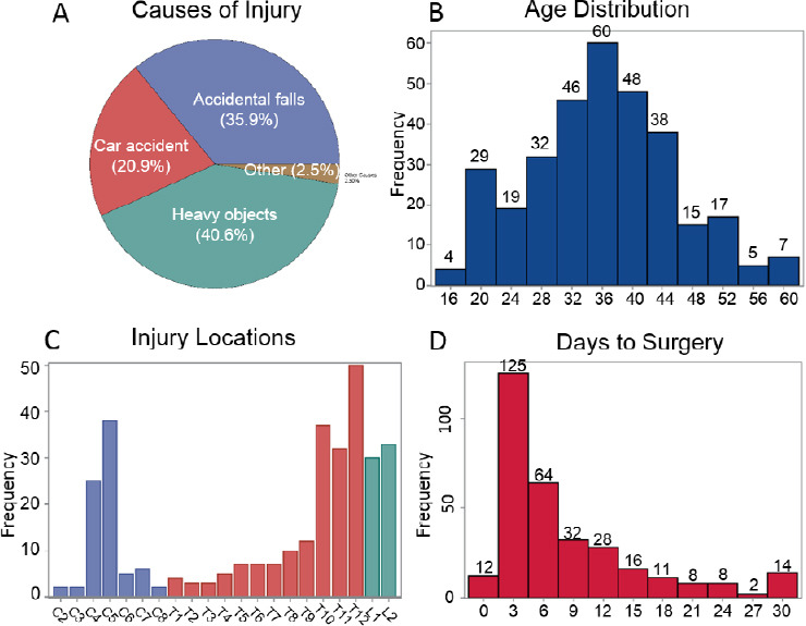
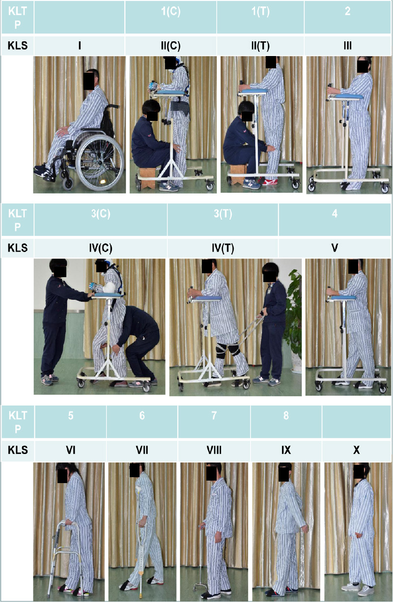
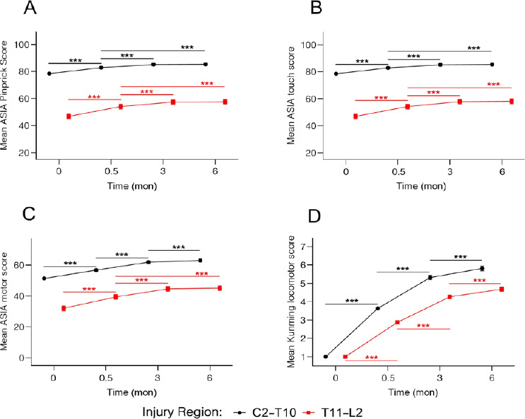
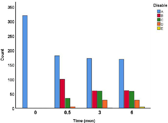
Although a large number of trials in the SCI field have been conducted, few proven gains have been realized for patients. In the present study, we determined the efficacy of a novel combination treatment involving surgical intervention and long-term weight-bearing walking training in spinal cord injury (SCI) subjects clinically diagnosed as complete or American Spinal Injury Association Impairment Scale (AIS) Class A (AIS-A). A total of 320 clinically complete SCI subjects (271 male and 49 female), aged 16-60 years, received early (≤ 7 days, n = 201) or delayed (8-30 days, n = 119) surgical interventions to reduce intraspinal or intramedullary pressure. Fifteen days post-surgery, all subjects received a weight-bearing walking training with the "Kunming Locomotion Training Program (KLTP)" for a duration of 6 months. The neurological deficit and recovery were assessed using the AIS scale and a 10-point Kunming Locomotor Scale (KLS). We found that surgical intervention significantly improved AIS scores measured at 15 days post-surgery as compared to the pre-surgery baseline scores. Significant improvement of AIS scores was detected at 3 and 6 months and the KLS further showed significant improvements between all pair-wise comparisons of time points of 15 days, 3 or 6 months indicating continued improvement in walking scores during the 6-month period. In conclusion, combining surgical intervention within 1 month post-injury and weight-bearing locomotor training promoted continued and statistically significant neurological recoveries in subjects with clinically complete SCI, which generally shows little clinical recovery within the first year after injury and most are permanently disabled. This study was approved by the Science and Research Committee of Kunming General Hospital of PLA and Kunming Tongren Hospital, China and registered at ClinicalTrials.gov (Identifier: NCT04034108) on July 26, 2019.

Keywords: American Spinal Injury Association Impairment Scale–A; functional recovery; human; intramedullary decompression; spinal cord injury; surgical intervention; walking training.


Conflict of interest statement

None

Figures

Figure 1
Figure 1
Demographics of 320 patients with severe traumatic spinal cord injury classified as American Spinal Injury Association Impair Scale-Class A (AIS-A). (A) Cause of injury. (B) Age distribution. (C) Injury locations. (D) Days between injury and surgery.
Figure 2
Figure 2
Tailored chest-waist cast for thoracic/lumbar injuries or a neck support for cervical injuries in a patient with severe traumatic spinal cord injury. (A–C) Front view (A), lateral view (B), and basic components (C) of a neck support. (D, E) Lateral view (D) and basic components (E) of a chest-waist cast.
Figure 3
Figure 3
Kunming locomotor training program (KLTP) and Kunming locomotor scale (KLS) in patients with severe traumatic spinal cord injury. The KLTP is made up of 8 progressive steps (1–8). All patients classified as American Spinal Injury Association Impairment Scale -Class A (AIS-A) were trained from step 1. Once the patient completed one step consistently, the training moved on to the next step. The 10-point KLS was used to assess locomotor recovery. The KLS is indicated by roman numerals I to X and is based on the KLTP. For each patient in the study, the KLS was evaluated and recorded before surgical treatment, 15 days after surgery (before rehabilitation), and 3 and 6 months after rehabilitation. During the KLTP training and KLS assessment, patients with cervical injuries received a neck support such as seen in 1(C), II(C) and 3(C) and IV(C). C: Cervical; T: thoracic.
Figure 4
Figure 4
Histology and immunohistochemistry of the necrotic spinal tissue from the injury center after severe traumatic spinal cord injury. (A) Dorsal view of the exposed spinal cord showing that, after a longitudinal incision of the spinal cord at the injury center, necrotic tissues were released out to the surface due to intraspinal pressure. (B, C) Hematoxylin and eosin (H & E) staining shows profound neutrophil infiltration (arrows), hemorrhage (open arrows) and degenerated segments of blood vessels (zigzag arrows) in the intraspinal necrotic tissue. (D–F) In the same region, fragments of degenerated axons (D, SMI-31-immunoreactive [IR], arrows) and myelin debris (E, Luxol fast blue [LFB] staining, arrows), along with degenerating astrocytes (F, glial fibrillary acidic protein [GFAP]-IR, arrows), were seen. (G) KP1/CD68 antibody staining further confirms that the necrotic tissue contains both infiltrated neutrophils (arrows) and reactive macrophages (open arrows).
Figure 5
Figure 5
Magnetic resonance (MR) images in a patient with severe traumatic spinal cord injury. (A) A T2-weighted MR image shows a sagittal image of a C3 spinal cord injury at 5 days post-injury. At the injury site, the cerebrospinal fluid signal became narrowed, indicating the obstruction of the cerebrospinal fluid flow at this region (red arrows). Tissue damage was found at the injury epicenter (yellow arrow). (B, C) Eleven years after surgery, T2- and T1-weighted MR images showed restored cerebrospinal fluid signal, particularly in the ventral subarachnoid space (red arrows). A defined cystic cavitation was found at the injury epicenter (yellow arrows). Note that spared cord tissue was also found surrounding the lesion cavity.
Figure 6
Figure 6
Functional assessments at cervical, thoracic, and lumbar injury levels over time after severe traumatic spinal cord. (A) American Spinal Injury Association Impairment Scaler (AIS) pinprick score. (B) AIS touch score. (C) AIS motor score. (D) Kunming locomotor score. Time points 0: before surgery; 0.5: 15 days after surgery, prior to the rehabilitation; 3.0: 3 months post-surgery and rehabilitation; 6.0: 6 months post-surgery and rehabilitation. **P < 0.01, ***P < 0.001 (see the Methods section for the sample size and statistical method).
Figure 7
Figure 7
Functional assessments at upper (C2–T10) and lower (T11–L2) spinal injury levels over time after severe traumatic spinal cord. (A) American Spinal Injury Association Impairment Scaler (AIS) pinprick score. (B) AIS touch score. (C) AIS motor score. (D) Kunming locomotor score. Time points 0: before surgery; 0.5: 15 days after surgery, prior to the rehabilitation; 3.0: 3 months post-surgery and rehabilitation; 6.0: 6 months post-surgery and rehabilitation. ***P < 0.001 (see the Methods section for the sample size and statistical method).
Figure 8
Figure 8
Distribution of American Spinal Injury Association Impairment Scale (AIS) disability over time after severe traumatic spinal cord. Time points 0: before surgery; 0.5: 15 days after surgery, prior to the rehabilitation; 3.0: 3 months post-surgery and rehabilitation; 6.0: 6 months post-surgery and rehabilitation. Count: The number of patients; A–E: AIS scales A to E.
Figure 9
Figure 9
Schematic illustration summarizes beneficial effects of the intradural/intramedullary decompression after severe traumatic spinal cord. (A) Laminectomy decompression allows to release the epidural pressure and remove epidural hematoma. (B) A dorsal longitudinal durotomy, alone or combined with a myelotomy, allows the removal of intradural hematoma, bone fragments, arachnoid adhesion, arachnoid cysts, and degenerated cord tissue, resulting in the reduction of swelled cord tissue and restoration of the cerebrospinal fluid (CSF) flow. (C) After suturing the dura, CSF flow is restored.

Similar articles

Cited by

References

    1. Ahuja CS, Wilson JR, Nori S, Kotter MRN, Druschel C, Curt A, Fehlings MG. Traumatic spinal cord injury. Nat Rev Dis Primers. 2017a;3:17018. - PubMed
    1. Ahuja CS, Nori S, Tetreault L, Wilson J, Kwon B, Harrop J, Choi D, Fehlings MG. Traumatic spinal cord injury-repair and regeneration. Neurosurgery. 2017b;80:S9–22. - PubMed
    1. Anderberg L, Aldskogius H, Holtz A. Spinal cord injury--scientific challenges for the unknown future. Ups J Med Sci. 2007;112:259–288. - PubMed
    1. Anderson KD. Targeting recovery: priorities of the spinal cord-injured population. J Neurotrauma. 2004;21:1371–1383. - PubMed
    1. Belanger M, Drew T, Provencher J, Rossignol S. A comparison of treadmill locomotion in adult cats before and after spinal transection. J Neurophysiol. 1996;76:471–491. - PubMed

Associated data