. 2021 May;16(5):899-904.
doi: 10.4103/1673-5374.290911.

Pannexin 1, a large-pore membrane channel, contributes to hypotonicity-induced ATP release in Schwann cells

Affiliations

Pannexin 1, a large-pore membrane channel, contributes to hypotonicity-induced ATP release in Schwann cells

Zhong-Ya Wei et al. Neural Regen Res. 2021 May.

Abstract

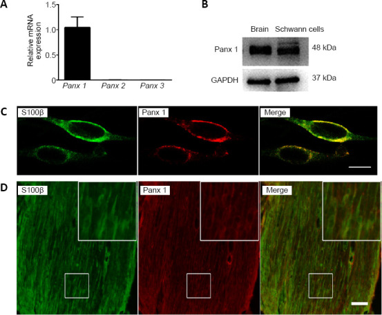
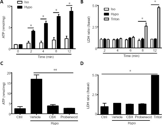
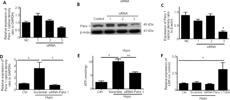
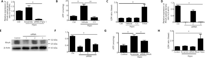
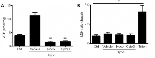
Pannexin 1 (Panx 1), as a large-pore membrane channel, is highly permeable to ATP and other signaling molecules. Previous studies have demonstrated the expression of Panx 1 in the nervous system, including astrocytes, microglia, and neurons. However, the distribution and function of Panx 1 in the peripheral nervous system are not clear. Blocking the function of Panx 1 pharmacologically (carbenoxolone and probenecid) or with small interfering RNA targeting pannexins can greatly reduce hypotonicity-induced ATP release. Treatment of Schwann cells with a Ras homolog family member (Rho) GTPase inhibitor and small interfering RNA targeting Rho or cytoskeleton disrupting agents, such as nocodazole or cytochalasin D, revealed that hypotonicity-induced ATP release depended on intracellular RhoA and the cytoskeleton. These findings suggest that Panx 1 participates in ATP release in Schwann cells by regulating RhoA and the cytoskeleton arrangement. This study was approved by the Animal Ethics Committee of Nantong University, China (No. S20180806-002) on August 5, 2018.

Keywords: ATP; Ras homolog family member A; Schwann cells; cytoskeleton; injury; neuron; pannexin 1; peripheral nerve.


Conflict of interest statement

None

Figures

Figure 1
Figure 1
Expression of Panx 1 in Schwann cells. (A) mRNA levels of Panx in Schwann cells. Data are expressed as the mean ± SEM (n = 4) and were analyzed by one-way analysis of variance or Student’s t-test. (B) The Panx 1 protein level in Schwann cells is shown; mouse brain tissue was used as a positive control. (C) Double immunostaining images show that Panx 1 (red, stained by Cy3) is expressed in cultured Schwann cells, as shown by the presence of S100β (green, stained by Alex-488). (D) Example images show double immunostaining with S100β (green, stained by Alex-488) and Panx 1 (red, stained by Cy3) in mouse sciatic nerves. The insets showing the boxed areas at higher magnification. Scale bars: 20 μm in C, 50 μm in D. GAPDH: Glyceraldehyde 3-phosphate dehydrogenase; Panx: pannexin.
Figure 2
Figure 2
Panx 1 blockers reduced ATP release induced by hypotonic challenge in Schwann cells. (A, B) Time course of ATP and LDH release from cultures treated in the Iso, Hypo, and Triton conditions. The Triton group was used as a positive control in the LDH assay. (C, D) ATP and LDH release were assessed under different conditions. Schwann cells were pretreated with carbenoxolone (100 μM) and probenecid (100 μM) for 60 minutes before exposure to the indicated solutions for 12 minutes. Dimethyl sulfoxide at the same volume in isotonic solution was used as a Ctrl. Data are expressed as the mean ± SEM (n = 4). *P < 0.05, **P < 0.01 (one-way analysis of variance or Student’s t-test). CBX: Carbenoxolone; Ctrl: control; Hypo: hypotonic; Iso: isotonic; LDH: lactate dehydrogenase; Vehicle: dimethyl sulfoxide.
Figure 3
Figure 3
siRNA knockdown of Panx 1 attenuates hypotonic solution-induced ATP release. (A) mRNA levels of Panx 1 in Schwann cells treated with scrambled siRNA (NC) and Panx 1-siRNA (1–3). (B, C) Panx 1 protein bands in RSC96 cells in the scrambled (control) and Panx 1-siRNA (1–3) groups. (D) The relative expression of Panx 1 in isotonic solution after transfection with scrambled siRNA (Ctrl) or in hypotonic (Hypo) solution after transfection with scrambled and siRNA-Panx 1 (#3) is shown. (E) ATP release was assessed using scrambled siRNA and Panx 1-siRNA-treated Schwann cells. (F) Lactate dehydrogenase (LDH) release was assessed under the same conditions as ATP release. Data are expressed as the mean ± SEM (n = 3). *P < 0.05, **P < 0.01. #P < 0.05, vs. control group (one-way analysis of variance or Student’s t-test). Panx 1: Pannexin 1; siRNA: small interfering RNA.
Figure 4
Figure 4
Inhibition of RhoA activation reduced hypotonic solution-induced ATP release. (A) Relative expression of RhoA upon treatment with isotonic (Ctrl) or hypotonic (Hypo) solution after transfection with scrambled and Panx 1 siRNA (3). (B) ATP release was assessed in Schwann cells treated with C3 transferase. Dimethyl sulfoxide at the same volume in isotonic solution was used as a control (Ctrl). (C) Lactate dehydrogenase (LDH) release was assessed under the same conditions as ATP release in B. (D) RhoA mRNA levels in Schwann cells treated with scrambled siRNA (NC) and RhoA-siRNA (1–3). (E, F) Western blot images and mean analysis showing the levels of RhoA protein in Hepa 1–6 cells from the scrambled (Ctrl) and Panx 1-siRNA (1–3) groups. (G) ATP was assessed in scrambled siRNA- and RhoA-siRNA (3)-treated Schwann cells. Cells were treated with isotonic solution after transfection with scrambled siRNA as a control. (H) LDH release was assessed under the same conditions as ATP release. Data are expressed as the mean ± SEM (n = 3). *P < 0.05, **P < 0.01 (one-way analysis of variance or Student’s t-test). GAPDH: Glyceraldehyde 3-phosphate dehydrogenase; LDH: lactate dehydrogenase; Panx 1: pannexin 1; RhoA: Ras homolog family member A; siRNA: small interfering RNA; Vehicle: dimethyl sulfoxide.
Figure 5
Figure 5
An intact cytoskeleton is required for ATP release during hypotonic challenge. (A, B) Schwann cells were incubated with nocodazole (Noco) or cytochalasin D (CytoD) for 2 hours before treatment with hypotonic (Hypo) solutions. ATP (A) and lactate dehydrogenase (LDH) (B) release were assessed under different conditions. Dimethyl sulfoxide at the same volume in isotonic solution was used as a control (Ctrl). Data are expressed as the mean ± SEM (n = 3). *P < 0.05. ##P < 0.01 (one-way analysis of variance or Student’s t-test). LDH: Lactate dehydrogenase; Vehicle: dimethyl sulfoxide.

References

    1. Baranova A, Ivanov D, Petrash N, Pestova A, Skoblov M, Kelmanson I, Shagin D, Nazarenko S, Geraymovych E, Litvin O, Tiunova A, Born TL, Usman N, Staroverov D, Lukyanov S, Panchin Y. The mammalian pannexin family is homologous to the invertebrate innexin gap junction proteins. Genomics. 2004;83:706–716. - PubMed
    1. Begum G, Song S, Wang S, Zhao H, Bhuiyan MIH, Li E, Nepomuceno R, Ye Q, Sun M, Calderon MJ, Stolz DB, St Croix C, Watkins SC, Chen Y, He P, Shull GE, Sun D. Selective knockout of astrocytic Na(+) /H(+) exchanger isoform 1 reduces astrogliosis, BBB damage, infarction, and improves neurological function after ischemic stroke. Glia. 2018;66:126–144. - PMC - PubMed
    1. Bhalla-Gehi R, Penuela S, Churko JM, Shao Q, Laird DW. Pannexin1 and pannexin3 delivery, cell surface dynamics, and cytoskeletal interactions. J Biol Chem. 2010;285:9147–9160. - PMC - PubMed
    1. Boudreault F, Grygorczyk R. Cell swelling-induced ATP release is tightly dependent on intracellular calcium elevations. J Physiol. 2004;561:499–513. - PMC - PubMed
    1. Boyce AK, Kim MS, Wicki-Stordeur LE, Swayne LA. ATP stimulates pannexin 1 internalization to endosomal compartments. Biochem J. 2015;470:319–330. - PubMed