

Mechanism underlying treatment of ischemic stroke using acupuncture: transmission and regulation
- PMID: 33229734
- PMCID: PMC8178780
- DOI: 10.4103/1673-5374.297061
Mechanism underlying treatment of ischemic stroke using acupuncture: transmission and regulation
Abstract
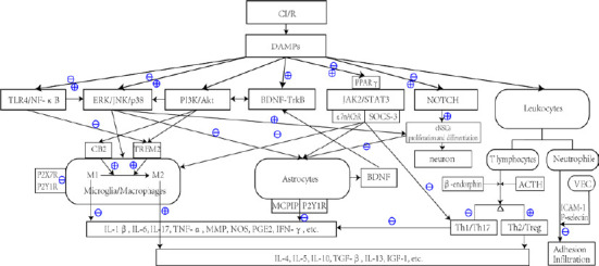
The inflammatory response after cerebral ischemia/reperfusion is an important cause of neurological damage and repair. After cerebral ischemia/reperfusion, microglia are activated, and a large number of circulating inflammatory cells infiltrate the affected area. This leads to the secretion of inflammatory mediators and an inflammatory cascade that eventually causes secondary brain damage, including neuron necrosis, blood-brain barrier destruction, cerebral edema, and an oxidative stress response. Activation of inflammatory signaling pathways plays a key role in the pathological process of ischemic stroke. Increasing evidence suggests that acupuncture can reduce the inflammatory response after cerebral ischemia/reperfusion and promote repair of the injured nervous system. Acupuncture can not only inhibit the activation and infiltration of inflammatory cells, but can also regulate the expression of inflammation-related cytokines, balance the effects of pro-inflammatory and anti-inflammatory factors, and interfere with inflammatory signaling pathways. Therefore, it is important to study the transmission and regulatory mechanism of inflammatory signaling pathways after acupuncture treatment for cerebral ischemia/reperfusion injury to provide a theoretical basis for clinical treatment of this type of injury using acupuncture. Our review summarizes the overall conditions of inflammatory cells, mediators, and pathways after cerebral ischemia/reperfusion, and discusses the possible synergistic intervention of acupuncture in the inflammatory signaling pathway network to provide a foundation to explore the multiple molecular mechanisms by which acupuncture promotes nerve function restoration.
Keywords: acupuncture; central nervous system; factor; inflammation; ischemic stroke; pathways; protein; stroke.
Conflict of interest statement
None
Figures
References
-
- Alberi L, Hoey SE, Brai E, Scotti AL, Marathe S. Notch signaling in the brain: in good and bad times. Ageing Res Rev. 2013;12:801–814. - PubMed
-
- Androutsellis-Theotokis A, Leker RR, Soldner F, Hoeppner DJ, Ravin R, Poser SW, Rueger MA, Bae SK, Kittappa R, McKay RD. Notch signalling regulates stem cell numbers in vitro and in vivo. Nature. 2006;442:823–826. - PubMed
-
- Arboleda-Velasquez JF, Manent J, Lee JH, Tikka S, Ospina C, Vanderburg CR, Frosch MP, Rodríguez-Falcón M, Villen J, Gygi S, Lopera F, Kalimo H, Moskowitz MA, Ayata C, Louvi A, Artavanis-Tsakonas S. Hypomorphic Notch 3 alleles link Notch signaling to ischemic cerebral small-vessel disease. Proc Natl Acad Sci U S A. 2011;108:E128–135. - PMC - PubMed
Publication types
LinkOut - more resources
Full Text Sources
Other Literature Sources
