. 2020 Nov 23;11(11):1003.
doi: 10.1038/s41419-020-03209-y.

Ionizing radiation results in a mixture of cellular outcomes including mitotic catastrophe, senescence, methuosis, and iron-dependent cell death

Affiliations

Ionizing radiation results in a mixture of cellular outcomes including mitotic catastrophe, senescence, methuosis, and iron-dependent cell death

Sandy Adjemian et al. Cell Death Dis. .

Abstract

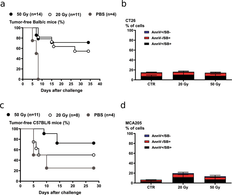
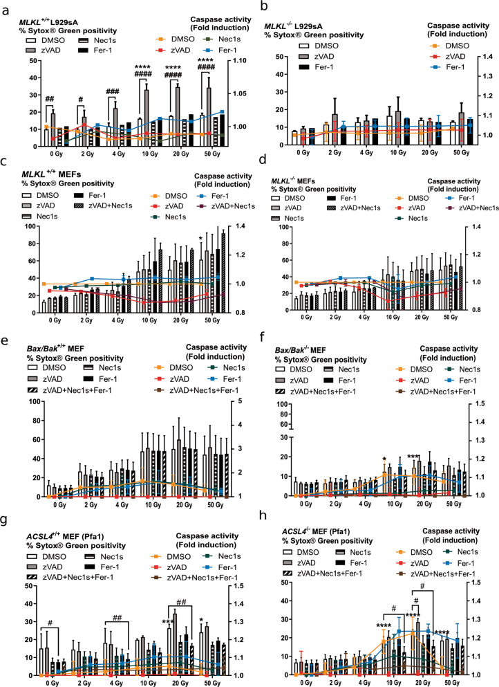
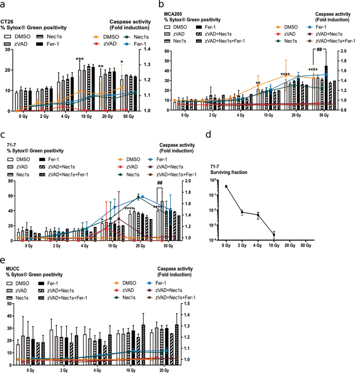
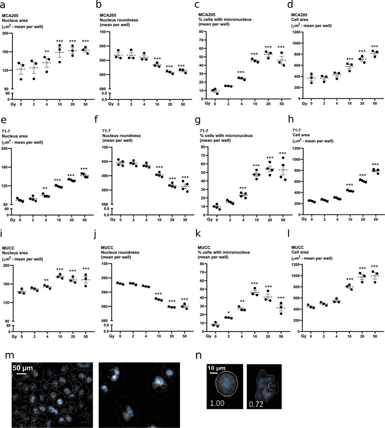
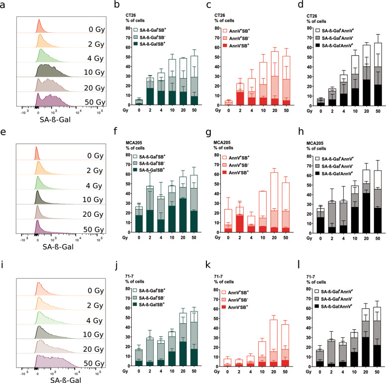
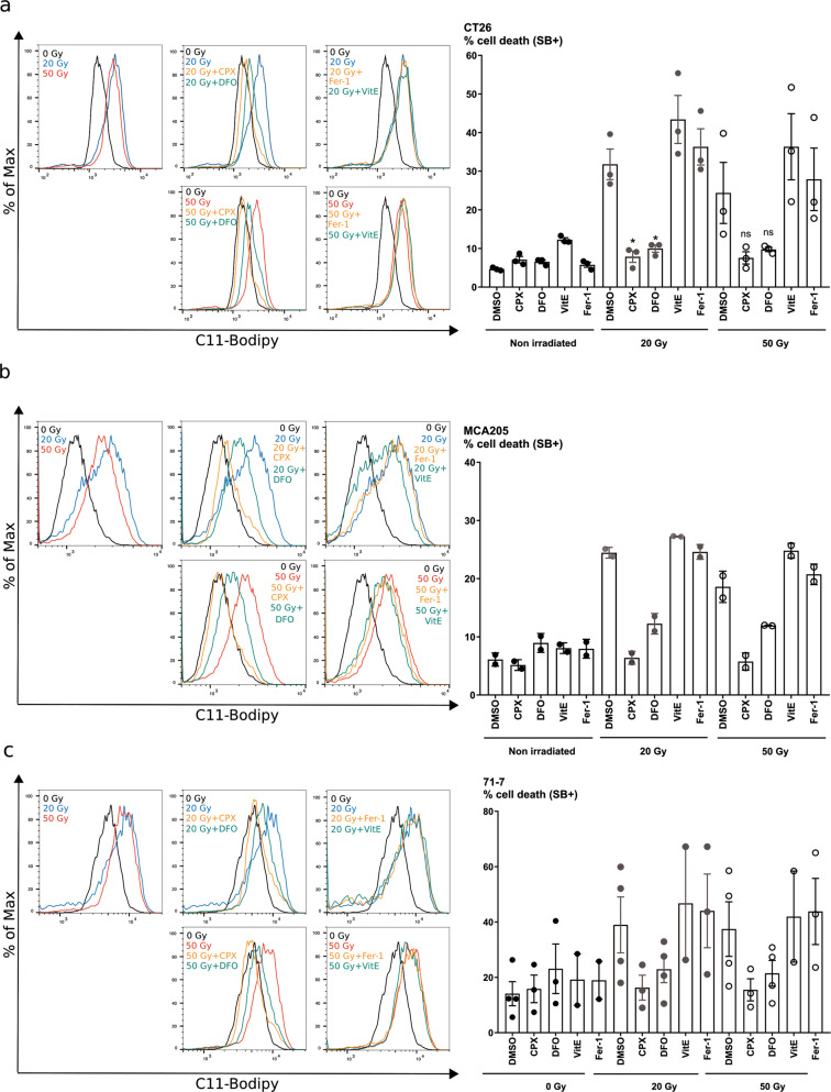
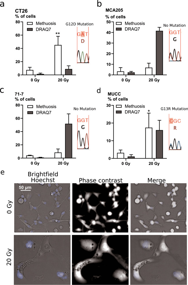
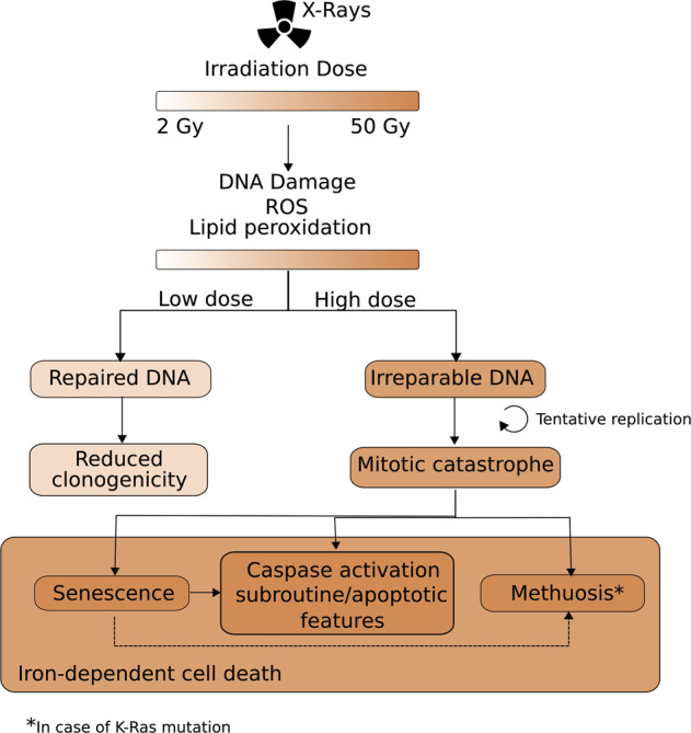
Radiotherapy is commonly used as a cytotoxic treatment of a wide variety of tumors. Interestingly, few case reports underlined its potential to induce immune-mediated abscopal effects, resulting in regression of metastases, distant from the irradiated site. These observations are rare, and apparently depend on the dose used, suggesting that dose-related cellular responses may be involved in the distant immunogenic responses. Ionizing radiation (IR) has been reported to elicit immunogenic apoptosis, necroptosis, mitotic catastrophe, and senescence. In order to link a cellular outcome with a particular dose of irradiation, we performed a systematic study in a panel of cell lines on the cellular responses at different doses of X-rays. Remarkably, we observed that all cell lines tested responded in a similar fashion to IR with characteristics of mitotic catastrophe, senescence, lipid peroxidation, and caspase activity. Iron chelators (but not Ferrostatin-1 or vitamin E) could prevent the formation of lipid peroxides and cell death induced by IR, suggesting a crucial role of iron-dependent cell death during high-dose irradiation. We also show that in K-Ras-mutated cells, IR can induce morphological features reminiscent of methuosis, a cell death modality that has been recently described following H-Ras or K-Ras mutation overexpression.


Conflict of interest statement

The authors declare that they have no conflict of interest.

Figures

Fig. 1
Fig. 1. Prophylactic vaccination with high-dose irradiated cells (20 or 50 Gy) induce immune protection in mice.
CT26 and MCA205 cells were irradiated with 20 or 50 Gy the day before the vaccination. PBS or cells irradiated with 20 and 50 Gy were injected subcutaneously in Balb/c. After 7 days, the mice were injected on the opposite flank with living cells. Tumor growth was monitored every 2 days up to 35 days post-challenge. Percentage of tumor-free mice after vaccination with irradiated CT26 cells and MCA205 cells are shown in a and c, respectively. The extent of cell death measured as Annexin V positivity (AnnV), Sytox Blue (SB) positivity, and a combination of the two was minimal at the time of injection and is shown in b (CT26 cells) and in d (MCA205 cells).
Fig. 2
Fig. 2. Cell death induction by single-dose ionizing radiation in cells with altered cell death pathways.
a–d Cell death and caspase activity were measured 72 h after IR at the indicated doses by Sytox Green and Ac-DEVD-amc fluorescence in MLKL+/+ L929sA (a), MLKL−/− L929sA (b), MLKL+/+ MEF (c), MLKL−/− MEF cells (d), Bax/Bak+/+ MEF (e), and Bax/Bak−/− MEF (f), ACSL4+/+ Pfa1 (g), and ACSL4−/− Pfa1 (h). When indicated, the cells were pre-treated for 1 h with zVAD-fmk, Nec1s or Fer-1, or a combination thereof. Histogram bars show Sytox Green positivity and lines indicate caspase activity (fold induction). Means ± SEM are shown (n = 2–3). A two-way ANOVA was performed with a Tukey’s multiple comparisons test. Asterisk (*) shows the comparison to 0 Gy DMSO. Hash (#) shows the comparison to the DMSO treatment for the respective irradiation dose. *,#p ≤ 0.05, ##p ≤ 0.01, ***, ###p ≤ 0.001, ****,####p ≤ 0.0001.
Fig. 3
Fig. 3. Cell death induction by single-dose ionizing radiation.
a–c, e Cell death and caspase activity were measured 72 h after IR at the indicated doses by Sytox Green and Ac-DEVD-amc fluorescence in CT26 (a), MCA205 (b), 71-7 (c), and MUCC (e). When indicated, the cells were pre-treated with zVAD-fmk, Nec1s or Fer-1, or a combination thereof. Histogram bars reflect Sytox Green intensity and lines describe caspase activity (fold induction). d Clonogenicity assay performed on 71-7 cells at different IR doses. Means ± SEM are shown (n = 2–3). A two-way ANOVA was performed with a Tukey’s multiple comparisons test. Asterisk (*) shows the comparison to 0 Gy DMSO. *p ≤ 0.05, **,##p ≤ 0.01, ***p ≤ 0.001, ****p ≤ 0.0001.
Fig. 4
Fig. 4. Induction of multinucleation and micronucleation by ionizing radiation.
Nuclear morphology was quantified through high-content imaging in MCA205 (a–d), 71-7 (e–h), MUCC (i–l) cells, 48 h after irradiation. Cells were stained with Hoechst and propidium iodide after permeabilization to allow segmentation of the nucleus and cytoplasm, respectively. Nuclear area was measured in MCA205 (a), 71-7 (e), and MUCC (i). The roundness of the nucleus was also assessed after irradiation in MCA205 (b), 71-7 (f), and MUCC (j). Percentage of cells with micronuclei is shown for MCA205 (c), 71-7 (g), and MUCC (k) cells. The area of the cells was also measured following irradiation in MCA205 (d), 71-7 (h), and MUCC (l) cells. m Representative picture of micronuclei segmentation. n Representative picture of segmentation mask for the assessment of the nuclear shape.
Fig. 5
Fig. 5. Ionizing radiation induces senescence.
Senescence induction was measured 72 h following irradiation at the indicated doses by flow cytometry with C12FDG, a β-galactosidase substrate emitting fluorescence upon its cleavage. Co-staining with Annexin V-APC and Sytox Blue was performed to indicate apoptosis and membrane permeabilization, respectively, in CT26 (a–d), MCA205 (e–h), and 71-7 (i–l) cells. Representative flow cytometry histograms are shown for C12FDG (SA-β-Gal) staining in CT26 (a), MCA205 (e), and 71-7 (i). Percentage of cells positive for C12FDG (SA-β-Gal) and/or Sytox Blue is shown for CT26 (b), MCA205 (f), and 71-7 (j) cells. Percentage of cells positive for Annexin V-APC and/or Sytox Blue is shown for CT26 (c), MCA205 (g), and 71-7 (k). Percentage of cells positive for C12FDG (SA-β-Gal) and/or Annexin V-APC is shown for CT26 (d), MCA205 (h), and 71-7 (l). Means ± SEM are shown (n = 2). SA-β-Gal senescence-associated β-Galactosidase.
Fig. 6
Fig. 6. Ionizing radiation induces lipid peroxidation and an iron-dependent type of cell death.
a–c Measurement of lipid peroxidation using C11-BODIPY probe and cell death using Sytox Blue in CT26 (a), MCA205 (b), and 71-7 (c) cells 24 h after irradiation with 20 or 50 Gy. Increase in lipid peroxidation can be seen in the first left quadrant by the shift of C11-BODIPY fluorescence measured after 20 Gy (blue) or 50 Gy (red) X-rays. When indicated, the cells were pre-treated with iron chelators CPX and DFO, or with antioxidants vitamin E and Fer-1 1 h before irradiation. Quantification of cell death is shown as percentage for CT26 (a), MCA205 (b), and 71-7 (c). Means ± SEM are shown (n = 2–3). A two-way ANOVA was performed with a Tukey’s multiple comparisons test. Asterisks (*) show the comparison to 20 Gy Methuosis. *p ≤ 0.05, **p ≤ 0.01.
Fig. 7
Fig. 7. Ionizing radiation induces features of methuosis.
Microscopic evaluation of the presence of phase-lucent vacuoles in CT26 (a), MCA205 (b), 71-7 (c), and MUCC (d) cells after 72 h irradiation with 20 Gy. An example of pictures for CT26 cells is shown in e. Means ± SEM are shown (n = 3). An unpaired t-test was performed for the methuosis data. Asterisks (*) show the comparison to 0 Gy. *p ≤ 0.05, **p ≤ 0.01.
Fig. 8
Fig. 8. Cellular responses to ionizing radiation.
Following X-ray irradiation with doses ranging from 2 to 50 Gy, there is a dose-dependent increase in ROS production, lipid peroxidation, and DNA damage. As a result, the DNA damage response is activated and will attempt to resolve the DNA breaks. In a case of low irradiation dose (<10 Gy), cells manage to repair their DNA, and still retain their capacity to proliferate. Cells with irreparable DNA that go into replication will be stuck in the mitotic cycle and undergo mitotic failure. Cells in a state of mitotic catastrophe will have several fates depending on the extent of the nuclear abnormalities and on the proteins expressed. Several outcomes of mitotic catastrophe seem to co-exist, such as senescence, methuosis, necrosis (membrane permeabilization), and activation of Bax, Bak, and caspases with presence of apoptotic features. A part of the senescent cells might become apoptotic (dotted arrow). K-Ras mutation can induce both senescence and methuosis; however, a link between the two cell states was never described. Here, we found that part of the large cells with flattened morphology also had methuotic vacuoles, which might indicate that methuosis could be an outcome of senescent cells (dotted arrow). Ultimately, the cells will die out of an iron-dependent type of cell death.

Similar articles

Cited by

References

    1. Prasanna PGS, et al. Normal tissue protection for improving radiotherapy: where are the gaps? Transl. Cancer Res. 2012;1:35–48. - PMC - PubMed
    1. Lee Y, et al. Therapeutic effects of ablative radiation on local tumor require CD8 ϩ T cells: changing strategies for cancer treatment. Blood. 2018;114:589–596. doi: 10.1182/blood-2009-02-206870. - DOI - PMC - PubMed
    1. Surace L, et al. Complement is a central mediator of radiotherapy-induced tumor-specific immunity and clinical response. Immunity. 2015;42:767–777. doi: 10.1016/j.immuni.2015.03.009. - DOI - PubMed
    1. Baskar R, Dai J, Wenlong N, Yeo R, Yeoh K-W. Biological response of cancer cells to radiation treatment. Front. Mol. Biosci. 2014;1:1–9. doi: 10.3389/fmolb.2014.00024. - DOI - PMC - PubMed
    1. Minchom, A., Aversa, C. & Lopez, J. Dancing with the DNA damage response: next-generation anti-cancer therapeutic strategies. Ther. Adv. Med. Oncol. 1–18, 10.1177/1758835918786658 (2018). - PMC - PubMed

Publication types