. 2021 Apr;15(4):1056-1072.
doi: 10.1038/s41396-020-00830-9. Epub 2020 Nov 23.

Limits to the cellular control of sequestered cryptophyte prey in the marine ciliate Mesodinium rubrum

Affiliations

Limits to the cellular control of sequestered cryptophyte prey in the marine ciliate Mesodinium rubrum

Andreas Altenburger et al. ISME J. 2021 Apr.

Erratum in

Abstract

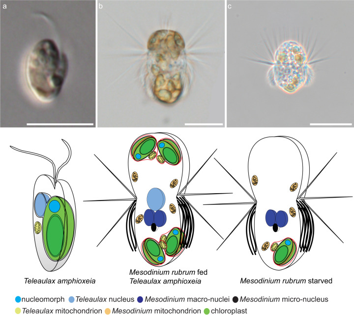
The marine ciliate Mesodinium rubrum is famous for its ability to acquire and exploit chloroplasts and other cell organelles from some cryptophyte algal species. We sequenced genomes and transcriptomes of free-swimming Teleaulax amphioxeia, as well as well-fed and starved M. rubrum in order to understand cellular processes upon sequestration under different prey and light conditions. From its prey, the ciliate acquires the ability to photosynthesize as well as the potential to metabolize several essential compounds including lysine, glycan, and vitamins that elucidate its specific prey dependency. M. rubrum does not express photosynthesis-related genes itself, but elicits considerable transcriptional control of the acquired cryptophyte organelles. This control is limited as light-dependent transcriptional changes found in free-swimming T. amphioxeia got lost after sequestration. We found strong transcriptional rewiring of the cryptophyte nucleus upon sequestration, where 35% of the T. amphioxeia genes were significantly differentially expressed within well-fed M. rubrum. Qualitatively, 68% of all genes expressed within well-fed M. rubrum originated from T. amphioxeia. Quantitatively, these genes contributed up to 48% to the global transcriptome in well-fed M. rubrum and down to 11% in starved M. rubrum. This tertiary endosymbiosis system functions for several weeks, when deprived of prey. After this point in time, the ciliate dies if not supplied with fresh prey cells. M. rubrum represents one evolutionary way of acquiring photosystems from its algal prey, and might represent a step on the evolutionary way towards a permanent tertiary endosymbiosis.


Conflict of interest statement

The authors declare that they have no conflict of interest.

Figures

Fig. 1
Fig. 1. Light micrographs of Teleaulax amphioxeia and Mesodinium rubrum with corresponding cartoons.
a Free-swimming T. amphioxeia with chloroplast, nucleomorph, mitochondrion, and nucleus. The outer membrane of the nucleus is connected to the outer membrane of the chloroplast. b Well-fed M. rubrum with two macronuclei, one micronucleus, and one enlarged cryptophyte nucleus. M. rubrum contains its own mitochondria, cryptophyte mitochondria, and cryptophyte chloroplasts that are arranged along the periphery of the cell. c Starved M. rubrum with two macronuclei, one micronucleus, and ciliate mitochondria. Starved M. rubrum was defined as cultures where at least 90% of cells had lost the cryptophyte nucleus. Well-fed cells of M. rubrum have one enlarged cryptophyte nucleus, which is always located in the center of the cell, termed CPN (centered prey nucleus) [24]. Well-fed cells might keep some extra prey nuclei in the periphery of the cell. Upon ciliate cell division, one of the two daughter cells receives the CPN, while in the other, one of the extra prey nuclei migrate close to the ciliate nuclei and enlarges [16]. Scale bar equals 5 µm in a, and 10 µm in b and c.
Fig. 2
Fig. 2. Workflow and transcriptome features of Teleaulax amphioxeia and Mesodinium rubrum.
a Sampling strategy. b Analysis workflow. c Summary of the nonredundant reference gene sets constructed from the de novo transcriptome assembly. nt nucleotides, aa amino acids, ORF open reading frame, KEGG Kyoto Encyclopedia of Genes and Genomes, GO gene ontology. d Comparison of gene length and GC content for M. rubrum and T. amphioxeia genes, respectively.
Fig. 3
Fig. 3. Global transcriptome features of M. rubrum.
a Percentage of T. amphioxeia genes identified in the starved M. rubrum DNA data by read alignment and k-mer screening methods. b Proportion of actively transcribed T. amphioxeia genes before and after sequestration. c Global transcriptome of M. rubrum with proportion of contributing T. amphioxeia and M. rubrum genes. d Global transcriptome of M. rubrum with proportion of transcript abundance originating from T. amphioxeia or M. rubrum.
Fig. 4
Fig. 4. Changes in gene expression of Teleaulax amphioxeia genes in response to sequestration.
a Principal component analysis of T. amphioxeia genes show a clear segregation between free-swimming, well-fed, and starved samples. b Amount of significantly differentially expressed genes (|log2FC| > 1.5 and FDR < 0.01) upon sequestration. c GO enrichment results for T. amphioxeia genes up-/downregulated after sequestration in well-fed samples visualized as an enrichment map. Nodes represent enriched gene-sets and edges represent mutual overlap between gene sets, thus clustering highly redundant gene-sets. d Changes in T. amphioxeia gene expression in the eukaryote replication complex pathway. Left part of each box shows log2 fold change in gene expression for free-swimming vs. well-fed samples. Right part of each box shows log2 fold change in gene expression for free-swimming vs. starved samples.
Fig. 5
Fig. 5. Changes in light- and time-controlled gene expression of free-swimming T. amphioxeia and after sequestration by M. rubrum.
a Pearson correlation analysis of T. amphioxeia genes among different samples show differences according to time and light condition. b Pearson correlation analysis of T. amphioxeia genes after sequestration by M. rubrum reveals an expression pattern that is independent of light and prey availability. c Amount of T. amphioxeia genes that were differentially expressed according to time and light condition in free-swimming cells and after sequestration by M. rubrum. d Small panels show expression fold change of light-dependent T. amphioxeia DEGs at night, night-versus-morning, and night-versus-day in free-swimming, inside well-fed M. rubrum and inside starved M. rubrum condition. The heat map shows T. amphioxeia genes that got differentially expressed according to time and light condition before sequestration by M. rubrum but maintained at high-expression levels at night after sequestration.
Fig. 6
Fig. 6. Transcriptional changes in M. rubrum upon sequestration in different light conditions.
a Comparison of the presence of genes from selected pathways in the ciliates M. rubrum, Paramecium tetraurelia, and Tetrahymena thermophile and the cryptophyte T. amphioxeia with differential expression of T. amphioxeia genes upon sequestration (free-swimming versus well-fed), showing services provided by T. amphioxeia to M. rubrum. b Principal component analysis of M. rubrum genes. c Barplot showing the amount of significantly differentially expressed M. rubrum genes (|log2FC| > 1.5 or |log2FC| > 1) according to prey and light conditions. d GO enrichment analysis of M. rubrum genes upregulated in the well-fed samples visualized as an enrichment map. e Heat map showing the differential expression (up/down fold change > 1.5) of active and passive transmembrane transporters in well-fed and starved M. rubrum cells.

References

    1. Altenburger A, Blossom HE, Garcia-Cuetos L, Jakobsen HH, Carstensen J, Lundholm N, et al. Dimorphism in cryptophytes—the case of Teleaulax amphioxeia/Plagioselmis prolonga and its ecological implications. Sci Adv. 2020;6:eabb1611. - PMC - PubMed
    1. Burki F, Kaplan M, Tikhonenkov DV, Zlatogursky V, Minh BQ, Radaykina LV, et al. Untangling the early diversification of eukaryotes: a phylogenomic study of the evolutionary origins of Centrohelida, Haptophyta and Cryptista. Proc R Soc B. 2016;283:20152802. - PMC - PubMed
    1. Cavalier-Smith T. Membrane heredity and early chloroplast evolution. Trends Plant Sci. 2000;5:174–82. - PubMed
    1. Douglas SE, Murphy CA, Spencer DF, Gray MW. Cryptomonad algae are evolutionary chimaeras of two phylogenetically distinct unicellular eukaryotes. Nature. 1991;350:148–51. - PubMed
    1. Cavalier-Smith T. Principles of protein and lipid targeting in secondary symbiogenesis: euglenoid, dinoflagellate, and sporozoan plastid origins and the eukaryote family tree. J Eukaryot Microbiol. 1999;46:347–66. - PubMed

Publication types