. 2021 Feb;7(1):20-32.
doi: 10.1002/cre2.341. Epub 2020 Nov 23.

Analysis of the mylohyoid nerve in elderly Japanese cadavers for dental implant surgery

Affiliations

Analysis of the mylohyoid nerve in elderly Japanese cadavers for dental implant surgery

Shintaro Koga et al. Clin Exp Dent Res. 2021 Feb.

Abstract

Objectives: Injury to the mandibular nerve (MN) branches may cause pain and irregular occlusal movement during mastication after mandibular dental treatments. Growing evidence indicates that the calcitonin gene-related peptide (CGRP) plays a key role in the development of peripheral sensitization and the associated enhanced pain, suggesting it may be a sign to ensure a safe and reliable dental implant treatment. Our focus was on the distribution of the MN branches and their communication with the lingual nerve (LN), the localized expression of CGRP, and the identification of a pain area related to the mylohyoid muscle (MM) fascia in the mandibular floor.

Material and methods: In this study, MM samples from 440 sides of 303 human cadavers aged 61-103 years were examined microscopically and immunohistochemically. These data were further evaluated by the use of principal component analysis.

Results: A complex but weak attachment site was identified for the fascia of the MM. CGRP expression was mainly located in small vessels and was scattered throughout the whole fascia of the MM. Communication between the MN and LN was found in 62.5% (275/440) of the samples. The results from the principal component analysis showed that the positive contributions were from the descending branch in the premolar region (correlation coefficient value R = 0.665), the ascending branch in the molar region (R = 0.709) and the intermediate branch of the digastric branch (R = 0.720) in component 1. In the fascia off the MM, strongly labeled CGRP-positive cells were also found around the blood vessels and the nerve.

Conclusions: The findings reported in this study indicate that there is a risk of damage when pulling the fascia off the MM at the border of the molar and premolar regions during dental implant surgery.

Keywords: communication; mandible; mylohyoid muscle; mylohyoid nerve; pain.


Conflict of interest statement

The authors declare no conflicts of interest.

Figures
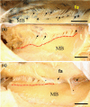
FIGURE 1
FIGURE 1
View of the fascia and some of the mylohyoid muscle (MM) attachments on the inner surface of the human mandibular body. The origin of the muscle fibers is shown by peeling the fascia (fa) off the MM from the mylohyoid line on the inner surface of the mandibular body (MB) (red dotted lines) (b,c). The site (red dotted lines) between the muscle fiber and thin serous membrane (*) is weakly attached at the mylohyoid line (arrows). (a) Attachment site of the MM; (b) Long site of attachment for the MM; (c) Short site of attachment for the MM; Ba = 200 mm; (a,b) dentulous samples; (c) edentulous samples. Bar = 1 cm
FIGURE 2
FIGURE 2
The attachment of the fascia covering the mylohyoid muscle (MM) in dentulous (Dent) and edentulous (Edn) samples. On the lingual side at the base of the mandible, the fascia of the MM appears as a wide and very thin sheet that covers the MM. The border of the MM origin is clearly different from the origin of the muscle, since that looks like a border line angled obliquely downward from the posterior region to the anterior regions (a). After peeling off the fascia of the MM from its origin along the mylohyoid line on the inner surface in the mandibular body, very fine and thin fascia‐composed fibers were found on the inner side of the mandible (b). The site between the muscle fiber and the thin serous membrane has a weak attachment at the mylohyoid line (arrows). Then, the periosteum (Ps) of the mandible body was removed from the mylohyoid line, and the small fine fibers were located between the periosteum and MM (c). On the lingual side at the base of the mandible, the border between the MM and the periosteum of the mandible body was almost indistinguishable at the site of the mylohyoid line (d). After peeling off the fascia of the MM from its origin along the mylohyoid line on the inner surface of the mandibular body, the periosteum of the mandible body was collected at this site, which is clearly not the fascia of the MM (e). Then, the periosteum of the mandible body was removed from the mylohyoid line, and the periosteum with thick connective tissue was collected between the periosteum and the MM (f). Red dotted line, origin of the MM; Bar = 1 cm. AR, alveolar ridge; fa, Fascia; MB, mandible
FIGURE 3
FIGURE 3
Schematic view of the mylohyoid muscle (MM) and the supply nerve branches on the outer surface of the MM. The incisor region (CIR), premolar region (PMR), and molar region (MR) were divided into three sections (lines): the branch of the mandibular nerve (MN) division points, such as the ascending (AB) and descending branch (GB) of the molar region; the ascending (BB) and descending branch (BH) of the premolar region; and the ascending (CB) and descending branch (IB) of the anterior canine and incisor regions (dotted line). We identified the digastric branch division points, such as the anterior branch (ANB), intermediate branch (INB), and posterior branch (PB) (lines)
FIGURE 4
FIGURE 4
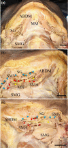
Microscopic features' view on the outer side of the mandibular floor with the submental nerve and artery and vein. (a) The submental nerve supplies the MM and runs along the submental artery and vein between the MM and the submandibular gland. (b) Right side and (c) left side, small fine MNs projected to the lateral surface of the MM. The submental nerves (yellow arrowheads, yellow dotted lines) ran along the submental artery (red arrowheads) and vein (blue arrowheads) between the MM and the submandibular gland. Some fine MNs run through a large slit between the muscle bundles of the MM and are connected to a branch of the LN. These fine MNs then projected to the lateral surface of the tongue. ABDM, anterior belly digastric muscle; MM, mylohyoid muscle; MN, mylohyoid nerve; SG, submandibular lymphatic ganglion; SMA, submental artery; SMG, submandibular gland; SMV, submental vein. Bar = 1 cm
FIGURE 5
FIGURE 5
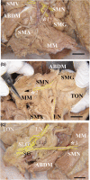
The communication between the MM and LN. The mandibular nerve (MN) is located at the most medial side of the submental vein (SMV) and artery (SMA) in the submandibular triangle. There were few MN branches except for the anterior region of the MM in this case (yellow dotted lines, *1–3). (a) Lateral view of the MM and anterior belly of the digastric muscle, (b) The inner side of the MM of (a) forming a communication pathway between the MM and LN (*2), (c) In the inner side of the MM, forming a clear communication pathway between the SMN and LN (*3). ABDM, anterior belly of the digastric anterior muscle; FA, facial artery; LN, lingual nerve; MM, mylohyoid muscle; SLG, sublingual gland; SMG, submandibular gland; SMN, submental nerve; TON, tongue. Bar = 1 cm
FIGURE 6
FIGURE 6
The submental nerve and submental vessels showing different innervations between the right and left sides in the same sample. The mandibular nerve (MN) is located at the most medial side of the submental vein (SMV) and artery (SMA) in the submandibular triangle. The MN branch projects to the MM. In the right and left views of the lateral side of the mandible, on the right side of the mandible (a,b), numerous branches of the MN (arrows) run along and innervate the MM in contrast to that of the left side of the MM (c,d). (a), Schematic of (b); (b) Lateral view of the right side of the MM and anterior belly of the digastric muscle. (c) Schematic of (d); (d) Lateral view of the left side of the MM and anterior belly of the digastric muscle. ABDM, anterior belly of the digastric anterior muscle; FA, facial artery; LN, lingual nerve; MM, mylohyoid muscle; SLG, sublingual gland; SMG, submandibular gland; TON, tongue. Bar = 1 cm
FIGURE 7
FIGURE 7
Microscopic features in the nonsublingual artery with a lateral view of the submental nerve, artery, and vein at the base of the mandible. In the lateral view of the basal region of the mandible, the submental nerve and artery and vein were located between the MM and anterior belly of the digastric muscle and the submandibular gland. The LN projected to the submandibular gland at the lateral surface of the MM. The main trunk of the submental vein and artery were clearly located in their basal regions near the hyoid bone (a,b). The MN was found in the upper region of the submandibular gland and extended many fine branches (arrowheads) to the MM. Various branches of the submental artery were mainly found in the posterior, medial, and anterior regions of the MM (a,b). On the medial side of the MM, the SMA branch (red arrowhead) extended to the lateral tongue through the MM. The LN (yellow arrowhead) was divided into branches in the lateral anterior region of the tongue (c,d). ABDM, anterior belly of the digastric muscle; APA, ascending pharyngeal artery; FA, facial artery; MM, mylohyoid muscle; MN, mylohyoid nerve; SD, sublingual duct; SG, submandibular lymphatic ganglion; SMA, submental artery; SMG, submandibular gland; SMV, submental vein. Bar = 1 cm
FIGURE 8
FIGURE 8
The dendrogram of variable factors obtained in all individual samples from the measured data, age, and sex by cluster analysis. The quality of the alignment of the three trees can be measured in this study (clusters 1–3; dotted line). The cluster 1 group was comprised of five elements (PB, IB, AMB, GB, and Com). The cluster 2 group was also comprised of five elements (INB, BH, AB, CB, and BB). The cluster 3 group was composed of three elements (LRD, age, and sex). MN division points such as the ascending (AB) and descending branch (GB) of the molar region, ascending (BB) and descending branch (BH) of the premolar region, and ascending (CB) and descending branch (IB) of the anterior canine and incisor regions are shown. We identified the branches of the digastric branch division points such as the anterior branch (ANB), intermediate branch (INB), and posterior branch (PB) in the incisor region, premolar region, and molar region. The quality of the alignment of the three trees can be measured in this study. Com, communication between the mandibular nerve and lingual nerve; LRD, the difference between the right and left
FIGURE 9
FIGURE 9
Variable factor map obtained in all individual samples from the measured data, age, and sex by principal component analysis (PCA). MN division points such as the ascending (AB) and descending branch (GB) of the molar region, ascending (BB) and descending branch (BH) of the premolar region, and ascending (CB) and descending branch (IB) of the anterior canine and incisor regions are shown. We identified the branches of the digastric branch division points such as the anterior branch (ANB), intermediate branch (INB), and posterior branch (PB) in the incisor region, premolar region and molar region. Com, communication between the mandibular nerve and lingual nerve; LRD, the difference between the right and left
FIGURE 10
FIGURE 10
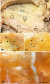
View of the reflection of the fascia of the mylohyoid muscle (MM) on the inner surface of the human mandibular body showing CGRP‐positive immunohistochemical staining in a whole mount sample. (a) The origin of the muscle fibers was seen by peeling off the fascia of the MM from the mylohyoid line of the inner surface of the mandibular body (MB). The sites (squares, b–d) are shown in the CGRP reaction areas in (a). The CGRP‐positive sites are found on some fine nerves of the fascia of the MM. The CGRP‐positive sites (arrows) were also mainly located in the large and small vessels (b–d). (b) In the posterior surface region of the mandibular body, small clusters of positive reactions (arrows) were concentrated in the middle zone of the fascia of the MM. (c) In the middle surface region of the mandibular body, small fine fibers that were positive for CGRP (arrows) were found on the fascia of the MM. (d) In the anterior surface region of the mandibular body, small fine fibers that were positive for CGRP (arrows) were found on the fascia of the MM. Ant, anterior region; ILS, inner lingual surface; Post, posterior region. Bar = 1 mm
FIGURE 11
FIGURE 11
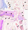
The localization of CGRP in immunohistochemically stained sections of the inferior lingual mucous membrane of the mylohyoid muscle (MM). Strongly labeled CGRP‐positive cells (arrows) were found in the mucosal lamina propria beneath the basal layer of the mucosal epithelium of the anterior and posterior regions of the mucosa of the MM (a,b). Fine positive fibers (arrows) were observed around the blood vessels in the anterior region of the mucosa of the MM (c). Small positive cells (arrows) were identified around mandibular glands and muscle fibers and in the fiber bundle of the nerves of the posterior region of the mucosa of the MM (d–f). (a) Anterior region of the mucosal epithelium, bar = 40 μm; (b) posterior region of the mucosal epithelium, the red dotted line indicates the border between the epithelial mucosa layer (EM) and the mucosal lamina propria layer (MLP), bar = 50 μm; (c) anterior region of the mucosal lamina propria, bar = 50 μm; (d) posterior region of the muscle fibers beneath the mucosal lamina propria, bar = 100 μm; (e) posterior region of the submandibular glands beneath the mucosal lamina propria, bar = 100 μm; (f) posterior region of the nerve bundles beneath the mucosal lamina propria, bar = 50 μm

Similar articles

Cited by

References

    1. Alhassani, A. A. , & AlGhamdi, A. S. (2010). Inferior alveolar nerve injury in implant dentistry: Diagnosis, causes, prevention, and management. Journal of Oral Implantology, 36, 401–407. 10.1563/AAID-JOI-D-09-00059 - DOI - PubMed
    1. Ambalavanar, R. , Dessem, D. , Moutanni, A. , Yallampalli, C. , Yallampalli, U. , Gangula, P. , & Bai, G. (2006). Muscle injury in rats induces upregulation of inflammatory cytokines in injured muscle and calcitonin gene‐related peptide in dorsal root ganglia innervating the injured muscle. Neuroscience, 143, 875–884. - PubMed
    1. Aoki, C. , Nara, T. , & Kageyama, I. (2011). Relative position of the mylohyoid line on dentulous and edentulous mandibles. Okajimas Folia Anatomica Japonica, 87, 71–76. - PubMed
    1. Azuma, Y. , Miwa, Y. , & Sato, I. (2016). Expression of CGRP in embryonic mouse masseter muscle. Annals of Anatomy, 206, 34–47. - PubMed
    1. Bell, D. , & McDermott, B. J. (1996). Calcitonin gene‐related peptide in the cardiovascular system: Characterization of receptor populations and their (patho)physiological significance. Pharmacological Reviews, 48, 253–288. - PubMed

Publication types

Substances