Review
. 2021;22(2):148-157.
doi: 10.2174/1389203721999201123200439.

Characterization of Phosphorylated Proteins Using Mass Spectrometry

Affiliations
Review

Characterization of Phosphorylated Proteins Using Mass Spectrometry

Li-Rong Yu et al. Curr Protein Pept Sci. 2021.

Abstract

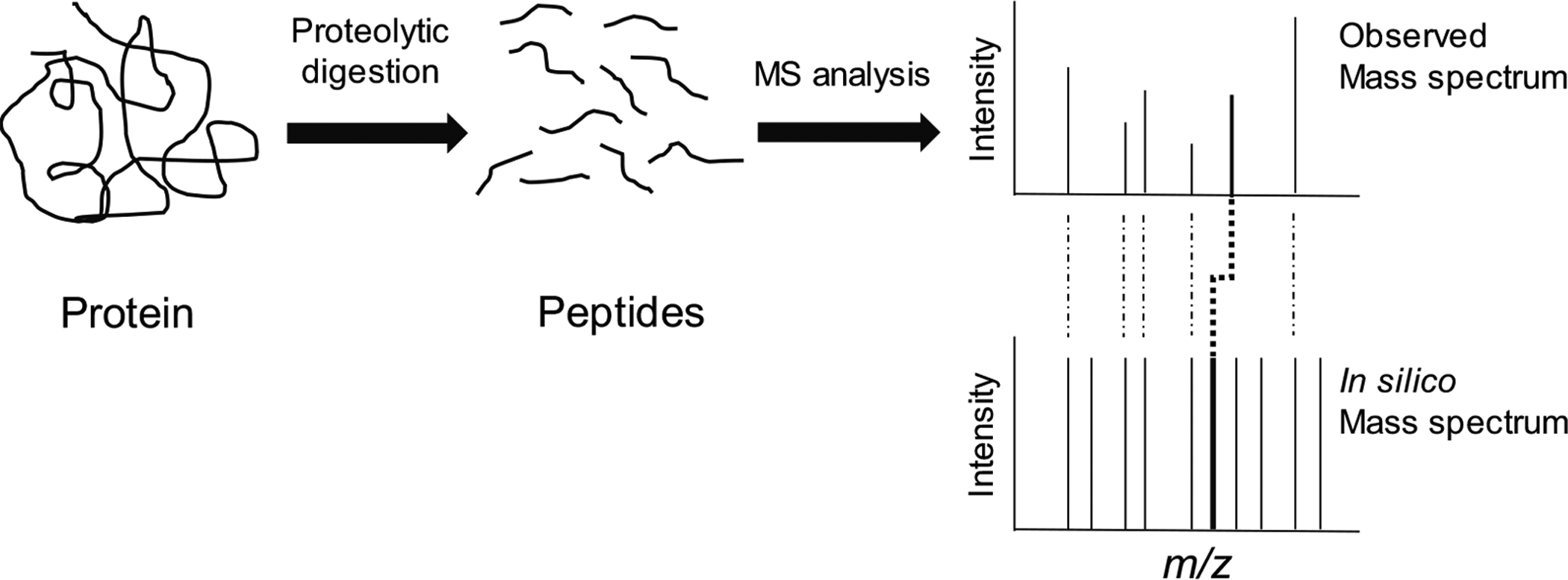
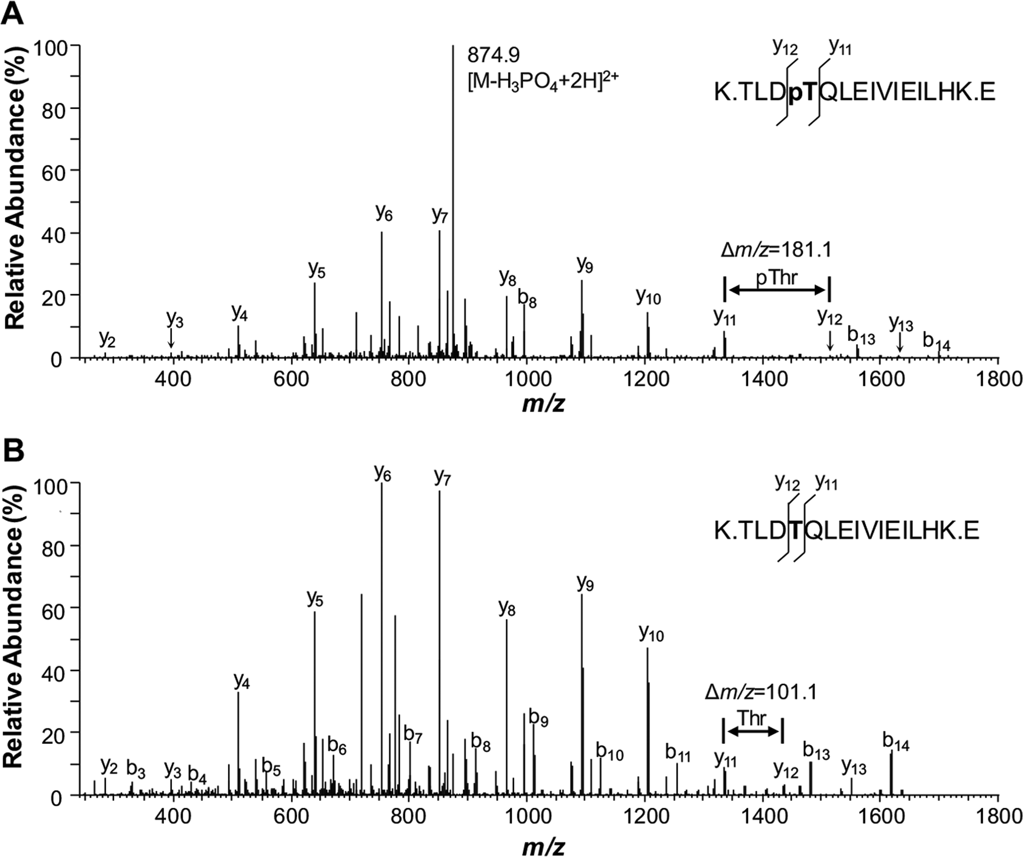
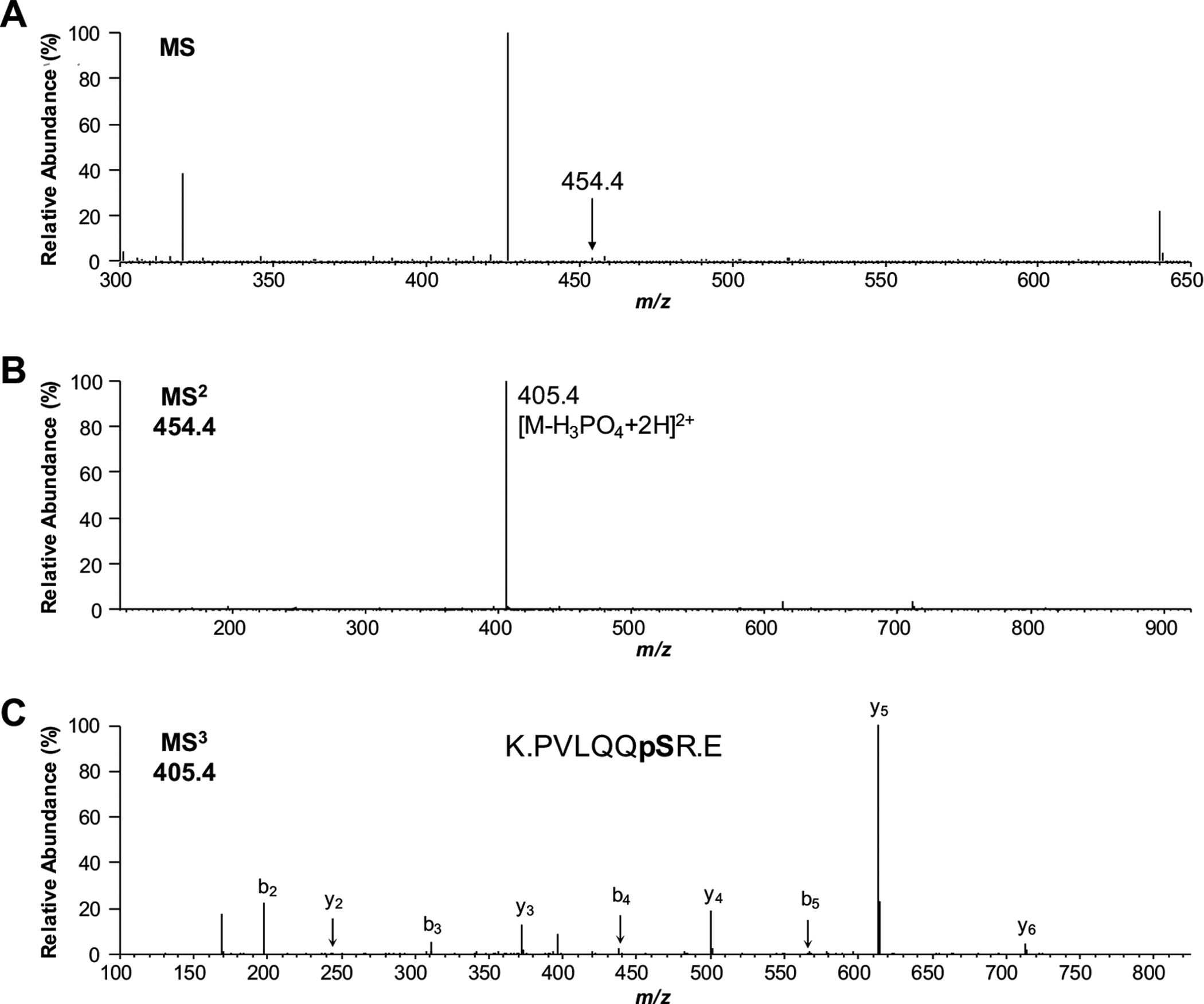
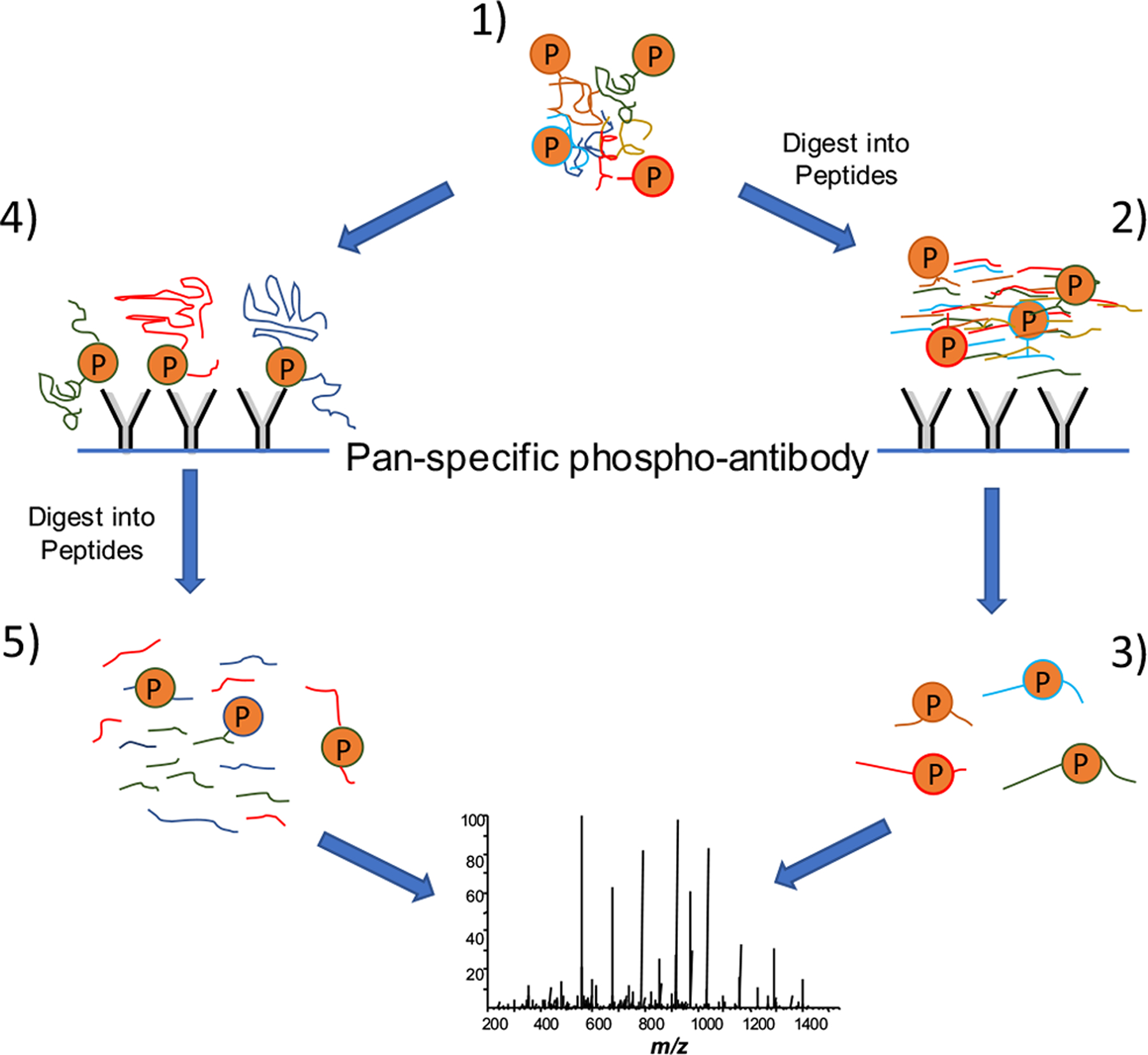
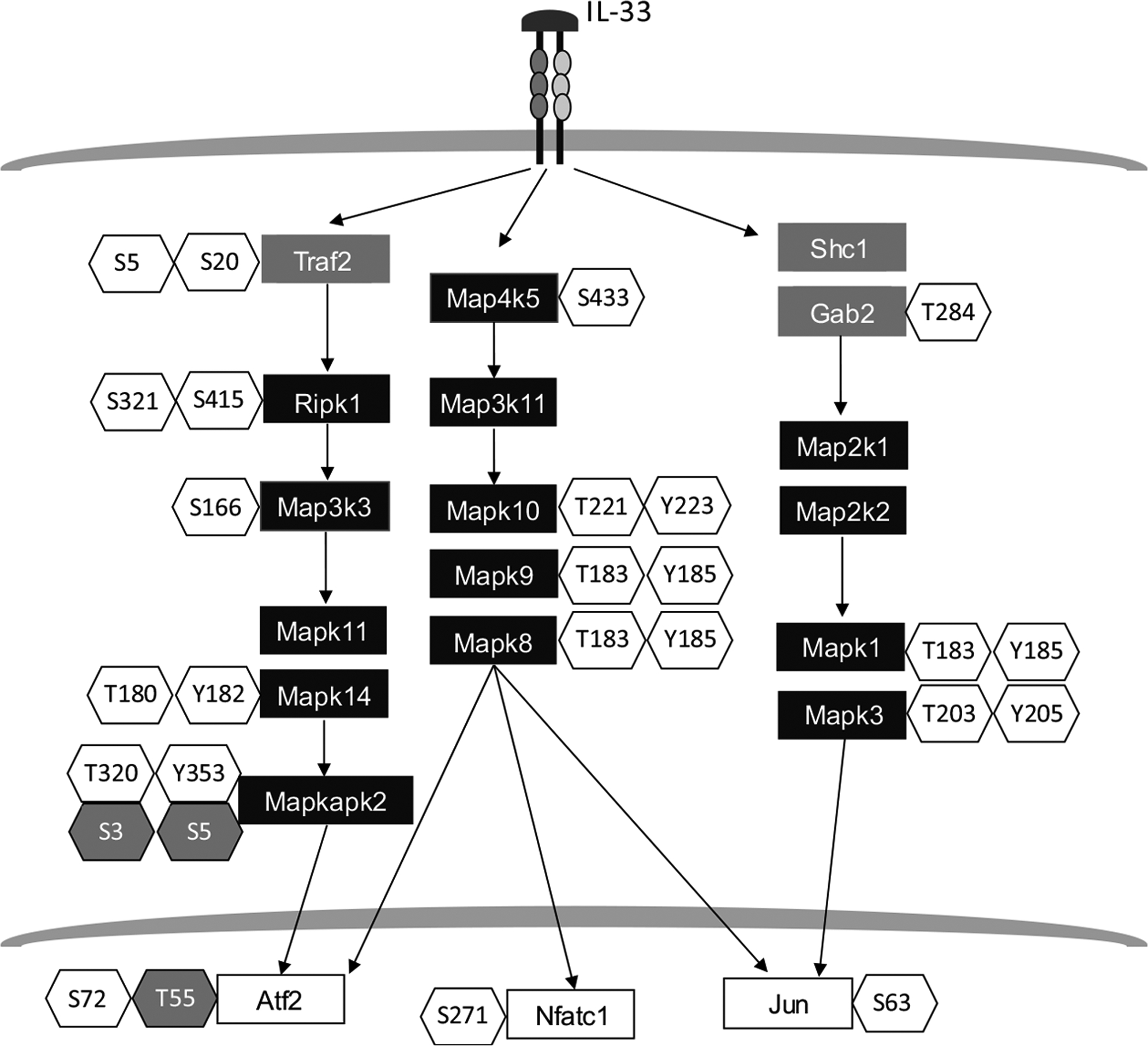
Phosphorylation is arguably the most important post-translational modification that occurs within proteins. Phosphorylation is used as a signal to control numerous physiological activities ranging from gene expression to metabolism. Identifying phosphorylation sites within proteins was historically a challenge as it required either radioisotope labeling or the use of phospho-specific antibodies. The advent of mass spectrometry (MS) has had a major impact on the ability to qualitatively and quantitatively characterize phosphorylated proteins. In this article, we describe MS methods for characterizing phosphorylation sites within individual proteins as well as entire proteome samples. The utility of these methods is illustrated in examples that show the information that can be gained using these MS techniques.

Keywords: Phosphorylation; immobilized metal affinity chromatography (IMAC); mass spectrometry; metal oxide affinity chromatography (MOAC); peptide mapping; phosphoproteomics; tandem mass spectrometry (MS2).


Conflict of interest statement

CONFLICT OF INTEREST

Declared none.

Figures

Fig. (1).
Fig. (1).
Identification of phosphorylated peptides using peptide mapping. In a peptide mapping experiment, a protein is proteolytically digested into peptides. The mass-to-charge (m/z) ratios of the peptides are recorded using mass spectrometry (MS). The observed m/z ratios are compared to those that arise from an in silico digest of proteins within a database with the same protease used to conduct the physical experiment. The protein is identified through the correlation between m/z ratios of unmodified peptides and their in silico ratios. Phosphorylated peptides are identified by virtue of their experimental m/z ratios being 80 (for singly charged peptide ions) higher than their predicted in silico ratios. Peptide mapping is most effective for identifying phosphorylated proteins in simple mixtures containing only a few (i.e., <5–10) proteins.
Fig. (2).
Fig. (2).
Phosphopeptide identification via tandem mass spectrometry (MS2). A comparison of the MS2 spectra of the phosphopeptide (A) with its unphosphorylated counterpart (B) shows excellent correspondence between the position of majority of fragment ions. The difference between the y11 and y12 ions in the phosphorylated peptide is 80 Da more than that for the non-phosphorylated version, indicating an addition of a phosphate group. Therefore, the MS2 spectrum not only indicates the peptide is phosphorylated, but also is able to pinpoint the location of the phosphorylation site.
Fig. (3).
Fig. (3).
Use of MS3 for phosphorylated peptide identification. (A) The peak at m/z 454.4 was automatically selected for MS2 from a MS spectrum showing several peptide signals. (B) The resulting MS2 spectrum showed a single dominant signal at m/z 405.4, suggesting that the peptide was phosphorylated; however, insufficient data was available to identify the peptide sequence. (C) Selection and fragmentation of this ion, however, was able to provide enough data to identify the peptide sequence and the exact site of phosphorylation.
Fig. (4).
Fig. (4).
Strategies for extraction and identification of phosphorylated peptides using immunoprecipitation. The extracted proteome sample (1) can be immediately digested into peptides, and phosphopeptides are extracted from this mixture using immunoprecipitation (2). The resultant mixture is highly enriched with phosphopeptides (3), from which a high percentage of the mass spectrometry (MS) identifications are phosphorylated peptides. Using an alternative strategy, intact phosphoproteins are extracted from the proteome sample (4) and then digested into peptides (5). The result is a mixture of peptides in which most of the species are unphosphorylated. As a result, only a small percentage of the MS identifications are phosphorylated.
Fig. (5).
Fig. (5).
Partial view of phosphoproteins identified in murine macrophage cells treated with interleukin-33 (IL-33). A total of 7,191 phosphorylated sites originated from 2,746 proteins were identified in IL-33 treated murine macrophages [39]. This figure shows a selection of kinases (black-filled rectangles), transcription factors (white-filled rectangles), and signaling molecules (grey-filled rectangles). Phosphorylated residues that were observed to be up-regulated are shown in unshaded hexagons, while those that were observed to be down-regulated are shown in gray-shaded hexagons.

References

    1. Wang Y; Wang L; Gong Z Regulation of acetylation in high mobility group protein B1 cytosol translocation. DNA Cell Biol, 2019, 38(5), 491–499. 10.1089/dna.2018.4592 - DOI - PubMed
    1. Mestermann K; Giavridis T; Weber J; Rydzek J; Frenz S; Nerreter T; Mades A; Sadelain M; Einsele H; Hudecek M The tyrosine kinase inhibitor dasatinib acts as a pharmacologic on/off switch for CAR T cells. Sci. Transl. Med, 2019, 11(499), 1–11. 10.1126/scitranslmed.aau5907 - DOI - PMC - PubMed
    1. Belman JP; Habtemichael EN; Bogan JS A proteolytic pathway that controls glucose uptake in fat and muscle. Rev. Endocr. Metab. Disord, 2014, 15(1), 55–66. 10.1007/s11154-013-9276-2 - DOI - PMC - PubMed
    1. Salzberg SL Open questions: How many genes do we have? BMC Biol., 2018, 16(1), 94. 10.1186/s12915-018-0564-x - DOI - PMC - PubMed
    1. Available from: nextprot.org/about/protein-existence

MeSH terms