

Visual Function in Pentosan Polysulfate Sodium Maculopathy
- PMID: 33231621
- PMCID: PMC7691795
- DOI: 10.1167/iovs.61.13.33
Visual Function in Pentosan Polysulfate Sodium Maculopathy
Abstract
Purpose: Individuals with pentosan polysulfate sodium (PPS) maculopathy commonly report symptoms of prolonged dark adaptation and difficulty reading. We hypothesize that PPS maculopathy causes degradation of visual function not fully captured with visual acuity testing.
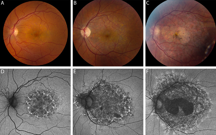
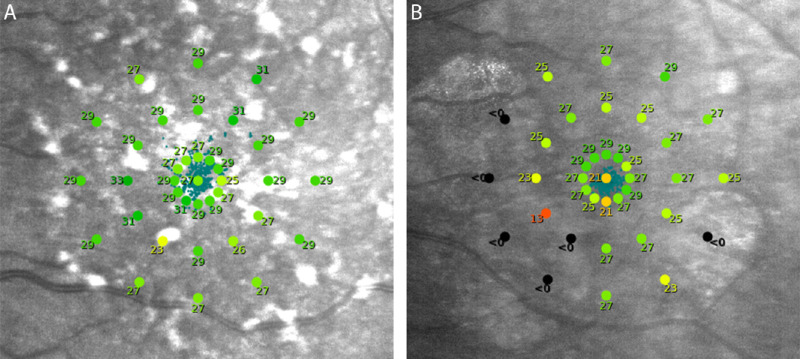
Methods: Subjects with PPS maculopathy underwent multimodal evaluation of retinal structure and function. Structural changes were graded as moderate or advanced. Patient-reported visual function was assessed with the National Eye Institute Visual Function Questionnaire 39 (NEI-VFQ-39) and Low Luminance Questionnaire (LLQ). Objective functional evaluations included Early Treatment of Diabetic Retinopathy Study (ETDRS) best-corrected visual acuity (BCVA), Pelli-Robson contrast sensitivity, mesopic microperimetry, and dark adaptometry. Functional testing results were correlated with structural disease category.
Results: Thirteen patients (26 eyes), median age 62 years (range, 37-76), completed the study. Median ETDRS letter score was 82 (Snellen equivalent 20/25). Median NEI-VFQ-39 and LLQ composite scores were 65 (range, 33-88) and 41 (range, 20-92), respectively. Median contrast sensitivity was 1.65 (range, 0.15-1.95), and median mesopic microperimetry average thresholds and percent reduced thresholds were 26 decibels (range, 0.4-28.6) and 21.6% (range, 0-100%), respectively. Median rod intercept time was 14.1 minutes (range, 4.4-20.0). Eyes with advanced disease based on retinal structure had significantly worse retinal function for several testing modalities.
Conclusions: PPS maculopathy causes considerable visual function degradation that is not fully captured with BCVA testing. There was good correlation between other measures of visual function and disease severity. These findings deepen our concern regarding this patient safety issue.
Conflict of interest statement
Disclosure:
Figures
References
-
- Hanno PM, Erickson D, Moldwin R, Faraday MM. Diagnosis and treatment of interstitial cystitis/bladder pain syndrome: AUA guideline amendment. J Urol. 2015; 193(5): 1545–1553. - PubMed
-
- Giusto LL, Zahner PM, Shoskes DA. An evaluation of the pharmacotherapy for interstitial cystitis. Expert Opin Pharmacother. 2018; 19(10): 1097–1108. - PubMed
-
- Pearce WA, Chen R, Jain N. Pigmentary maculopathy associated with chronic exposure to pentosan polysulfate sodium. Ophthalmology. 2018; 125(11): 1793–1802. - PubMed
-
- Hanif A, Shah R, Yan J, et al. .. Strength of association between pentosan polysulfate and a novel maculopathy. Ophthalmology 2019; 126(10): 1464–1466. - PubMed
Publication types
MeSH terms
Substances
LinkOut - more resources
Full Text Sources
Medical
