. 2020 Dec 2;48(21):12252-12268.
doi: 10.1093/nar/gkaa1069.

The expression of Rpb10, a small subunit common to RNA polymerases, is modulated by the R3H domain-containing Rbs1 protein and the Upf1 helicase

Affiliations

The expression of Rpb10, a small subunit common to RNA polymerases, is modulated by the R3H domain-containing Rbs1 protein and the Upf1 helicase

Małgorzata Cieśla et al. Nucleic Acids Res. .

Abstract

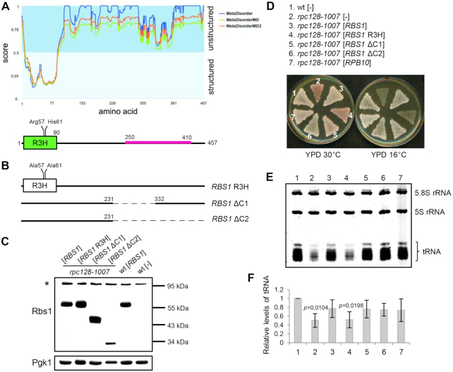
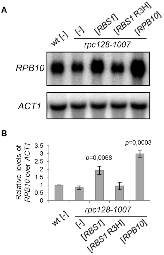
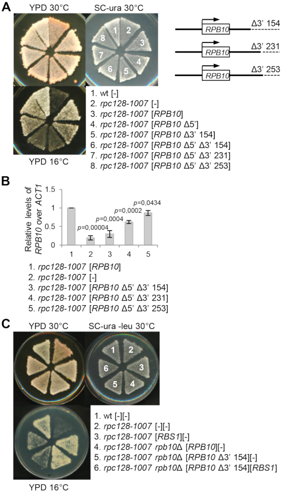
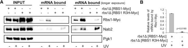
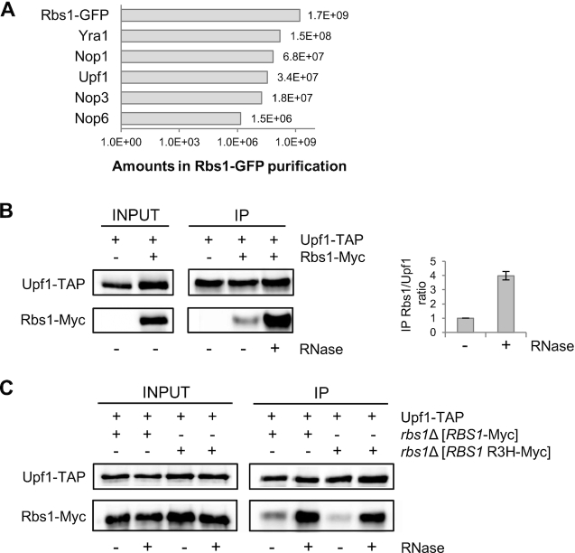
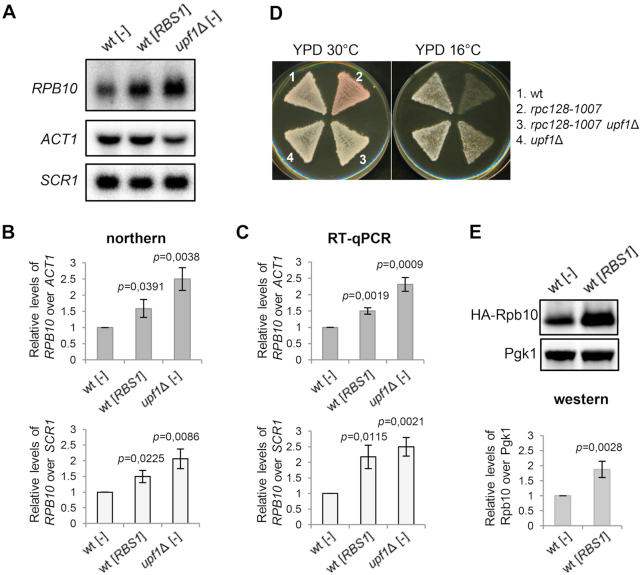
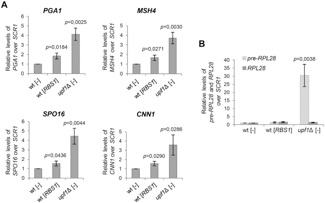
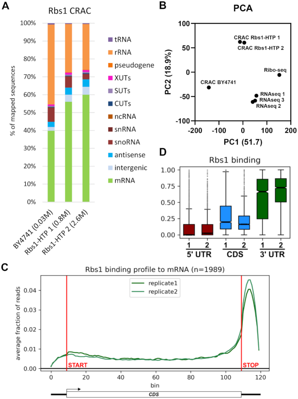
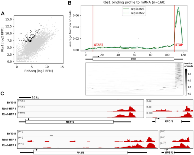
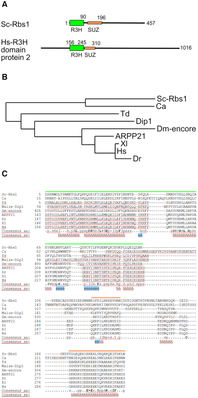
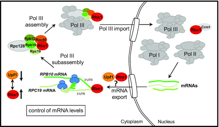
The biogenesis of eukaryotic RNA polymerases is poorly understood. The present study used a combination of genetic and molecular approaches to explore the assembly of RNA polymerase III (Pol III) in yeast. We identified a regulatory link between Rbs1, a Pol III assembly factor, and Rpb10, a small subunit that is common to three RNA polymerases. Overexpression of Rbs1 increased the abundance of both RPB10 mRNA and the Rpb10 protein, which correlated with suppression of Pol III assembly defects. Rbs1 is a poly(A)mRNA-binding protein and mutational analysis identified R3H domain to be required for mRNA interactions and genetic enhancement of Pol III biogenesis. Rbs1 also binds to Upf1 protein, a key component in nonsense-mediated mRNA decay (NMD) and levels of RPB10 mRNA were increased in a upf1Δ strain. Genome-wide RNA binding by Rbs1 was characterized by UV cross-linking based approach. We demonstrated that Rbs1 directly binds to the 3' untranslated regions (3'UTRs) of many mRNAs including transcripts encoding Pol III subunits, Rpb10 and Rpc19. We propose that Rbs1 functions by opposing mRNA degradation, at least in part mediated by NMD pathway. Orthologues of Rbs1 protein are present in other eukaryotes, including humans, suggesting that this is a conserved regulatory mechanism.


Figures

Figure 1.
Figure 1.
The R3H domain is essential for the function of Rbs1 protein in Pol III assembly. (A) Predicted disorder of Rbs1 protein according to Metadisorder server (30). Localization of the R3H domain (green) and the prionogenic sequence (pink) is shown below. (B) Schematic presentation of modified versions of RBS1 that were constructed on corresponding plasmids: [RBS1 R3H], [RBS1 ΔC1] and [RBS1 ΔC2]. See the Materials and Methods section for details. (C-F) Examination of transformants of control strain (wild type [wt]) and rpc128-1007 mutant with plasmids that encoded modified versions of Rbs1 protein, [RBS1] and [RPB10] control plasmids, and the empty vector [–]. (C) The modified versions of Rbs1 protein were efficiently expressed. Yeast cells were analyzed by western blot. Antibody specific for Rbs1 detects only overproduced protein. Determination of Pgk1 levels served as loading control. (D) Inactivation of the R3H domain prevented genetic suppression of the Pol III assembly mutant. Cells that were grown on an SC-ura plate were replicated on YPD plates and incubated for 3 days at the respective temperatures. (E) Inactivation of the R3H domain prevented the correction of low tRNA levels in the Pol III assembly mutant. Small RNA species were separated on a 7 M urea–6% polyacrylamide gel using equal amounts of RNA per lane (5 μg) and stained with ethidium bromide. (F) Bands corresponding to total tRNAs were quantified. Bars represent tRNA levels normalized to 5.8S rRNA which served as loading control. Standard deviations were estimated on the basis of three independent experiments. The P value calculated for ratio of tRNAs (rpc128-1007[-]/wt[-], rpc128-1007[RBS1R3H]/wt[-] showed statistical significance (P < 0.02). P values were calculated using a two-tailed t-test.
Figure 2.
Figure 2.
Effect of RBS1 overexpression on steady-state levels of RPB10 mRNA. (A) RNA that was isolated from the control strain (wt) and transformants of the rpc128-1007 mutant with the [RBS1 R3H] plasmid that encoded the mutated Rbs1 R3H protein, [RBS1] and [RPB10] control plasmids, and the empty vector [–] was analyzed by northern blot using probes that were specific to RPB10 mRNA and ACT1 mRNA that encodes actin (loading control). (B) The levels of RPB10 mRNA were normalized to the loading control and calculated relative to levels in the wt strain, which was set to 1. Bars represent the mean ± standard deviation of three independent experiments. P values were calculated using a two-tailed t-test.
Figure 3.
Figure 3.
The role of Rbs1 in controlling Pol III assembly is supported by the 3′ regulatory region of the RPB10 gene. The modified versions of the RPB10 gene that lacks designated sequences in the 5′ and/or 3′ regulatory regions were constructed on the plasmids as described in Supplementary data. (A) Effect of deletions in the 3′ UTR on suppression of the rpc128-1007 Pol III assembly mutant by RPB10. Δ3′ 154, Δ3′ 231 and Δ3′ 253, respectively, limited 3′UTR to 154, 231 and 253 nucleotides downstream a stop codon. The control strain (wt) and transformants of the rpc128-1007 mutant with derivatives of [RPB10] containing designated deletions, the [RPB10] control plasmid, and the empty vector [–] were grown on SC-ura plates, replicated on YPD plates, and incubated for 3 days at the respective temperatures. (B) RNA isolated from transformants of the rpc128-1007 mutant with derivatives of [RPB10] containing designated deletions and the [RPB10] control plasmid was used to synthesis of cDNA samples that were analyzed by RT-qPCR. mRNA levels were normalized to ACT1 mRNA and calculated relative to amounts in the strain harboring the [RPB10] control plasmid, which was set to 1. Bars represent the mean ± standard deviation of three independent experiments. P values were calculated using a two-tailed t-test. (C) Δ3′154 deletion in 3′ UTR of RPB10 negatively affected suppression of the Pol III assembly mutant rpc128-1007 by RBS1. A double rpb10Δ rpc128-1007 mutant that harbored the [RPB10 Δ3′154] plasmid was additionally transformed with the [RBS1] plasmid or empty vector [–]. A double rpb10Δ rpc128-1007 mutant that harbored [RPB10], a single rpc128-1007 mutant that harbored [RBS1], and the wild type strain (wt) were additionally transformed with the respective empty vectors and served as controls. Yeast cells that were grown on an SC-ura-leu plate were replicated on YPD plates and incubated for 3 days at the respective temperatures.
Figure 4.
Figure 4.
The binding of Rbs1 with poly(A) mRNA in living cells requires the R3H domain. (A) Poly(A) mRNA was isolated from cells that expressed Myc-tagged Rbs1 or Rbs1 R3H without or after RNA-protein cross-linking by UV irradiation. Input and poly(A) mRNA-bound fractions were analyzed by western blot with antibodies that were specific to Myc, Nab2 (positive control) and Pgk1 (loading control). mRNA bound proteins were visualized by shorter and longer gel exposure. Band intensities from western blot images were quantified using MultiGauge 3.0 software (Fujifilm). (B) The relative amount of mRNA bound in Rbs1-Myc was set to 1. Bars represent the mean ± standard deviation of three independent experiments. P values were calculated using a two-tailed t-test.
Figure 5.
Figure 5.
Rbs1 interacts with Upf1 helicase. (A) Upf1 is an RNA-interacting protein that was co-purified with Rbs1. The affinity purification of green fluorescent protein-tagged Rbs1 was followed by quantitative mass spectrometry. The relative amounts of co-purified proteins were determined by MaxQuant software, expressed as arbitrary units (11). (B, C) Upf1 and Rbs1 interaction, determined by co-immunoprecipitation. RNase treatment enhanced the Rbs1-Upf1 interaction. Total cell extracts (INPUT) were isolated from (B) a strain that expressed endogenous Myc-tagged Rbs1 and TAP-tagged Upf1 and from a control strain that expressed only Upf1-TAP or (C) the rbs1Δ strain that expressed TAP-tagged Upf1 and overexpressed Myc-tagged Rbs1 or Rbs1 R3H, encoded by multicopy plasmids. Extracts were subjected to immunoprecipitation using IgG-coated magnetic beads. This protocol was based on affinity of the protein A-containing TAP tag to IgG. Immunoprecipitated proteins were eluted and analyzed by western blot using peroxidase anti-peroxidase (PAP) and anti-Myc antibodies. RNase treatment of the extracts and beads is designated at the bottom of the western blot images. Band intensities were quantified using MultiGauge 3.0 software (Fujifilm). The ratio of immunoprecipitated Rbs1-Myc to Upf1-TAP from probes that were or were not treated with RNase was calculated. The ratio in the probe that was not treated with RNase was set to 1. Bars represent the mean ± standard deviation of two independent experiments.
Figure 6.
Figure 6.
Effects of Upf1 and Rbs1 on RPB10 expression and Pol III assembly. (A–C) Opposite effects of Rbs1 and Upf1 on RPB10 mRNA levels. RNA was isolated from the upf1Δ mutant and a control strain (wt) that carried an empty vector [–] or the [RBS1] plasmid. RNA was independently analyzed by northern blot (A, B) and RT-qPCR (C). RPB10 mRNA levels were normalized to ACT1 mRNA (upper panel) or SCR1 (lower panel RPB10 mRNA levels were calculated relative to amounts in the wt strain, which was set to 1. (D) The phenotype of the Pol III assembly mutant rpc128-1007 was suppressed by upf1Δ. Cells that were grown on YPD plates were replicated on fresh YPD plates and incubated for 3 days at the respective temperatures. (E) Protein extracts were prepared from a control strain (wt) that carried an empty vector [–] or the [RBS1] plasmid additionally transformed with a centromeric [HA-RPB10] plasmid. The band corresponding to HA-tagged Rpb10 was quantified, normalized to Pgk1 signal used as a loading control and calculated relative to amounts in the wt strain, which was set to 1. Bars represent the mean ± standard deviation of three independent experiments. P values were calculated using a two-tailed t-test (B, C and E).
Figure 7.
Figure 7.
The RT-qPCR analysis indicated effects of Rbs1 on the expression of yeast genes that are controlled by 3′-UTR NMD decay (A) but no influence of Rbs1 on the level of RPL28 pre-mRNA, a direct NMD target (B). RNAs that were isolated from the upf1Δ mutant and control strain (wt) that carried an empty vector [–] or [RBS1] plasmid were analyzed by RT-qPCR with probes that were specific to PGA1 mRNA, MSH4 mRNA, SPO16 mRNA, and CNN1 mRNA (A) or intron-containing RPL28 pre-mRNA and mature RPL28 mRNA (B). The levels of the tested mRNAs were normalized to SCR1 RNA and calculated relative to levels in the wt strain, which was set to 1. Bars represent the mean ± standard deviation of three independent experiments. P values were calculated using a two-tailed t-test.
Figure 8.
Figure 8.
Rbs1 preferentially binds 3′UTR regions in mRNAs. (A) Transcriptome-wide binding profiles for Rbs1-HTP and for the control BY4741 strain expressing untagged Rbs1. Bar diagrams illustrate the percentage of all sequences mapped to each of the RNA classes indicated on the right of the figure. (B) Principal component analysis (PCA) showing differences between Rbs1 CRAC, RNA-seq and Ribo-seq. Axis titles show the extent of variation explained by a given principal component. (C) Metagene representation of read density over mature mRNAs (n = 1989). Data were separated into 120 bins: 10 for the 5′ UTR, 100 for the CDS and 10 for the 3′ UTR. Horizontal lines indicate were CDS starts and stops. Metagene analysis performed for mRNA containing at least 100 reads in Rbs1 CRAC data (n = 1989). (D) Boxplot of two Rbs1 CRAC replicates presenting binding to mature mRNA. Centre lines of box plots show the medians; box limits indicate the 25th and 75th percentiles; whiskers extend 1.5 times the interquartile range from the 25th and 75th percentiles, data points are plotted for outliers.
Figure 9.
Figure 9.
mRNAs enriched in Rbs1 binding. (A) Scatter plot comparing the Rbs1 RNA binding to RNA-seq with marked high confidence targets of Rbs1 (black). The targets were selected using following criteria: P value <0.01, ratio between Rbs1 binding and RNA-seq >1.5 and >128 uniquely mapped RPM. (B) Upper panel: Metagene representation of read density over high confidence Rbs1 target mature mRNAs (n = 160). Data were separated as panel 9C. Bottom panel: Heatmap representation of read density over high confidence Rbs1 target mature mRNAs (n = 160). (C) Binding of Rbs1-HTP across the MET13, RPC19, NAM8 and RPB10 mRNAs. Each track presents raw number of uniquely mapped reads, with the exact value indicated in the left part of each track. A scale bar is shown at the top.
Figure 10.
Figure 10.
Evolutionary conservation of potential Rbs1 orthologues. (A) Schematic diagram of the domain composition of Sc-Rbs1 and human R3H domain-containing protein 2. Green rectangles indicate R3H domains. Orange rectangles indicate the SUZ domain. The taller rectangles for R3H indicate a more structured nature of this domain over less structured SUZ (see Figure 1A). (B) Phylogenetic tree of selected potential orthologues of Rbs1 generated with phylogeny.fr (25,26). (C) Sequence alignment of the R3H-SUZ region of selected Rbs1 homologues (output from Promals3D (24)). Green lines indicate the R3H domain. Orange lines indicate the SUZ domain. The following symbols apply, with sequence IDs in parentheses: Sc-Rbs1, Saccharomyces cerevisiae Rbs1; Ca, hypothetical protein (Candida albicans, KHC63810.1); Td, putative R3H domain protein (Taphrina deformans, CCG82637.1); Maize Dip1 (DIP1 Zea mays, AAZ73119.1); Dm-encore, encore protein (Drosophila melanogaster, NP_995992.1); ARPP21, (H. hapiens, XP_016861070), Hs, R3H domain-containing protein 2 (H. sapiens, XP_011536342.1); Dr, R3H domain-containing protein 2 (Danio rerio, XP_021333011.1); Xl, R3H domain-containing protein 2-like (Xenopus leavis, XP_018105556.1). Consensus amino acid residues in bold are conserved in all sequences: l, aliphatic; @, aromatic; h, hydrophobic; o, alcohol; p, polar residues; t, tiny; s, small; b, bulky residues; +, positively charged; –, negatively charged; c, charged. Predicted/determined secondary structure (consensus ss): h, helices; e, strands
Figure 11.
Figure 11.
Early steps in Pol III biogenesis in the yeast Saccharomyces cerevisiae that connect the control of Rpb10 expression and its role in assembly of the Pol III complex through a regulatory loop that involves Rbs1 protein. See explanation in text.

Similar articles

Cited by

References

    1. Hanske J., Sadian Y., Müller C.W.. The cryo-EM resolution revolution and transcription complexes. Curr. Opin. Struct. Biol. 2018; 52:8–15. - PMC - PubMed
    1. Werner F., Grohmann D.. Evolution of multisubunit RNA polymerases in the three domains of life. Nat. Rev. Microbiol. 2011; 9:85–98. - PubMed
    1. Abel C.-B., Verónica M.-F., Ana I G.-G., Francisco N.. Subunits common to RNA polymerases. The Yeast Role in Medical Applications. 2017; doi:10.5772/intechopen.70936.
    1. Wild T., Cramer P.. Biogenesis of multisubunit RNA polymerases. Trends Biochem. Sci. 2012; 37:99–105. - PubMed
    1. Khatter H., Vorländer M.K., Müller C.W.. RNA polymerase I and III: similar yet unique. Curr. Opin. Struct. Biol. 2017; 47:88–94. - PubMed

Publication types

MeSH terms