

Compartmentalized GPCR Signaling from Intracellular Membranes
- PMID: 33231722
- PMCID: PMC8141539
- DOI: 10.1007/s00232-020-00158-7
Compartmentalized GPCR Signaling from Intracellular Membranes
Abstract
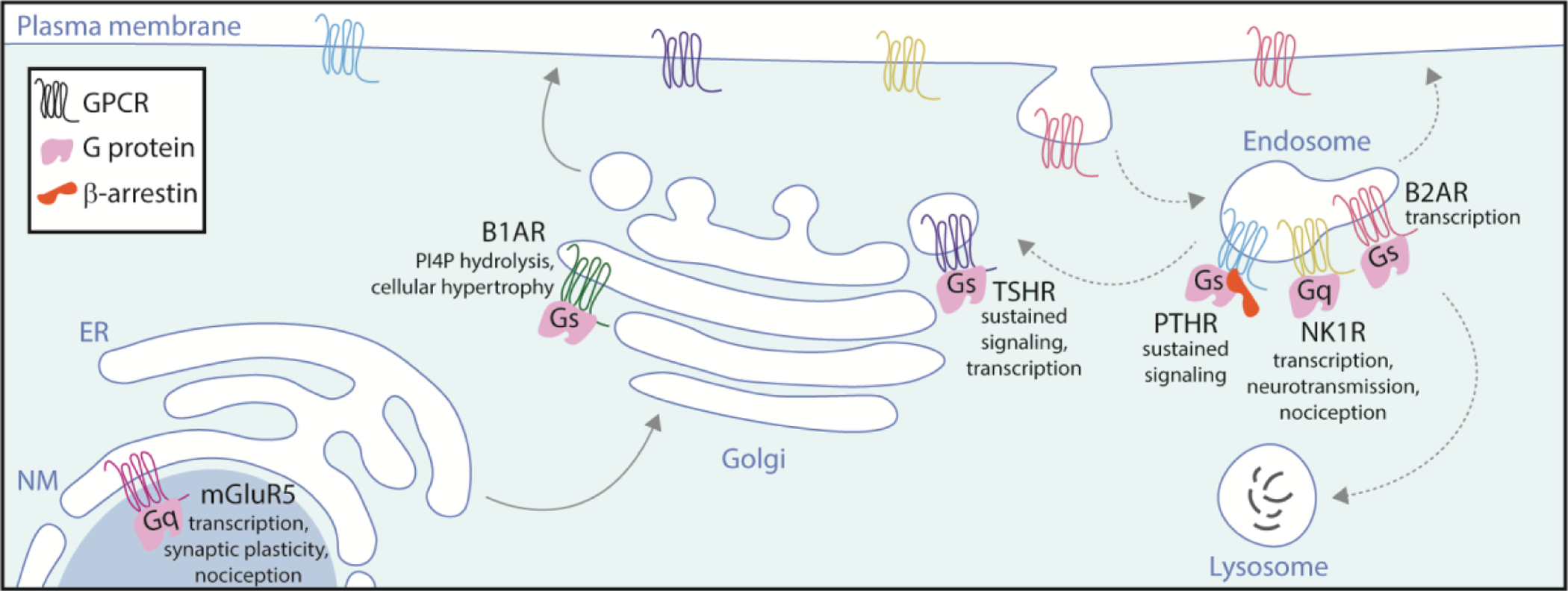
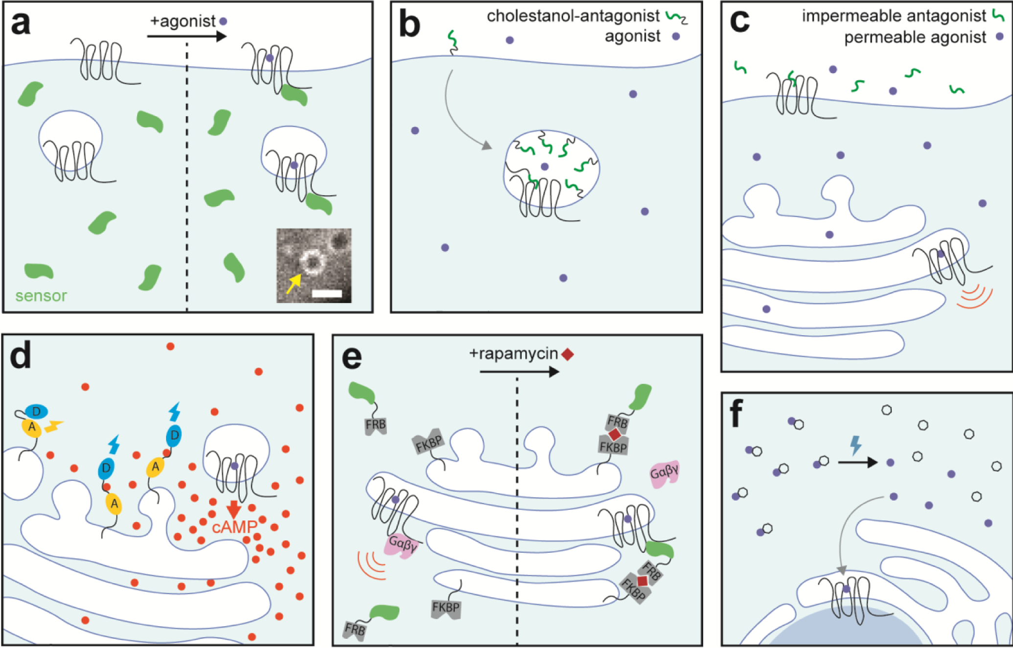
G protein-coupled receptors (GPCRs) are integral membrane proteins that transduce a wide array of inputs including light, ions, hormones, and neurotransmitters into intracellular signaling responses which underlie complex processes ranging from vision to learning and memory. Although traditionally thought to signal primarily from the cell surface, GPCRs are increasingly being recognized as capable of signaling from intracellular membrane compartments, including endosomes, the Golgi apparatus, and nuclear membranes. Remarkably, GPCR signaling from these membranes produces functional effects that are distinct from signaling from the plasma membrane, even though often the same G protein effectors and second messengers are activated. In this review, we will discuss the emerging idea of a "spatial bias" in signaling. We will present the evidence for GPCR signaling through G protein effectors from intracellular membranes, and the ways in which this signaling differs from canonical plasma membrane signaling with important implications for physiology and pharmacology. We also highlight the potential mechanisms underlying spatial bias of GPCR signaling, including how intracellular membranes and their associated lipids and proteins affect GPCR activity and signaling.
Keywords: Endosomes; GPCR; Golgi; Nuclear membrane; Signaling; Trafficking.
Conflict of interest statement
Conflicts of interest
The authors declare no conflicts of interest.
Figures
Similar articles
-
Spatial encryption of G protein-coupled receptor signaling in endosomes; Mechanisms and applications.Biochem Pharmacol. 2017 Nov 1;143:1-9. doi: 10.1016/j.bcp.2017.04.028. Epub 2017 Apr 27. Biochem Pharmacol. 2017. PMID: 28456515 Review.
-
Spatial encoding of GPCR signaling in the nervous system.Curr Opin Cell Biol. 2019 Apr;57:83-89. doi: 10.1016/j.ceb.2018.12.006. Epub 2019 Jan 29. Curr Opin Cell Biol. 2019. PMID: 30708280 Free PMC article. Review.
-
Subcellular Organization of GPCR Signaling.Trends Pharmacol Sci. 2018 Feb;39(2):200-208. doi: 10.1016/j.tips.2017.11.009. Epub 2018 Jan 28. Trends Pharmacol Sci. 2018. PMID: 29478570 Free PMC article. Review.
-
Endomembrane GPCR signaling: 15 years on, the quest continues.Trends Biochem Sci. 2025 Jan;50(1):46-60. doi: 10.1016/j.tibs.2024.10.006. Epub 2024 Nov 11. Trends Biochem Sci. 2025. PMID: 39532582 Review.
-
Advances in Membrane Trafficking and Endosomal Signaling of G Protein-Coupled Receptors.Int Rev Cell Mol Biol. 2018;339:93-131. doi: 10.1016/bs.ircmb.2018.03.001. Epub 2018 Apr 30. Int Rev Cell Mol Biol. 2018. PMID: 29776606 Review.
Cited by
-
GPR161 structure uncovers the redundant role of sterol-regulated ciliary cAMP signaling in the Hedgehog pathway.bioRxiv [Preprint]. 2023 May 24:2023.05.23.540554. doi: 10.1101/2023.05.23.540554. bioRxiv. 2023. Update in: Nat Struct Mol Biol. 2024 Apr;31(4):667-677. doi: 10.1038/s41594-024-01223-8. PMID: 37292845 Free PMC article. Updated. Preprint.
-
Ca2+ Signaling in Cardiovascular Fibroblasts.Biomolecules. 2024 Oct 27;14(11):1365. doi: 10.3390/biom14111365. Biomolecules. 2024. PMID: 39595542 Free PMC article. Review.
-
Charged Small Molecule Binding to Membranes in MD Simulations Evaluated against NMR Experiments.J Phys Chem B. 2022 Sep 15;126(36):6955-6963. doi: 10.1021/acs.jpcb.2c05024. Epub 2022 Sep 5. J Phys Chem B. 2022. PMID: 36063117 Free PMC article.
-
Location-biased activation of the proton-sensor GPR65 is uncoupled from receptor trafficking.Proc Natl Acad Sci U S A. 2023 Sep 26;120(39):e2302823120. doi: 10.1073/pnas.2302823120. Epub 2023 Sep 18. Proc Natl Acad Sci U S A. 2023. PMID: 37722051 Free PMC article.
-
Bitter Taste Receptor Agonists Induce Apoptosis in Papillary Thyroid Cancer.Head Neck. 2025 Aug;47(8):2101-2113. doi: 10.1002/hed.28120. Epub 2025 Mar 5. Head Neck. 2025. PMID: 40040415 Free PMC article.
References
-
- Allen JA, Yu JZ, Donati RJ, and Rasenick MM (2005). Beta-adrenergic receptor stimulation promotes G(alpha)s internalization through lipid rafts: A study in living cells. Mol Pharmacol 67, 1493–1504. - PubMed
Publication types
MeSH terms
Substances
Grants and funding
LinkOut - more resources
Full Text Sources
