Review
. 2021 May 12;121(9):5336-5358.
doi: 10.1021/acs.chemrev.0c00644. Epub 2020 Nov 24.

Toward a Molecular Basis of Cellular Nucleoside Transport in Humans

Affiliations
Review

Toward a Molecular Basis of Cellular Nucleoside Transport in Humans

Nicholas J Wright et al. Chem Rev. .

Abstract

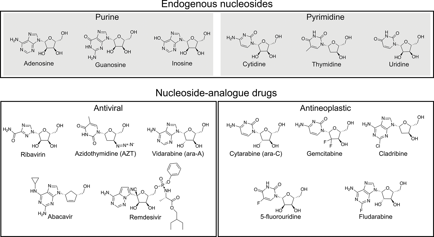
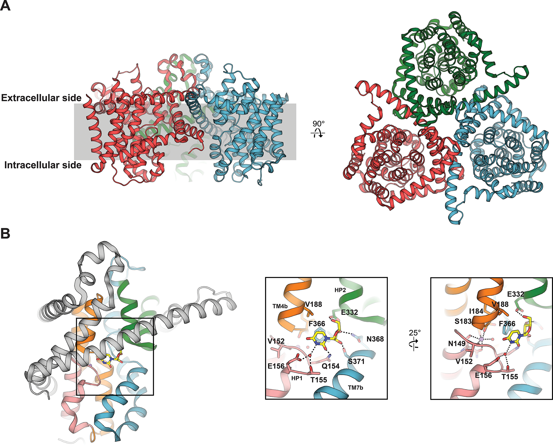
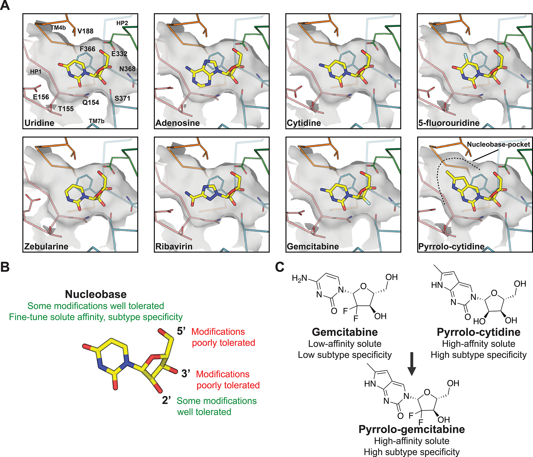
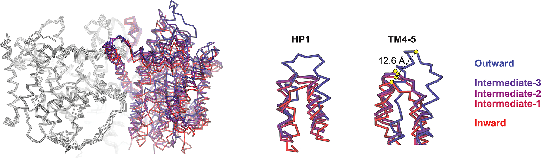
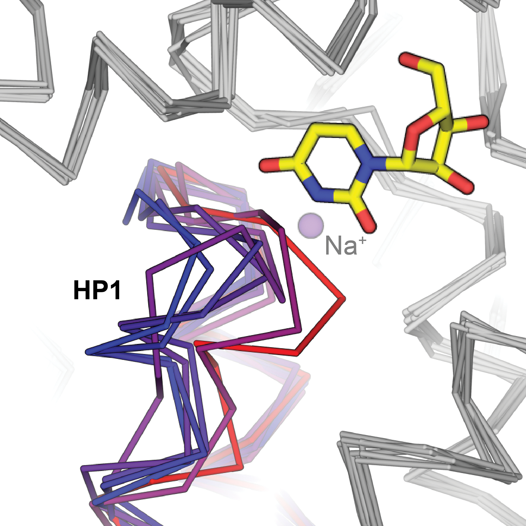
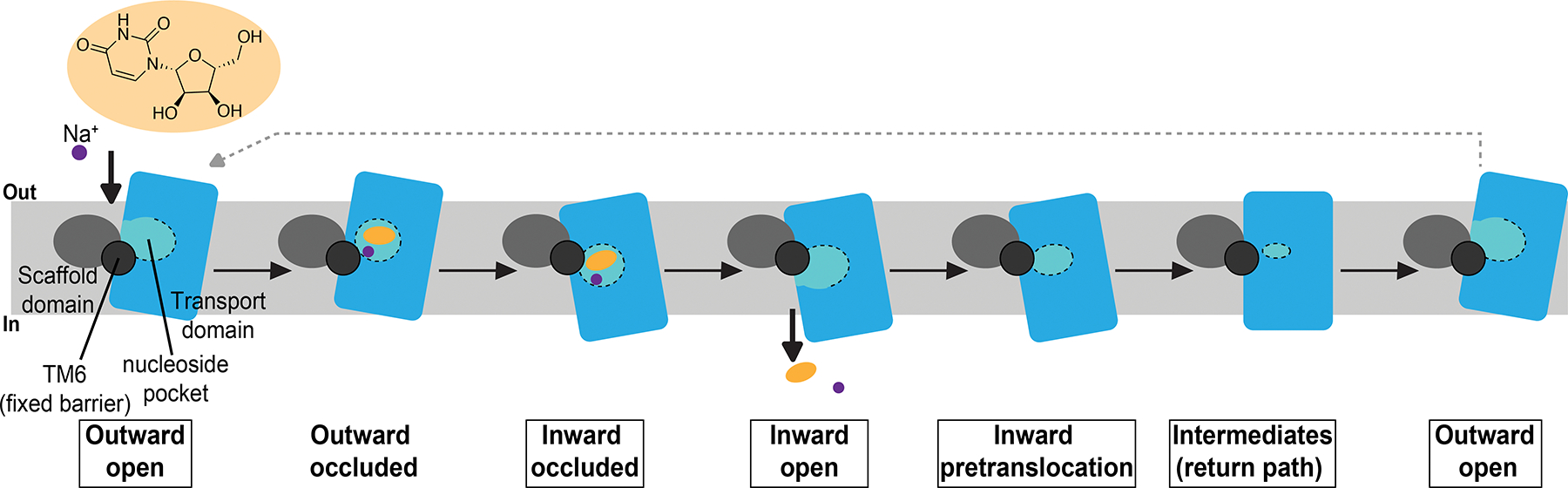
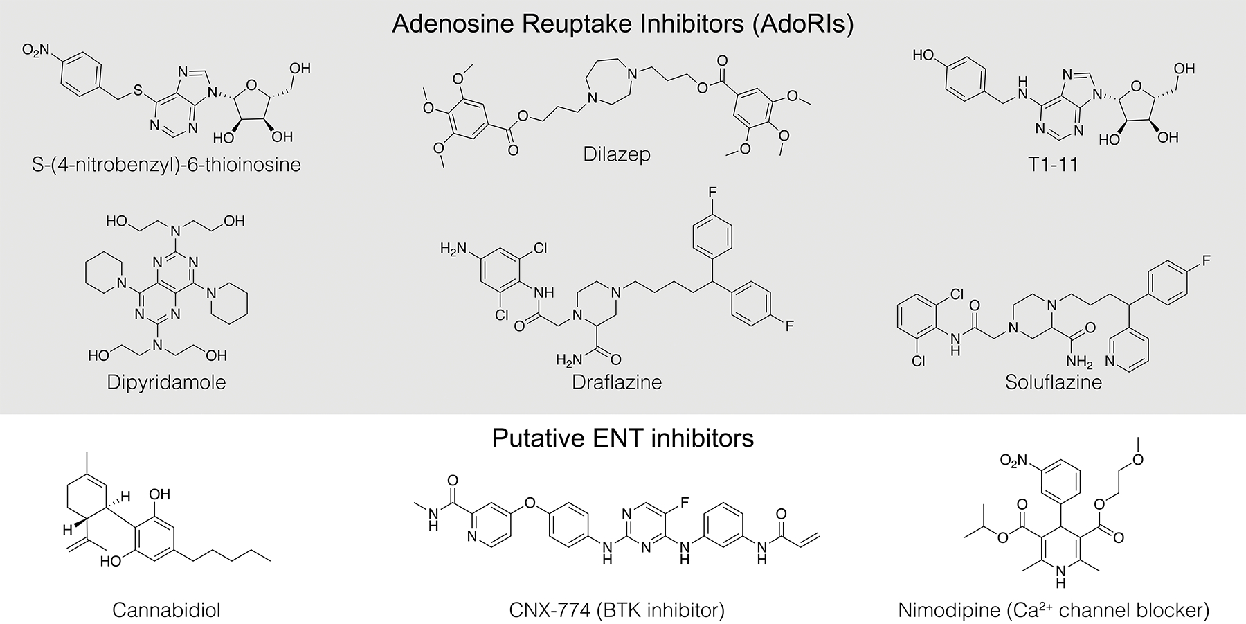
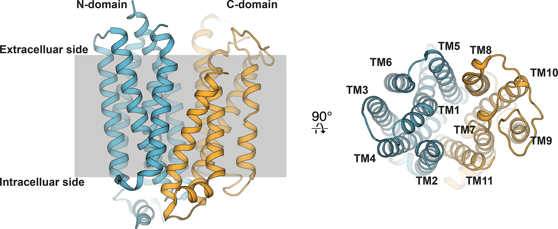
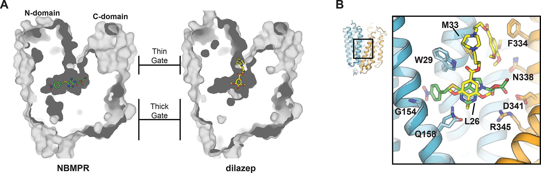
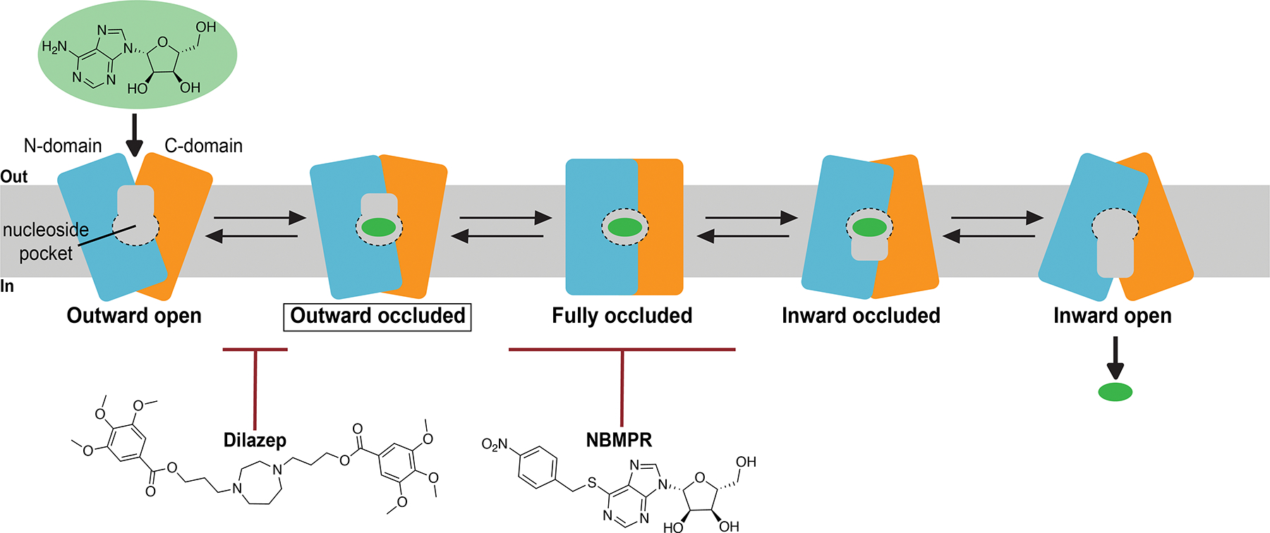
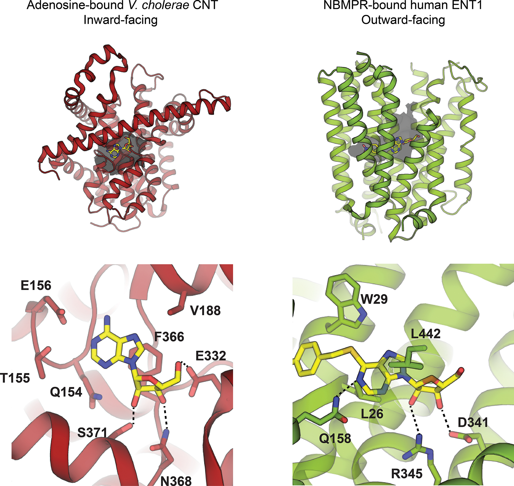
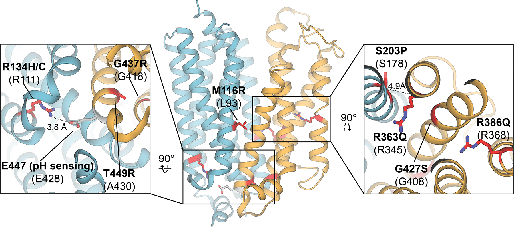
Nucleosides play central roles in all facets of life, from metabolism to cellular signaling. Because of their physiochemical properties, nucleosides are lipid bilayer impermeable and thus rely on dedicated transport systems to cross biological membranes. In humans, two unrelated protein families mediate nucleoside membrane transport: the concentrative and equilibrative nucleoside transporter families. The objective of this review is to provide a broad outlook on the current status of nucleoside transport research. We will discuss the role played by nucleoside transporters in human health and disease, with emphasis placed on recent structural advancements that have revealed detailed molecular principles of these important cellular transport systems and exploitable pharmacological features.


Conflict of interest statement

Author Information The authors declare no competing financial interests.

Figures

Figure 1
Figure 1
Select endogenous nucleosides (top) and nucleoside/nucleotide antiviral analog drugs (bottom).
Figure 2
Figure 2
A) Overview of the vcCNT trimeric structure (PDB ID 3TIJ). B) Detailed interactions of substrate nucleoside (yellow sticks) and coordinated coupled sodium ion (purple sphere) with vcCNT. Scaffold domain depicted in grey, HP1 in salmon, HP2 in green, TM4/5 in orange and TM7/8 in blue.
Figure 3
Figure 3
A) Detailed interactions of nucleosides and nucleoside analog drugs with vcCNT (PDB IDs, top left to bottom right: 4PD6, 4PD9, 4PDA, 4PB2, 4PD7, 4PB1, 4PD5, 4PD8). B) Summary of chemical determinants of solute recognition by vcCNT C) Rational chemical modifications that resulted in a chemotherapeutic drug with enhanced transport properties.
Figure 4
Figure 4
Structural superposition of crystal structures of CNTNW in different conformational states (structures aligned with reference to protomers A and B, protomers A and B colored in grey, color key for protomer C at right). Large displacements in HP1 and TM4-5 due to conformational transitions are highlighted at right (PDB IDs: 5L26, 5L27, 5L24, 5U9W, 5L2A for inward, intermediate-1, intermediate-2, intermediate-3 and outward, respectively).
Figure 5
Figure 5
Conformational changes in the transport domain mediates collapse of the nucleoside binding pocket during the elevator-like transitions. Surface representation of each transporter conformer shown, with the nucleoside pocket circled in red (PDB IDs: 5L26, 5L27, 5L24, 5U9W, 5L2A for inward, intermediate-1, intermediate-2, intermediate-3 and outward, respectively. Protomer C depicted for each conformer, except for inward pre-translocation which is depicted from chain A of PDB ID 5U9W).
Figure 6
Figure 6
HP1 of CNTNW adopts a variety of conformations in the absence of sodium. Inward-facing sodium- and substrate-bound CNTNW HP1 depicted in red, with substrate uridine shown as sticks and sodium as a purple sphere (PDB ID 5L26). HP1 conformers from protomers A and/or B from sodium free conditions shown in shades of violet to blue (PDB IDs 5L27, 5L24, 5UW9, 5L2A).
Figure 7
Figure 7
Current working model for the mechanism of nucleoside transport mediated by CNTs. Structural states in which experimental structures are available are boxed. Scaffold domain depicted in grey, transport domain in blue. The nucleoside binding pocket (light blue) is demarcated with dotted line.
Figure 8
Figure 8
Chemical structures of select Adenosine Reuptake Inhibitors (AdoRIs) and other putative ENT inhibitors.
Figure 9
Figure 9
High-resolution crystal structure of the dilazep human ENT1 co-crystal structure (PDB ID 6OB7). The first 6 transmembrane helices colored blue (N-domain), and last 5 transmembrane helices colored orange (C-domain).
Figure 10
Figure 10
A) Dilazep and NBMPR binding sites in human ENT1 (dilazep depicted in yellow and NBMPR green, PDB IDs 6OB6 and 6OB7, respectively). B) Detailed depiction of inhibitor-transporter interactions. Protein portion of N-domain colored blue and C-domain colored orange (lighter shades of either color used for the NBMPR human ENT1 cocrystal structure). Interacting residues depicted as sticks.
Figure 11
Figure 11
Current working model for the mechanism of nucleoside transport mediated by human ENT1. Currently, only the outward occluded transporter conformer has been structurally elucidated (boxed in diagram). Adenosine reuptake inhibitors dilazep and NBMPR exhibit distinct mechanisms of inhibition by perturbing two different conformational transitions in the transport cycle.
Figure 12
Figure 12
Comparison of inward-facing adenosine-bound V. cholerae CNT (left, PDB ID 4PD9) with outward-facing NBMPR-bound human ENT1 (right, PDB ID 6OB6).
Figure 13
Figure 13
Mapping of slc29a3 mutations associated with a broad spectrum of genetic disorders onto their corresponding conserved positions in the human ENT1 crystal structure (PDB ID 6OB7; mutation sites highlighted in red, pH sensing residue highlighted in grey). Mutations in human ENT3 are labeled in bold, and the corresponding conserved positions in human ENT1 are listed below in parentheses.
Figure 14
Figure 14
Human ENT1 residue I216 lines the distal end of the cavity within the transporter N-domain. Surface electrostatic potential map calculated for the dilazep human ENT1 cocrystal structure (PDB ID 6OB7) using the Adaptive Poisson-Boltzmann Solver. Well-ordered waters observed in the proximal end of the N-domain cavity in the high-resolution structure are depicted as red spheres.

References

    1. Baldwin SA; Mackey JR; Cass CE; Young JD, Nucleoside transporters: Molecular biology and implications for therapeutic development. Mol Med Today 1999, 5, 216–224. - PubMed
    1. King AE; Ackley MA; Cass CE; Young JD; Baldwin SA, Nucleoside transporters: From scavengers to novel therapeutic targets. Trends Pharmacol Sci 2006, 27, 416–425. - PubMed
    1. Parkinson FE; Damaraju VL; Graham K; Yao SYM; Baldwin SA; Cass CE; Young JD, Molecular biology of nucleoside transporters and their distributions and functions in the brain. Curr Top Med Chem 2011, 11, 948–972. - PubMed
    1. Young JD; Yao SYM; Baldwin JM; Cass CE; Baldwin SA, The human concentrative and equilibrative nucleoside transporter families, SLC28 and SLC29. Molecular Aspects of Medicine 2013, 34, 529–547. - PubMed
    1. Young JD; Yao SYM; Sun L; Cass CE; Baldwin SA, Human equilibrative nucleoside transporter (ENT) family of nucleoside and nucleobase transporter proteins. Xenobiotica 2008, 38, 995–1021. - PubMed

Publication types