Top-down proteomics: challenges, innovations, and applications in basic and clinical research
- PMID: 33232185
- PMCID: PMC7864889
- DOI: 10.1080/14789450.2020.1855982
Top-down proteomics: challenges, innovations, and applications in basic and clinical research
Abstract
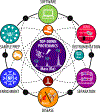
Introduction- A better understanding of the underlying molecular mechanism of diseases is critical for developing more effective diagnostic tools and therapeutics toward precision medicine. However, many challenges remain to unravel the complex nature of diseases. Areas covered- Changes in protein isoform expression and post-translation modifications (PTMs) have gained recognition for their role in underlying disease mechanisms. Top-down mass spectrometry (MS)-based proteomics is increasingly recognized as an important method for the comprehensive characterization of proteoforms that arise from alternative splicing events and/or PTMs for basic and clinical research. Here, we review the challenges, technological innovations, and recent studies that utilize top-down proteomics to elucidate changes in the proteome with an emphasis on its use to study heart diseases. Expert opinion- Proteoform-resolved information can substantially contribute to the understanding of the molecular mechanisms underlying various diseases and for the identification of novel proteoform targets for better therapeutic development . Despite the challenges of sequencing intact proteins, top-down proteomics has enabled a wealth of information regarding protein isoform switching and changes in PTMs. Continuous developments in sample preparation, intact protein separation, and instrumentation for top-down MS have broadened its capabilities to characterize proteoforms from a range of samples on an increasingly global scale.
Keywords: Heart Diseases; Mass Spectrometry; Post-translational Modifications; Proteoforms; Top-down Proteomics.
Conflict of interest statement
Financial & competing interest disclosure
The University of Wisconsin–Madison has filed a provisional patent application P180335US01, US serial number 62/682027 (June, 7 2018) on the basis of this work. Y.G. and K.A.B. are named as inventors on the provisional patent application. The University of Wisconsin—Madison has filed a provisional patent application serial No. 62/949,869 (December 18, 2019) on the basis of this work. Y.G. and D.S.R. are named as the inventors on the provisional patent application. The other authors declare no competing interests.
Figures


Similar articles
-
Top-down Proteomics: Technology Advancements and Applications to Heart Diseases.Expert Rev Proteomics. 2016 Aug;13(8):717-30. doi: 10.1080/14789450.2016.1209414. Epub 2016 Jul 26. Expert Rev Proteomics. 2016. PMID: 27448560 Free PMC article. Review.
-
Novel Strategies to Address the Challenges in Top-Down Proteomics.J Am Soc Mass Spectrom. 2021 Jun 2;32(6):1278-1294. doi: 10.1021/jasms.1c00099. Epub 2021 May 13. J Am Soc Mass Spectrom. 2021. PMID: 33983025 Free PMC article. Review.
-
Improving Proteoform Identifications in Complex Systems Through Integration of Bottom-Up and Top-Down Data.J Proteome Res. 2020 Aug 7;19(8):3510-3517. doi: 10.1021/acs.jproteome.0c00332. Epub 2020 Jul 10. J Proteome Res. 2020. PMID: 32584579 Free PMC article.
-
Characterization of Proteoforms with Unknown Post-translational Modifications Using the MIScore.J Proteome Res. 2016 Aug 5;15(8):2422-32. doi: 10.1021/acs.jproteome.5b01098. Epub 2016 Jul 1. J Proteome Res. 2016. PMID: 27291504 Free PMC article.
-
Mass spectrometry-intensive top-down proteomics: an update on technology advancements and biomedical applications.Anal Methods. 2024 Jul 18;16(28):4664-4682. doi: 10.1039/d4ay00651h. Anal Methods. 2024. PMID: 38973469 Free PMC article. Review.
Cited by
-
Top-Down Proteomics of Mouse Islets With Beta Cell CPE Deletion Reveals Molecular Details in Prohormone Processing.Endocrinology. 2023 Nov 2;164(12):bqad160. doi: 10.1210/endocr/bqad160. Endocrinology. 2023. PMID: 37967211 Free PMC article.
-
Pilot Evaluation of the Long-Term Reproducibility of Capillary Zone Electrophoresis-Tandem Mass Spectrometry for Top-Down Proteomics of a Complex Proteome Sample.J Proteome Res. 2024 Apr 5;23(4):1399-1407. doi: 10.1021/acs.jproteome.3c00872. Epub 2024 Feb 28. J Proteome Res. 2024. PMID: 38417052 Free PMC article.
-
Bench to Bedside: Proteomic Biomarker Analysis of Cerebrospinal Fluid in Patients With Spondylomyelopathy.Cureus. 2021 Jun 28;13(6):e16003. doi: 10.7759/cureus.16003. eCollection 2021 Jun. Cureus. 2021. PMID: 34336494 Free PMC article. Review.
-
Mass Spectrometry Strategies for O-Glycoproteomics.Cells. 2024 Feb 25;13(5):394. doi: 10.3390/cells13050394. Cells. 2024. PMID: 38474358 Free PMC article. Review.
-
Emerging chemistry and biology in protein glutathionylation.Curr Opin Chem Biol. 2022 Dec;71:102221. doi: 10.1016/j.cbpa.2022.102221. Epub 2022 Oct 9. Curr Opin Chem Biol. 2022. PMID: 36223700 Free PMC article. Review.
References
-
- Uhlén M, Fagerberg L, Hallström BM et al. Proteomics. Tissue-based map of the human proteome. Science, 347(6220), 1260419 (2015). - PubMed
Publication types
MeSH terms
Grants and funding
LinkOut - more resources
Full Text Sources
Medical
Miscellaneous
