. 2020 Nov 24;12(23):24270-24287.
doi: 10.18632/aging.202143. Epub 2020 Nov 24.

Metformin protects against myocardial ischemia-reperfusion injury and cell pyroptosis via AMPK/NLRP3 inflammasome pathway

Affiliations

Metformin protects against myocardial ischemia-reperfusion injury and cell pyroptosis via AMPK/NLRP3 inflammasome pathway

Jing Zhang et al. Aging (Albany NY). .

Abstract

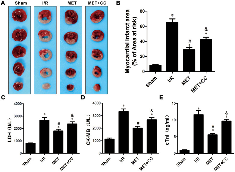
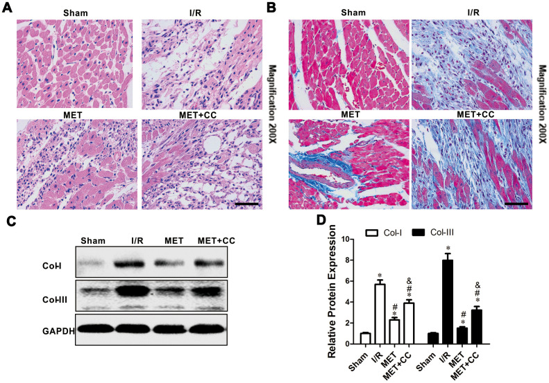
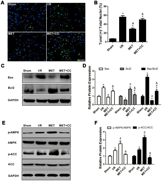
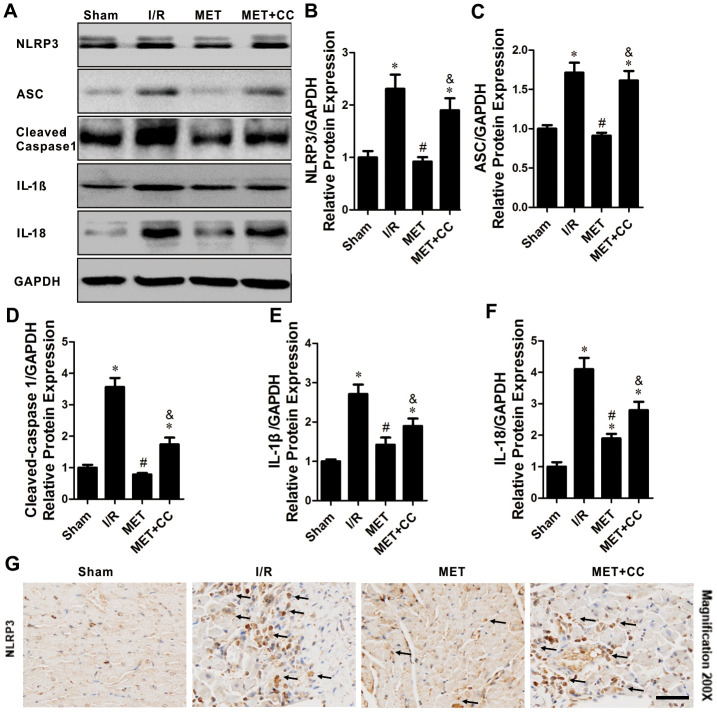
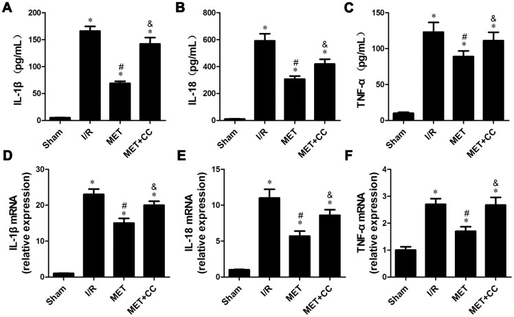
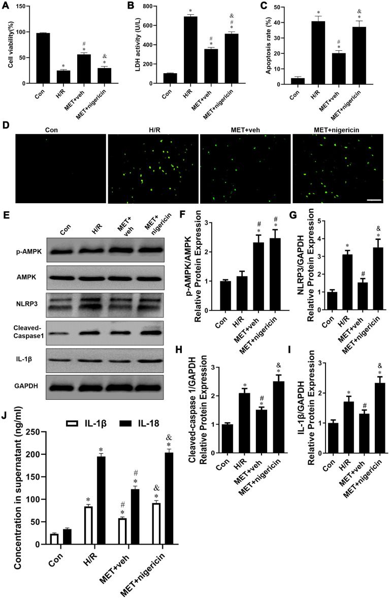
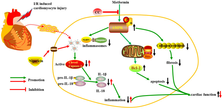
Ischemia/reperfusion (I/R) injury is a life-threatening vascular emergency following myocardial infarction. Our previous study showed cardioprotective effects of metformin against myocardial I/R injury. In this study, we further examined the involvement of AMPK mediated activation of NLRP3 inflammasome in this cardioprotective effect of metformin. Myocardial I/R injury was simulated in a rat heart Langendorff model and neonatal rat ventricle myocytes (NRVMs) were subjected to hypoxi/reoxygenation (H/R) to establish an in vitro model. Outcome measures included myocardial infarct size, hemodynamic monitoring, myocardial tissue injury, myocardial apoptotic index and the inflammatory response. myocardial infarct size and cardiac enzyme activities. First, we found that metformin postconditioning can not only significantly alleviated myocardial infarct size, attenuated cell apoptosis, and inhibited myocardial fibrosis. Furthermore, metformin activated phosphorylated AMPK, decreased pro-inflammatory cytokines, TNF-α, IL-6 and IL-1β, and decreased NLRP3 inflammasome activation. In isolated NRVMs metformin increased cellular viability, decreased LDH activity and inhibited cellular apoptosis and inflammation. Importantly, inhibition of AMPK phosphorylation by Compound C (CC) resulted in decreased survival of cardiomyocytes mainly by inducing the release of inflammatory cytokines and increasing NLRP3 inflammasome activation. Finally, in vitro studies revealed that the NLRP3 activator nigericin abolished the anti-inflammatory effects of metformin in NRVMs, but it had little effect on AMPK phosphorylation. Collectively, our study confirmed that metformin exerts cardioprotective effects by regulating myocardial I/R injury-induced inflammatory response, which was largely dependent on the enhancement of the AMPK pathway, thereby suppressing NLRP3 inflammasome activation.

Keywords: APMK; NLRP3; metformin; myocardial ischemia reperfusion injury; pyroptosis.


Conflict of interest statement

CONFLICTS OF INTEREST: The authors declare that they have no conflicts of interest.

Figures

Figure 1
Figure 1
Metformin ameliorated IR-induced cardiac tissue damage in a rat I/R injury model. (A) Representative images of myocardial infarct size stained by TTC staining. (B) Myocardial infarct volumes presented as percentage of infarct area/area at risk (n = 6–8 per group). (C) Mean levels of lactate dehydrogenase (LDH) in all groups (n = 6 per group). (D) Mean levels of creatine kinase-MB (CK-MB) in all groups (n = 6 per group). (E) Mean levels of cardiac troponin I (cTnI) in all groups (n = 6 per group). Values are expressed as the mean ± SEM. * P < 0.05 vs. Sham. # P < 0.05 vs. I/R. & P < 0.05 vs. MET.
Figure 2
Figure 2
Metformin protected I/R injury-induced myocardial injury by suppressing collagen synthesis. (A) Representative pictures of H&E-stained cardiac sections and (B) representative images of myocardial fibrosis stained with the Masson trichrome method (n = 4 per group). Magnification 200x, Scale bar = 100μm; (C) Collagen-related proteins in the ischemic area, including COL-I, COL-III and GAPDH were examined by Western blot analysis. (D) Quantitative analysis of COL-I and COL-III expression (n = 4 per group). Values are expressed as the mean ± SEM. * P < 0.05 vs. Sham. # P < 0.05 vs. I/R. & P < 0.05 vs. MET.
Figure 3
Figure 3
Activation of AMPK with Metformin protected against myocardial I/R injury induced apoptosis. (A) Top representative TUNEL-stained (green fluorescence) and DAPI-stained (blue fluorescence) photomicrographs are shown (Magnification 200x, Scale bar = 50 μm). (B) Bar graph represents the quantification of apoptotic cells (green fluorescence)/the total number of nucleated cells (blue fluorescence, n = 6 per group). (C) Apoptosis-related proteins in the ischemic area, including Bax, Bcl2 and GAPDH were examined by Western blot analysis. (D) Quantitative analysis of Bax, Bcl2 and calculate the ratio of Bax/Bcl2 (n = 4 per group). (E) AMPK pathway-related proteins in the ischemic area, including p-AMPK, AMPK, p-ACC, ACC and GAPDH were examined by Western blot analysis. (F) Quantitative analysis of p-AMPK, AMPK, p-ACC, ACC expression and calculate the ratio of p-AMPK/AMPK and p-ACC/ACC (n = 4 per group). Values are expressed as the mean ± SEM. * P < 0.05 vs. Sham. # P < 0.05 vs. I/R. & P < 0.05 vs. MET.
Figure 4
Figure 4
The inhibiting effects of Metformin on NLRP3 inflammasome activation within the infarct area following myocardial I/R injury. (A) Apoptosis-related proteins in the ischemic area, including NLRP3, ASC, cleaved-caspase 1, IL-1β, IL-18 and GAPDH were examined by Western blot analysis. (BF) Quantitative analysis of NLRP3, ASC, cleaved-caspase 1, IL-1β and IL-18 expression (n = 4 per group). (G) Bottom representative immunohistochemical-stained NLRP3 in cardiac tissue of each group are shown (n = 4 per group). Magnification 200x, Scale bar = 100 μm; Values are expressed as the mean ± SEM. * P < 0.05 vs. Sham. # P < 0.05 vs. I/R. & P < 0.05 vs. MET.
Figure 5
Figure 5
Metformin inhibited inflammatory cytokines release following myocardial I/R injury. (A) The IL-1β content, (B) IL-18 content and (C) TNF-α content were detected by ELISA (n = 6–7 per group). (D) The mRNA levels of IL-1β, (E) IL-18 and (F) TNF-α were measured using quantitative RT-PCR (n = 6–7 per group). The housekeeping gene β-actin was used for normalization. Values are expressed as the mean ± SEM. * P < 0.05 vs. Sham. # P < 0.05 vs. I/R. & P < 0.05 vs. MET.
Figure 6
Figure 6
Activation of NLRP3 with nigericin abolished Metformin-inhibited the release of pro-inflammatory factor in vitro. (A) Cell viability was measured by MTT assay (n = 6 per group). (B) The supernatant was collected and used to determine the LDH activity (n = 6 per group). (C) Statistical results of TUNEL-positive cells per field. (D) Tunel assay (apoptotic cells stained in green fluorescence) was performed to assess examine the apoptosis rate of NRVM cells in all groups (n = 6 per group). Magnification 200x, Scale bar = 100 μm; (E) Western blots were performed to determine p-AMPK, AMPK, NLRP3, cleaved-caspase 1, IL-1β and GAPDH in the total cell lysates. (FI) Quantitative analysis of p-AMPK, AMPK, NLRP3, cleaved-caspase 1, IL-1β expression and calculate the ratio of p-AMPK/AMPK (n = 4 per group). (J) The levels of IL-1β, IL-18 and TNF-α in the supernatant from NCVMs were detected by ELISA (n = 6 per group). Values are expressed as the mean ± SEM. * P < 0.05 vs. Con. # P < 0.05 vs. H/R. & P < 0.05 vs. MET+veh.
Figure 7
Figure 7
The schematic diagram of the protective properties of Metformin against myocardial I/R injury via the AMPK/NLRP3 inflammasome pathway.

Similar articles

Cited by

References

    1. Tamis-Holland JE, Jneid H, Reynolds HR, Agewall S, Brilakis ES, Brown TM, Lerman A, Cushman M, Kumbhani DJ, Arslanian-Engoren C, Bolger AF, Beltrame JF, and American Heart Association Interventional Cardiovascular Care Committee of the Council on Clinical Cardiology, and Council on Cardiovascular and Stroke Nursing, and Council on Epidemiology and Prevention, and Council on Quality of Care and Outcomes Research. Contemporary diagnosis and management of patients with myocardial infarction in the absence of obstructive coronary artery disease: a scientific statement from the American heart association. Circulation. 2019; 139:e891–908. 10.1161/CIR.0000000000000670 - DOI - PubMed
    1. Okwuosa IS, Lewsey SC, Adesiyun T, Blumenthal RS, Yancy CW. Worldwide disparities in cardiovascular disease: challenges and solutions. Int J Cardiol. 2016; 202:433–40. 10.1016/j.ijcard.2015.08.172 - DOI - PubMed
    1. Pepine CJ. New concepts in the pathophysiology of acute myocardial infarction. Am J Cardiol. 1989; 64:2B–8B. 10.1016/s0002-9149(89)80002-5 - DOI - PubMed
    1. Neri M, Riezzo I, Pascale N, Pomara C, Turillazzi E. Ischemia/reperfusion injury following acute myocardial infarction: a critical issue for clinicians and forensic pathologists. Mediators Inflamm. 2017; 2017:7018393. 10.1155/2017/7018393 - DOI - PMC - PubMed
    1. Sposito AC, Berwanger O, de Carvalho LS, Saraiva JF. Correction to: GLP-1RAs in type 2 diabetes: mechanisms that underlie cardiovascular effects and overview of cardiovascular outcome data. Cardiovasc Diabetol. 2019; 18:23. 10.1186/s12933-019-0825-1 - DOI - PMC - PubMed

Publication types

MeSH terms