

Deep-learning algorithms for the interpretation of chest radiographs to aid in the triage of COVID-19 patients: A multicenter retrospective study
- PMID: 33232368
- PMCID: PMC7685476
- DOI: 10.1371/journal.pone.0242759
Deep-learning algorithms for the interpretation of chest radiographs to aid in the triage of COVID-19 patients: A multicenter retrospective study
Abstract
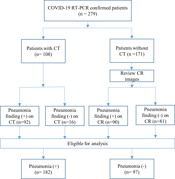
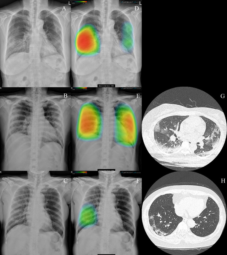
The recent medical applications of deep-learning (DL) algorithms have demonstrated their clinical efficacy in improving speed and accuracy of image interpretation. If the DL algorithm achieves a performance equivalent to that achieved by physicians in chest radiography (CR) diagnoses with Coronavirus disease 2019 (COVID-19) pneumonia, the automatic interpretation of the CR with DL algorithms can significantly reduce the burden on clinicians and radiologists in sudden surges of suspected COVID-19 patients. The aim of this study was to evaluate the efficacy of the DL algorithm for detecting COVID-19 pneumonia on CR compared with formal radiology reports. This is a retrospective study of adult patients that were diagnosed as positive COVID-19 cases based on the reverse transcription polymerase chain reaction among all the patients who were admitted to five emergency departments and one community treatment center in Korea from February 18, 2020 to May 1, 2020. The CR images were evaluated with a publicly available DL algorithm. For reference, CR images without chest computed tomography (CT) scans classified as positive for COVID-19 pneumonia were used given that the radiologist identified ground-glass opacity, consolidation, or other infiltration in retrospectively reviewed CR images. Patients with evidence of pneumonia on chest CT scans were also classified as COVID-19 pneumonia positive outcomes. The overall sensitivity and specificity of the DL algorithm for detecting COVID-19 pneumonia on CR were 95.6%, and 88.7%, respectively. The area under the curve value of the DL algorithm for the detection of COVID-19 with pneumonia was 0.921. The DL algorithm demonstrated a satisfactory diagnostic performance comparable with that of formal radiology reports in the CR-based diagnosis of pneumonia in COVID-19 patients. The DL algorithm may offer fast and reliable examinations that can facilitate patient screening and isolation decisions, which can reduce the medical staff workload during COVID-19 pandemic situations.
Conflict of interest statement
The authors have declared that no competing interests exist.
Figures


References
-
- Organization WH. WHO Director-General’s remarks at the media briefing on 2019-nCoV on 11 February 2020. World Health Organization, Geneva: Available via https://wwwwhoint/dg/speeches/detail/who-director-general-s-remarks-at-t... Accessed. 2020;10.
-
- Dennie C, Hague C, Lim RS, Manos D, Memauri BF, Nguyen ET, et al. The Canadian Society of Thoracic Radiology (CSTR) and Canadian Association of Radiologists (CAR) Consensus Statement Regarding Chest Imaging in Suspected and Confirmed COVID-19. Can Assoc Radiol J. 2020. - PubMed
-
- Radiology ACo. ACR recommendations for the use of chest radiography and computed tomography (CT) for suspected COVID-19 infection. ACR website Advocacy-and Economics/ACR-Position-Statements/Recommendations-for-Chest-Radiography-and-CTfor-Suspected-COVID19-Infection Updated March. 2020;22.
-
- Chung HS, Lee DE, Kim JK, Yeo IH, Kim C, Park J, et al. Revised Triage and Surveillance Protocols for Temporary Emergency Department Closures in Tertiary Hospitals as a Response to COVID-19 Crisis in Daegu Metropolitan City. J Korean Med Sci. 2020;35(19):e189 Epub 2020/05/19. 10.3346/jkms.2020.35.e189 - DOI - PMC - PubMed
Publication types
MeSH terms
LinkOut - more resources
Full Text Sources
Medical
