. 2020 Nov 24;16(11):e1009018.
doi: 10.1371/journal.ppat.1009018. eCollection 2020 Nov.

Defensin-driven viral evolution

Affiliations

Defensin-driven viral evolution

Karina Diaz et al. PLoS Pathog. .

Abstract

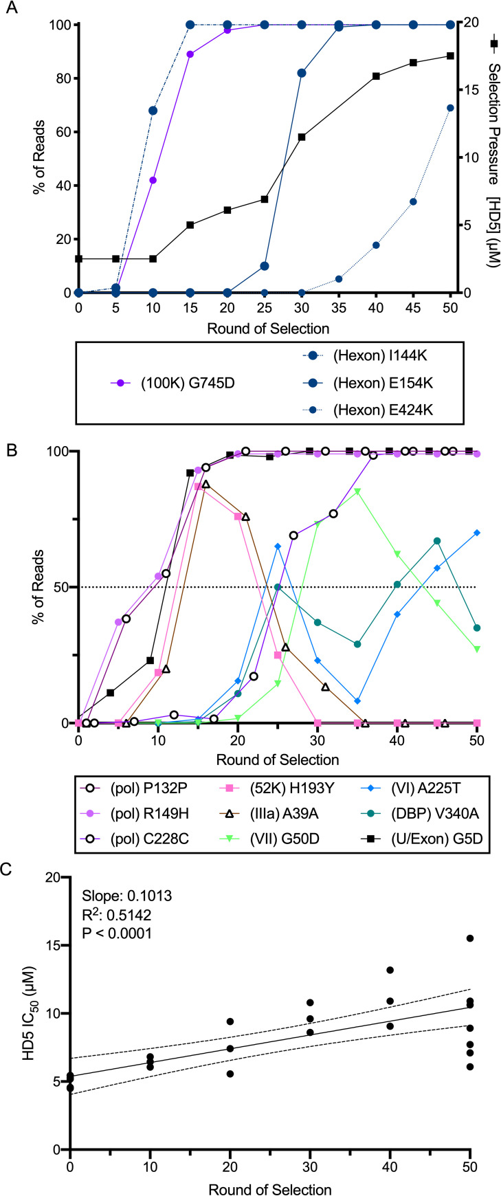
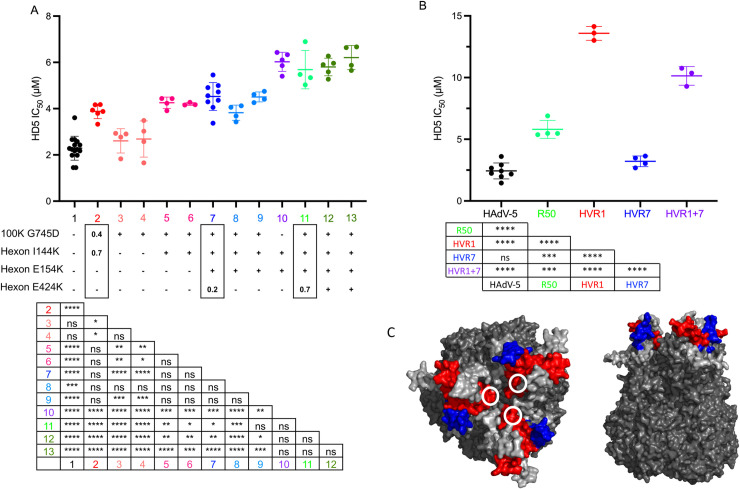
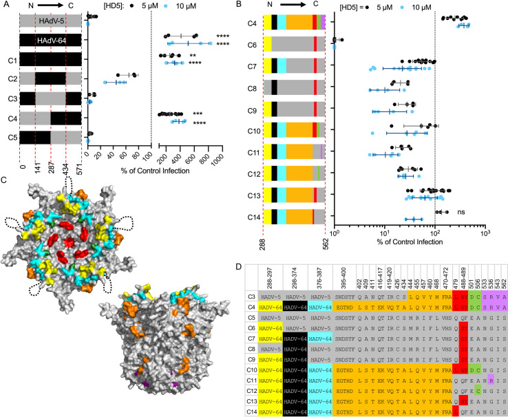
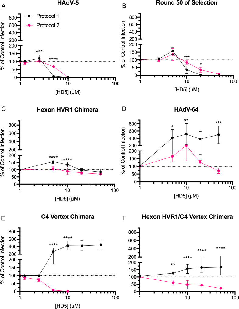
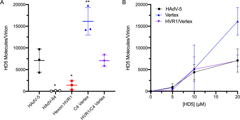
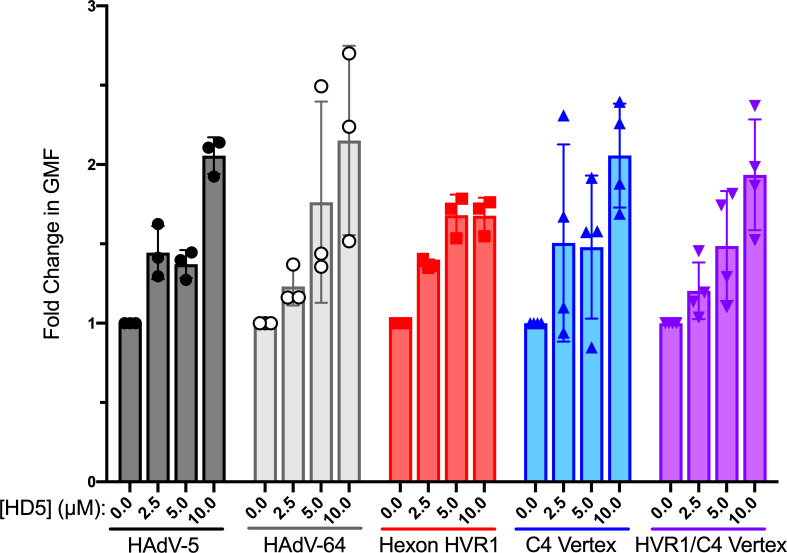
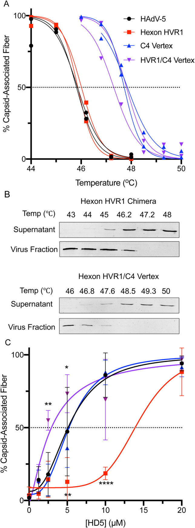
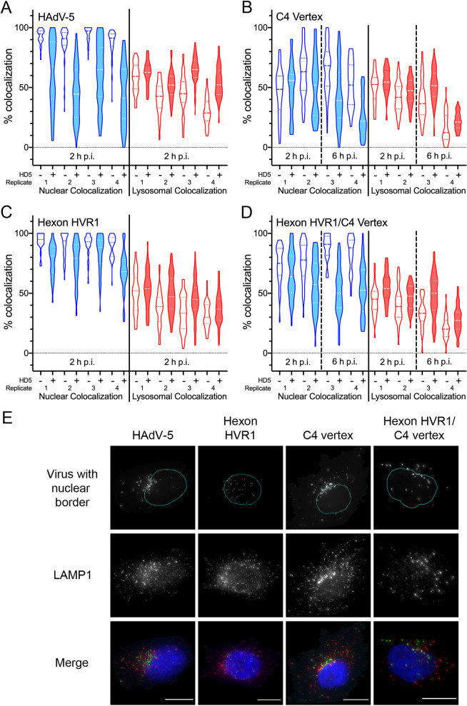
Enteric alpha-defensins are potent effectors of innate immunity that are abundantly expressed in the small intestine. Certain enteric bacteria and viruses are resistant to defensins and even appropriate them to enhance infection despite neutralization of closely related microbes. We therefore hypothesized that defensins impose selective pressure during fecal-oral transmission. Upon passaging a defensin-sensitive serotype of adenovirus in the presence of a human defensin, mutations in the major capsid protein hexon accumulated. In contrast, prior studies identified the vertex proteins as important determinants of defensin antiviral activity. Infection and biochemical assays suggest that a balance between increased cell binding and a downstream block in intracellular trafficking mediated by defensin interactions with all of the major capsid proteins dictates the outcome of infection. These results extensively revise our understanding of the interplay between defensins and non-enveloped viruses. Furthermore, they provide a feasible rationale for defensins shaping viral evolution, resulting in differences in infection phenotypes of closely related viruses.


Conflict of interest statement

The authors have declared that no competing interests exist.

Figures

Fig 1
Fig 1. Selection of HD5-resistant HAdV-5.
HAdV-5 “mutator” virus was pre-bound to 293β5 cells and then incubated with HD5 to select for resistant viruses. (A) The right y-axis indicates the concentration of HD5 used for selection (solid black line). The left y-axis indicates the percentage of reads containing the denoted mutations in hexon (blue) and L4-100K (purple) in pools of virus expanded from the bulk selected population. (B) All mutations in other proteins that were found in at least 50% of the population during selection. (C) HD5 IC50s of pools of virus expanded from the bulk selected population were determined on 293β5 cells. Each point is an independent experiment, and linear regression with 95% confidence bands is graphed.
Fig 2
Fig 2. Hexon HVR1 is a determinant of HD5-mediated neutralization.
(A) The HD5 IC50s of HAdV-5 “mutator” virus (column 1, black); pools of virus expanded from rounds 10 (column 2, red), 40 (column 7, dark blue), and 50 (column 11, light green); plaque purified viruses from rounds 10 (columns 3–4, salmon; columns 5–6, pinks), 40 (columns 8 and 9, blues), and 50 (columns 12 and 13, greens) of the selection; and an engineered virus containing only the indicated hexon mutations (column 10, purple) were determined on 293β5 cells. The presence of each hexon and L4-100K mutation is denoted below the graph. For the pooled viruses, the fraction of reads containing each mutation is indicated within the rectangles. Each point is an independent experiment, and bars are the mean ± SD. The results of one-way ANOVA with Tukey’s multiple comparison test are indicated by asterisks below. (B) The HD5 IC50s of the indicated viruses were determined on A549 cells. Each point is an independent experiment, and bars are the mean ± SD. Virus expanded from the 50th round of selection (R50) is equivalent to sample 11 in panel A. Results of one-way ANOVA with Tukey’s multiple comparisons test are shown by asterisks below. (C) Space-filling model of the structure of a HAdV-5 hexon trimer (PDBID:6CGV [44]) from top and side views. HVR1 (blue) is modeled using the structure of the shorter HVR1 from species D HAdV-26 (PDBID:5TX1 [45]). White circles indicate the position of E424 in HVR7 (red). All other HVRs are light gray.
Fig 3
Fig 3. Determinants of HD5-mediated neutralization are located within the C-terminal half of penton base.
(A and B) HAdV-5, HAdV-64, and chimeric viruses were incubated with 5 μM (black) or 10 μM (blue) HD5 and then assessed for infectivity on A549 cells. In (A), colors denote sequences derived from HAdV-5 (grey) or HAdV-64 (black), and amino acid residue numbers refer to HAdV-5 PB. In (B), colors correspond to the chart in (D) of the differential amino acid residues between HAdV-5 and HAdV-64 in the C-terminal half of PB, numbered according to HAdV-5. Each point is an independent experiment, and bars are the mean ± SD of the percent infectivity compared to control cells infected with each virus in the absence of HD5. Note that the same data for C4 are plotted on both graphs. For (A), results of two-way ANOVA with Dunnett’s multiple comparison to HAdV-5 are indicated by asterisks. For (B), all comparisons to chimera C4 are significant (P ≤ 0.01) except where indicated as not significant (ns). (C) Space-filling model of the crystal structure of a pentamer of HAdV-5 PB (PDBID:6CGV [44]) in top and side views, with differential residues colored as in (D). The unresolved RGD loop (residues 298–374) is denoted by a dotted black line.
Fig 4
Fig 4. Hexon and vertex play different roles during virus-defensin interactions.
Purified (A) HAdV-5, (B) virus expanded from round 50 of selection, (C) hexon HVR1 chimera, (D) HAdV-64, (E) C4 vertex chimera and (F) hexon HVR1/C4 vertex chimera were either incubated with HD5 and then added to A549 cells (protocol 1 –black) or bound to A549 cells prior to HD5 addition (protocol 2 –pink). Data are the mean of 3 to 11 independent experiments ± SD. Results of two-way ANOVA with Sidak’s multiple comparisons at each HD5 concentration are indicated by asterisks.
Fig 5
Fig 5. Both hexon HVR1 and vertex contribute to HD5 binding.
(A) HAdV-5, HAdV-64, and chimeric viruses were incubated in the presence of 20 μM HD5. The amount of HD5 bound per virion was quantified from 3 independent experiments. (B) HD5 molecules bound per virion of HAdV-5 and chimeras C4 vertex and hexon HVR1/C4 vertex were also determined in the presence of 5 μM and 10 μM HD5. Note that the data for 20 μM HD5 for these viruses in (A) are reproduced in (B). Lines are the mean ± SD. The results of ordinary one-way ANOVA with Dunnett’s multiple comparisons to HAdV-5 is denoted by asterisks.
Fig 6
Fig 6. HD5 increases the binding of viruses to cells regardless of infection phenotype.
AF488-labeled HAdV-5, HAdV-64, and chimeric viruses were incubated with or without HD5 and then allowed to bind to A549 cells in the cold (as in Fig 4, protocol 1). Data are the fold change in geometric mean fluorescence (GMF) of cells bound by the respective labeled virus incubated with the indicated HD5 concentrations relative to no HD5 for each virus. Each point is an independent experiment, and bars are the mean ± SD of 3–4 independent experiments. Differences between each genotype and HAdV-5 at each HD5 concentration are not significant by ordinary two-way ANOVA with Dunnett’s multiple comparison test.
Fig 7
Fig 7. Fiber thermostability does not correlate with infection phenotype.
The percent of fiber that remains capsid associated was determined (A) as a function of temperature in the absence of HD5 or (C) as a function of HD5 concentration for HAdV-5 (black) and hexon HVR1 chimera (red) at 48°C and for C4 vertex chimera (blue) and hexon HVR1/C4 vertex chimera (purple) at 49.3°C. (B) Representative immunoblots from the temperature gradients of hexon HVR1 and hexon HVR1/C4 vertex chimeras are shown. In (A) each point and line is an individual replicate. In (C), each point is the mean ± SD of 3 independent experiments, and the results of two-way ANOVA with Dunnett’s multiple comparisons to HAdV-5 are denoted by asterisks.
Fig 8
Fig 8. HD5 neutralization correlates with a reduction in nuclear localization.
AF488-labeled HAdV-5 (A), C4 vertex (B), hexon HVR1 (C), and hexon HVR1/C4 vertex (D) were bound to A549 cells in the cold and then incubated with (shaded plots) or without (unshaded plots) 10 μM HD5 (as in Fig 4, protocol 2). Cells were then warmed to 37°C and fixed at the indicated times post-infection (p.i.). Images obtained by epifluorescence microscopy were analyzed for percent colocalization of the virus with the nucleus (DAPI, blue) or lysosome (LAMP1, red) on a per cell basis. Violin plots marked with the median value (dashed lines) and interquartile ranges (dotted lines) for 23 to 106 cells for each of four independent experimental replicates are shown. The difference in mean colocalization of virus with the nucleus and with lysosomes in the presence and absence of HD5 was calculated for each replicate for each genotype. For the C4 vertex and hexon HVR1/C4 vertex chimeras, only data from 6 h p.i. was used. The calculated values for each chimera were then compared to HAdV-5 by ordinary ANOVA with Sidak’s multiple comparisons test. All comparisons are not significant except for hexon HVR1 nuclear colocalization, where p = 0.0014. (E) Representative images of cells at 2 h p.i. (HAdV-5 and hexon HVR1 chimera) or 6 h p.i. (C4 vertex and hexon HVR1/C4 vertex chimera) in the presence of HD5 are shown. In the top panels, nuclear borders from the CellProfiler analysis (cyan) are superimposed on AF488 fluorescence (gray). The merge data includes DAPI (blue), AF488 (green), and LAMP1 (red). Note that the AF488 fluorescence was individually adjusted in these images to account for the differences in brightness between genotypes. Scale bars are 10 μm.

References

    1. Holly MK, Diaz K, Smith JG. Defensins in Viral Infection and Pathogenesis. Annu Rev Virol. 2017;4(1):369–91. Epub 2017/07/19. 10.1146/annurev-virology-101416-041734 . - DOI - PubMed
    1. Selsted ME, Ouellette AJ. Mammalian defensins in the antimicrobial immune response. Nat Immunol. 2005;6(6):551–7. Epub 2005/05/24. 10.1038/ni1206 . - DOI - PubMed
    1. Wilson SS, Wiens ME, Smith JG. Antiviral mechanisms of human defensins. J Mol Biol. 2013;425(24):4965–80. Epub 2013/10/08. 10.1016/j.jmb.2013.09.038 - DOI - PMC - PubMed
    1. Brice DC, Diamond G. Antiviral Activities of Human Host Defense Peptides. Curr Med Chem. 2020;27(9):1420–43. Epub 2019/08/07. 10.2174/0929867326666190805151654 . - DOI - PMC - PubMed
    1. Wiens ME, Smith JG. Alpha-defensin HD5 inhibits furin cleavage of human papillomavirus 16 L2 to block infection. J Virol. 2015;89(5):2866–74. Epub 2014/12/30. 10.1128/JVI.02901-14 - DOI - PMC - PubMed

Publication types