Review
. 2021 May 5;45(3):fuaa060.
doi: 10.1093/femsre/fuaa060.

The impact of the Fungus-Host-Microbiota interplay upon Candida albicans infections: current knowledge and new perspectives

Affiliations
Review

The impact of the Fungus-Host-Microbiota interplay upon Candida albicans infections: current knowledge and new perspectives

Christophe d'Enfert et al. FEMS Microbiol Rev. .

Abstract

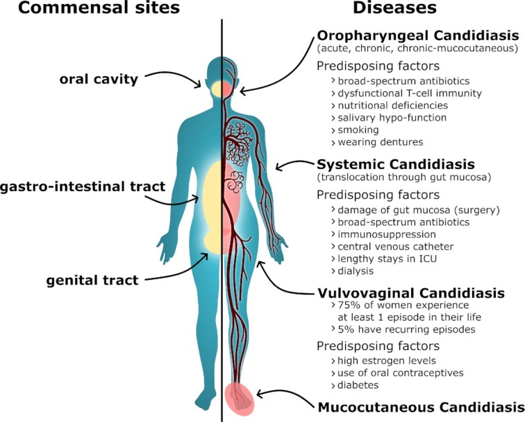
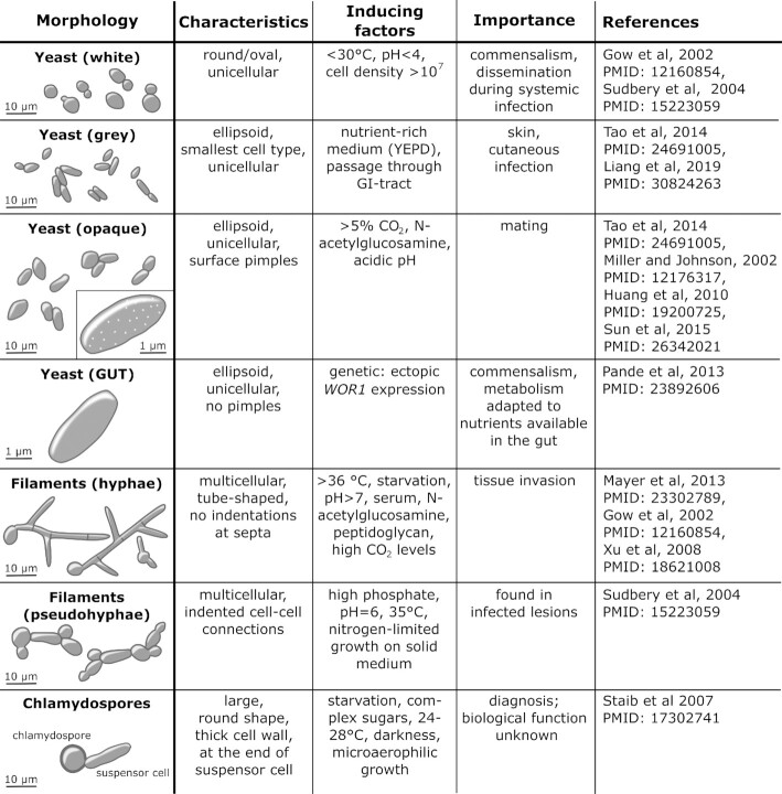
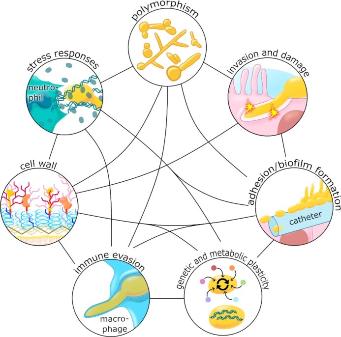
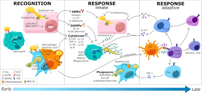
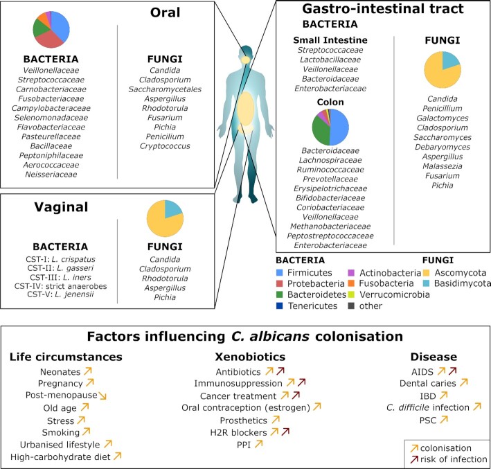
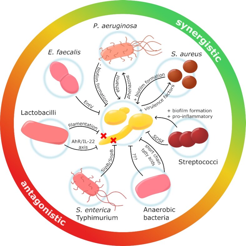
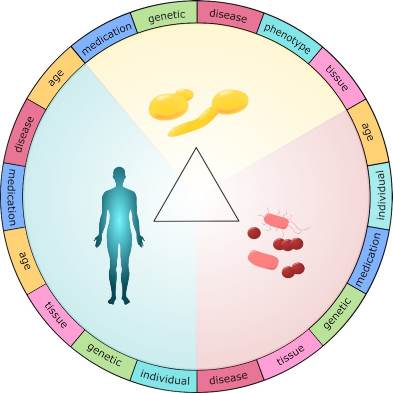
Candida albicans is a major fungal pathogen of humans. It exists as a commensal in the oral cavity, gut or genital tract of most individuals, constrained by the local microbiota, epithelial barriers and immune defences. Their perturbation can lead to fungal outgrowth and the development of mucosal infections such as oropharyngeal or vulvovaginal candidiasis, and patients with compromised immunity are susceptible to life-threatening systemic infections. The importance of the interplay between fungus, host and microbiota in driving the transition from C. albicans commensalism to pathogenicity is widely appreciated. However, the complexity of these interactions, and the significant impact of fungal, host and microbiota variability upon disease severity and outcome, are less well understood. Therefore, we summarise the features of the fungus that promote infection, and how genetic variation between clinical isolates influences pathogenicity. We discuss antifungal immunity, how this differs between mucosae, and how individual variation influences a person's susceptibility to infection. Also, we describe factors that influence the composition of gut, oral and vaginal microbiotas, and how these affect fungal colonisation and antifungal immunity. We argue that a detailed understanding of these variables, which underlie fungal-host-microbiota interactions, will present opportunities for directed antifungal therapies that benefit vulnerable patients.

Keywords: Candida; Candida infections; antifungal immunity; fungal variability; fungus-host-microbiota interactions; microbiota; microbiota variability; mycobiota; patient variability.


Figures

Figure 1.
Figure 1.
Three-way interactions between the fungus, the host and the local microbiota strongly influence the likelihood and severity of C. albicans infections. See text.
Figure 2.
Figure 2.
Sites of C. albicans commensalism and disease on the human body. Sites of C. albicans commensalism (left side) include the oral cavity, gastrointestinal tract (gut) and the genital tract. C. albicans can infect these sites (right side) to cause oropharyngeal or vulvovaginal candidiasis. C. albicans can also cause systemic infections of the blood and internal organs, which often arise via translocation of C. albicans from the gut into the bloodstream. Candida albicans also causes mucocutaneous infections of the skin and nails. Factors that predispose individuals to such infections are listed. See text.
Figure 3.
Figure 3.
Candida albicans is polymorphic, displaying a range of cellular growth forms. C. albicans yeast cells can undergo phenotypic switching between white, grey and opaque growth forms that present with different shapes and cell surface characteristics (Gow, Brown and Odds ; Sudbery, Gow and Berman ; Xu et al. ; Huang et al. ; Mayer, Wilson and Hube ; Tao et al. ; Sun et al. 2015). These forms are induced in response to different environmental inputs, and hence are associated with different types of infection (Gow, Brown and Odds 2002). Significantly, the opaque form is associated with efficient mating in C. albicans (Miller and Johnson 2002), with grey cells displaying an intermediate mating competence between opaque and white cells (Tao et al. 2014). The gastrointestinally induced transition (GUT) phenotype is observed in C. albicans cells that ectopically express WOR1 (Pande, Chen and Noble 2013), a key regulator of commensalism. The transition from (white) yeast cells to pseudohyphae or hyphae is stimulated by a wide variety of environmental inputs, which include elevated temperatures, pH and peptidoglycan. Pseudohyphae can be distinguished from hyphae on the basis of the position of the septal junction between a mother yeast cell and its filamentous daughter, and by the presence of invaginations at these septal junctions in pseudohyphae, but not hyphae (Merson-Davies and Odds ; Sudbery ; Sudbery, Gow and Berman 2004). Candida albicans can be induced to form chlamydospores under specific environmental conditions (Jansons and Nickerson 1970), but the biological significance of this growth form remains obscure (Staib and Morschhäuser 2007). See text.
Figure 4.
Figure 4.
A combination of virulence factors and fitness attributes promote C. albicans virulence. Polymorphism: The ability of C. albicans to undergo morphological transitions allows it to adapt to different growth conditions, adhere to biotic and abiotic surfaces, invade cells and tissue, and escape from immune cells. Invasion and damage: A combination of induced endocytosis and active penetration promote fungal invasion of host tissues, and the accumulation of the toxin, candidalysin, in the invasion pocket leads to pore formation and host cell damage. Adhesion/biofilm formation: The battery of adhesins promotes fungal adhesions to biological and abiotic surfaces, which can lead to the development of biofilms, for example on medical devices such as catheters. Genetic and metabolic plasticity: Candida albicans displays a high degree of metabolic flexibility, which allows it to adapt rapidly to diverse host niches. This fungus also displays great genetic plasticity, which permits rapid evolutionary adaptation to selective pressures and stresses such as exposure to antifungal drugs. Stress responses: Candida albicans activates robust stress responses following exposure to host imposed stresses, including ROS and RNS, which enhances fungal survival following immune attack, for example. Cell wall: As well as maintaining cell morphology, the robust cell wall provides protection against host-imposed stresses including changes in osmolarity. Immune evasion: Candida albicans has evolved a variety of immune evasion strategies that include the modulation of PAMP exposure at the cell surface to evade immune recognition, and phagocytic escape mechanisms to evade killing by innate immune cells. See text.
Figure 5.
Figure 5.
Immune recognition of, and immune responses against, C. albicans. Candida albicans yeast and hyphal cells are recognised by neutrophils, macrophages and dendritic cells via pattern recognition receptors (see key). This recognition activates the expression and release of proinflammatory cytokines and chemokines that promote the recruitment of macrophages and neutrophils to the site of infection. Epithelial cells respond to hypha formation and the subsequent secretion of candidalysin by the fungus, by activating the expression and release of AMPs, DAMPs, chemokines and cytokines via p38/cFos and ERK/MKP1 signalling. The AMPs attenuate fungal growth and invasion, while DAMPs and cytokines promote inflammation. Myeloid cells promote fungal killing and clearance through a combination of phagocytosis and NETosis in the case of neutrophils. Fungal recognition leads to the maturation of dendritic cells, and their surface presentation of fungal antigens to naïve T-cells, which stimulates adaptive immunity. The interactions between antigen-presenting dendritic cells and naïve T-cells induces T-cell activation and differentiation into various effector T cell subsets that regulate mucosal immunity largely via IL-17 and IL-22 secretion, and stimulate macrophages via IFN-γ. See text.
Figure 6.
Figure 6.
Oral, vaginal and GI microbiota, and factors that influence C. albicans colonisation of these body sites. The major microbial groups (family level for bacteria and genus level for fungi) of the healthy oral cavity (only for bacteria) (Bik et al. ; Dewhirst et al. 2010), GI tract (Booijink et al. ; Arumugam et al. ; Zhou et al. ; Villmones et al. 2018) and vagina (Human Microbiome Project Consortium 2012) are listed in decreasing order of abundance. Pie charts indicate the relative abundance of the phyla in a representative healthy oral cavity and colon (see key for colour code). The fungal component of the oral microbiota is extremely variable, and many fungi present in this compartment are likely to be transient (see text). Therefore, for the oral cavity, the fungal genera are not presented in descending order of abundance, and no pie chart is provided. The lower panel summarises factors that influence the degree of C. albicans colonisation (yellow) and likelihood of infection (brown arrows) of these mucosal surfaces: arrows up, increased likelihood of colonisation/infection; arrows down, decreased likelihood of colonisation/infection. See text.
Figure 7.
Figure 7.
The interplay between C. albicans and certain bacterial present in human microbiotas. The growth of C. albicans in mucosal niches is generally constrained by the local bacterial microbiota via colonisation resistance. However, specific interactions with certain bacteria present in the vagina, oral cavity and/or GI tract influence the growth and/or virulence of C. albicans more directly. These interactions can be antagonistic (i.e. reduce the growth and virulence of the fungus) or synergistic (i.e. enhance the growth or virulence of the fungus). Anaerobic bacteria antagonise C. albicans colonisation by mechanisms that include the production of short chain fatty acids. S. enterica Typhimurium kills C. albicans hyphae by injecting effectors into the fungus via the SopB translocase. Lactobacilli antagonise C. albicans colonisation acidifying the local environment which reduces filamentation, and by generating metabolites that enhance IL-22-mediated immunity. E. faecalis blocks yeast-hypha morphogenesis and biofilm formation using EntV. Interactions between C. albicans and P. aeruginosa mutually enhance their virulence via cross-talk involving ethanol production by the fungus, which promotes toxic phenazine production by the bacterium, and this in turn promotes alcohol production by C. albicans. S. aureus binds C. albicans hyphae and promotes biofilm formation and antimicrobial resistance. Furthermore, co-infection with S. aureus significantly enhances the lethality of C. albicans. Streptococci block the formation of C. albicans hyphae via the diffusible factor, SDSF, but potentially co-exist as commensals with C. albicans. See text.
Figure 8.
Figure 8.
The complexity of fungus-host-microbiota interactions is dramatically increased by variability between C. albicans clinical isolates, between individuals, and in their microbiotas. Fungal variability arises through significant genetic and phenotypic variation between clinical isolates of C. albicans. The immune-competence of individuals varies significantly depending on their genetics, age and lifestyle. Furthermore, the microbiotas of the GI tract, oral cavity and vagina can each vary dramatically, depending on the age and health of the individual, and their diet, possible medications and life circumstances. Therefore, variation in each of the three elements of the fungus-host-microbiota interplay strongly influences the susceptibility of an individual to C. albicans infection as well as the outcome of that infection. See text.

Similar articles

Cited by

References

    1. Aagaard K, Riehle K, Ma Ket al. . A metagenomic approach to characterization of the vaginal microbiome signature in pregnancy. PLoS One. 2012;7:e36466. - PMC - PubMed
    1. Acosta-Rodriguez EV, Rivino L, Geginat Jet al. . Surface phenotype and antigenic specificity of human interleukin 17-producing T helper memory cells. Nat Immunol. 2007;8:639–46. - PubMed
    1. Adler CJ, Dobney K, Weyrich LSet al. . Sequencing ancient calcified dental plaque shows changes in oral microbiota with dietary shifts of the Neolithic and Industrial revolutions. Nat Genet. 2013;45:450–5., 455e1. - PMC - PubMed
    1. Aggor FEY, Break TJ, Trevejo-Nuñez Get al. . Oral epithelial IL-22/STAT3 signaling licenses IL-17-mediated immunity to oral mucosal candidiasis. Sci Immunol. 2020;5. - PMC - PubMed
    1. Ahrné S, Nobaek S, Jeppsson Bet al. . The normal Lactobacillus flora of healthy human rectal and oral mucosa. J Appl Microbiol. 1998;85:88–94. - PubMed

Publication types

MeSH terms