. 2021 Apr;36(4):895-904.
doi: 10.1002/mds.28399. Epub 2020 Nov 24.

Immunohistochemical Detection of Synuclein Pathology in Skin in Idiopathic Rapid Eye Movement Sleep Behavior Disorder and Parkinsonism

Affiliations

Immunohistochemical Detection of Synuclein Pathology in Skin in Idiopathic Rapid Eye Movement Sleep Behavior Disorder and Parkinsonism

Ahmed Al-Qassabi et al. Mov Disord. 2021 Apr.

Abstract

Background: Recent studies reported abnormal alpha-synuclein deposition in biopsy-accessible sites of the peripheral nervous system in Parkinson's disease (PD). This has considerable implications for clinical diagnosis. Moreover, if deposition occurs early, it may enable tissue diagnosis of prodromal PD.

Objective: The aim of this study was to develop and test an automated bright-field immunohistochemical assay of cutaneous pathological alpha-synuclein deposition in patients with idiopathic rapid eye movement sleep behavior disorder, PD, and atypical parkinsonism and in control subjects.

Methods: For assay development, postmortem skin biopsies were taken from 28 patients with autopsy-confirmed Lewy body disease and 23 control subjects. Biopsies were stained for pathological alpha-synuclein in automated stainers using a novel dual-immunohistochemical assay for serine 129-phosphorylated alpha-synuclein and pan-neuronal marker protein gene product 9.5. After validation, single 3-mm punch skin biopsies were taken from the cervical 8 paravertebral area from 79 subjects (28 idiopathic rapid eye movement sleep behavior disorder, 20 PD, 10 atypical parkinsonism, and 21 control subjects). Raters blinded to clinical diagnosis assessed the biopsies.

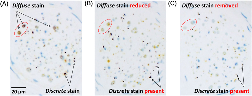
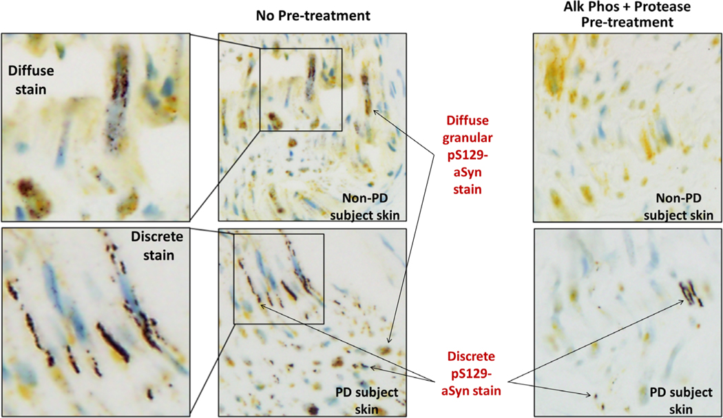
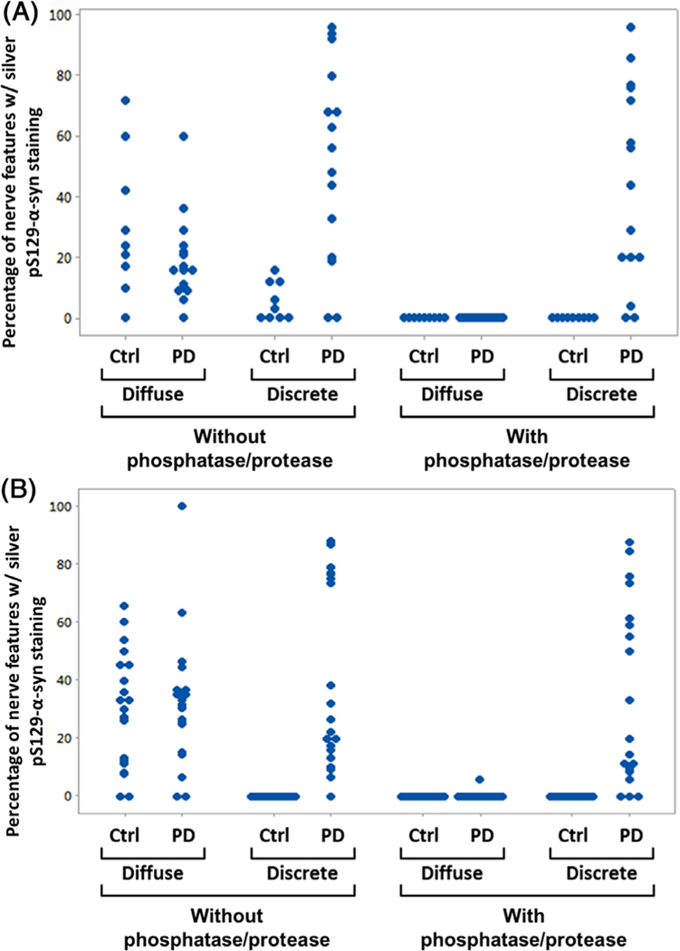
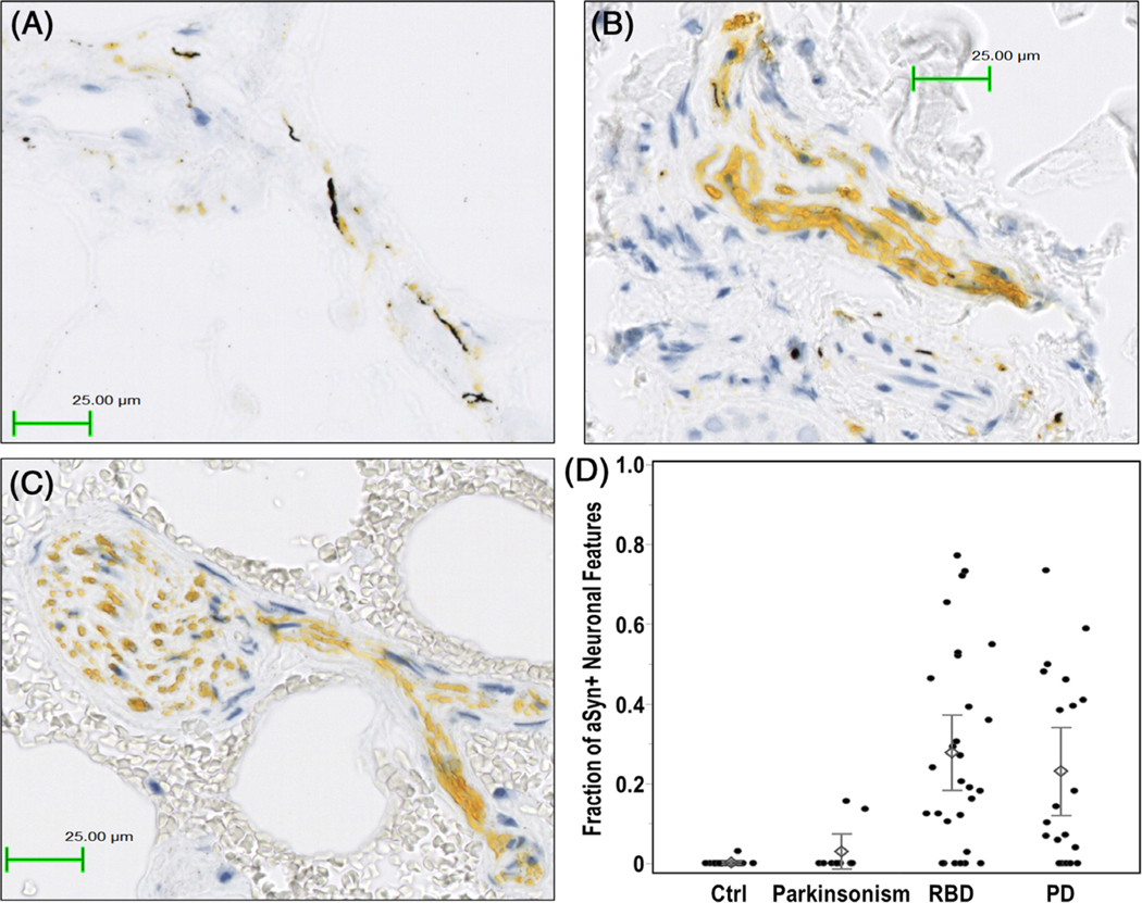
Results: The immunohistochemistry assay differentiated alpha-synuclein pathology from nonpathological-appearing alpha-synuclein using combined phosphatase and protease treatments. Among autopsy samples, 26 of 28 Lewy body samples and none of the 23 controls were positive. Among living subjects, punch biopsies were positive in 23 (82%) subjects with idiopathic rapid eye movement sleep behavior disorder, 14 (70%) subjects with PD, 2 (20%) subjects with atypical parkinsonism, and none (0%) of the control subjects. After a 3-year follow-up, eight idiopathic rapid eye movement sleep behavior disorder subjects phenoconverted to defined neurodegenerative syndromes, in accordance with baseline biopsy results.

Conclusion: Even with a single 3-mm punch biopsy, there is considerable promise for using pathological alpha-synuclein deposition in skin to diagnose both clinical and prodromal PD. © 2020 International Parkinson and Movement Disorder Society.

Keywords: PGP9.5; Parkinson's disease; RBD; UCHL1; alpha-synuclein; skin biopsy.


Conflict of interest statement

Relevant conflicts of interest/financial disclosures: T.-S.T., A.R., M.A.S., R.C.B., H.Z., J.M., J.P., F.L., M.D.R., L.P.-D., and L.T. performed the work associated with this manuscript as employees or contractors of Roche Tissue Diagnostics. M.R. performed the work associated with this manuscript as employee of Roche Centralised and Point of Care Solutions. T.K., C.C., M.C., K.I.T., A.B., and S.D. performed the work associated with this manuscript as employees of Hoffmann-La Roche. W.Z. performed the work associated with this manuscript as an employee of Prothena Biosciences. T.G.B. was a paid consultant to Roche Tissue Diagnostics for advice contributing to the development of the methodology reported herein.

Figures

FIG. 1.
FIG. 1.
Skin formalin-fixed, paraffin-embedded (FFPE) sections from patients with Parkinson’s disease (PD) stained with pS129-alpha-synuclein (aSyn) and protein gene product 9.5 (PGP9.5) silver/yellow dual-immunohistochemistry (IHC) assay showing (A) two distinct types of silver pS129-aSyn stain and their differential sensitivities to (B) protease and (C) phosphatase. The pS129-aSyn stain with diffuse and granular morphology was observed mostly in the larger nerve bundles as a circumferential ring around a nerve fiber. This type of pS129-aSyn stain was either reduced or apparently removed (reduced below detectable levels) after pretreatment at 37°C with, respectively, protease 3 for 4 minutes or recombinant bovine alkaline phosphatase for 8 minutes. In contrast, the discrete type of pS129-aSyn stain with smooth contours was resistant to either protease or phosphatase pretreatment. Nerve fibers innervating skin structures were detected using a rabbit monoclonal antibody against PGP9.5 and alkaline phosphatase-activated QM-Dabcyl yellow chromogen as described in Subjects and Methods.
FIG. 2.
FIG. 2.
Morphologies of pS129-alpha-synuclein (aSyn) stain produced by pS129-aSyn and protein gene product 9.5 (PGP9.5) silver/yellow dual-immunohistochemistry (IHC) assay in scalp sections from autopsied non-PD and PD subjects with and without combined protease and phosphatase pretreatments. In non-PD subject skin, only diffuse and granular pS129-aSyn stain was observed. The diffuse pS129-aSyn stain exhibited pipelike appearance sheathing nerve fibers sectioned horizontally along the length of the fibers. Discrete pS129 stain was not observed in skin sections of subjects without PD. Both diffuse and discrete stains were seen in skin of subjects with PD. Discrete pS129-aSyn stain appeared as tracks running along the length of nerve fibers. In both non-PD and PD subjects, diffuse and granular pS129-aSyn stain was removed on sequential pretreatments at 37°C with recombinant bovine alkaline phosphatase (Alk Phos) and protease 3 for, respectively, 8 and 4 minutes.
FIG. 3.
FIG. 3.
(A) Individual value plot showing percentages of nerve features containing either diffuse or discrete pS129-alpha-synuclein (aSyn) stain in 4-μm sections of postmortem scalp skin biopsies from 15 subjects with PD and 9 control (Ctrl) subjects without PD. All formalin-fixed, paraffin-embedded (FFPE) skin sections were stained with pS129-aSyn and protein gene product 9.5 (PGP9.5) silver/yellow dual-immunohistochemistry (IHC) assay without and with phosphatase/protease pretreatment as described in Subjects and Methods. (B) Individual value plot showing percentages of nerve features containing either diffuse or discrete pS129-aSyn stain in 4-μm sections of postmortem abdomen skin biopsies from 20 subjects with PD and 20 Ctrl subjects without PD. All FFPE skin sections were stained with pS129-aSyn and PGP9.5 silver/yellow dual-IHC assay without and with phosphatase/protease pretreatment as described in Subjects and Methods. Nerve features are defined as innervated skin structures with yellow chromogens staining neuronal marker PGP9.5. Examples of these are shown in Supporting Information Figures S5 and S6.
FIG. 4.
FIG. 4.
Example of colocalization of pathological alpha-synuclein (aSyn) deposition (silver black stain) in neuronal structures (dabcyl yellow stain) from skin biopsies of (A) a subject with PD and (B) a subject with idiopathic RBD. In contrast, skin biopsy from (C) a control subject exhibited only dabcylstained protein gene product 9.5 (PGP9.5) without silver-stained pathological aSyn. (D) Scatterplot of the proportion of pathological aSyn positivity in PGP9.5-positive neuronal features in each biopsy sample (using all eight sections per subject) classified according to diagnostic status. Diamonds represent average proportion of pathological aSyn-positive neuronal features. Error bars correspond to ±1 standard error from the corresponding mean value. Ctrl, control; PD, Parkinson’s disease; RBD, rapid eye movement sleep behavior disorder.

References

    1. Lindberg I, Shorter J, Wiseman RL, Chiti F, Dickey CA, McLean PJ. Chaperones in neurodegeneration. J Neurosci 2015;35(41):13853–13859. - PMC - PubMed
    1. Witt SN. Molecular chaperones, alpha-synuclein, and neurodegeneration. Mol Neurobiol 2013;47(2):552–560. - PMC - PubMed
    1. Doppler K, Ebert S, Uceyler N, et al. Cutaneous neuropathy in Parkinson’s disease: a window into brain pathology. Acta neuropathol 2014;128(1):99–109. - PMC - PubMed
    1. Beach TG, Adler CH, Serrano G, et al. Prevalence of submandibular gland Synucleinopathy in Parkinson’s disease, dementia with Lewy bodies and other Lewy body disorders. Parkinson’s Dis 2016;6(1):153–163. - PMC - PubMed
    1. Donadio V, Incensi A, Leta V, et al. Skin nerve alpha-synuclein deposits: a biomarker for idiopathic Parkinson disease. Neurology 2014;82(15):1362–1369. - PubMed

Publication types

Substances