. 2021 Feb 3;109(3):530-544.e6.
doi: 10.1016/j.neuron.2020.11.001. Epub 2020 Nov 23.

Sleep Promotes Downward Firing Rate Homeostasis

Affiliations

Sleep Promotes Downward Firing Rate Homeostasis

Alejandro Torrado Pacheco et al. Neuron. .

Abstract

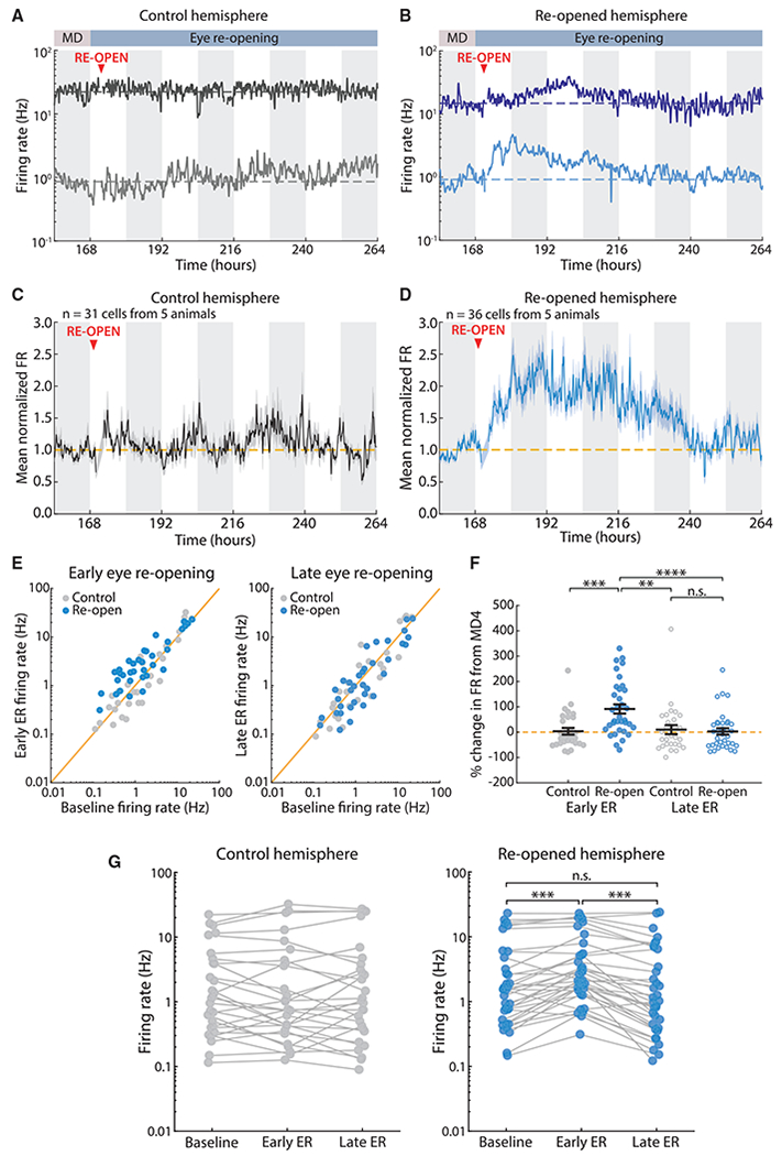
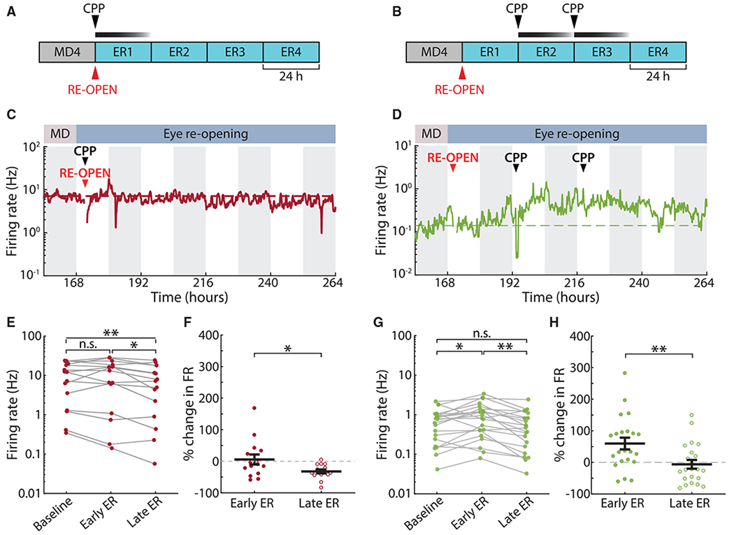
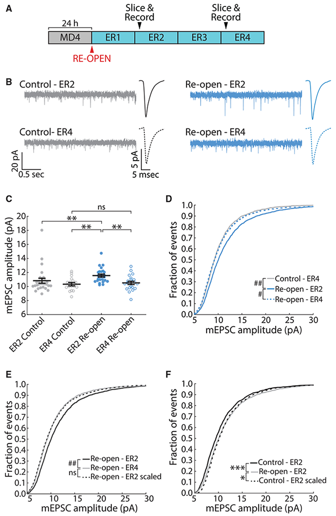
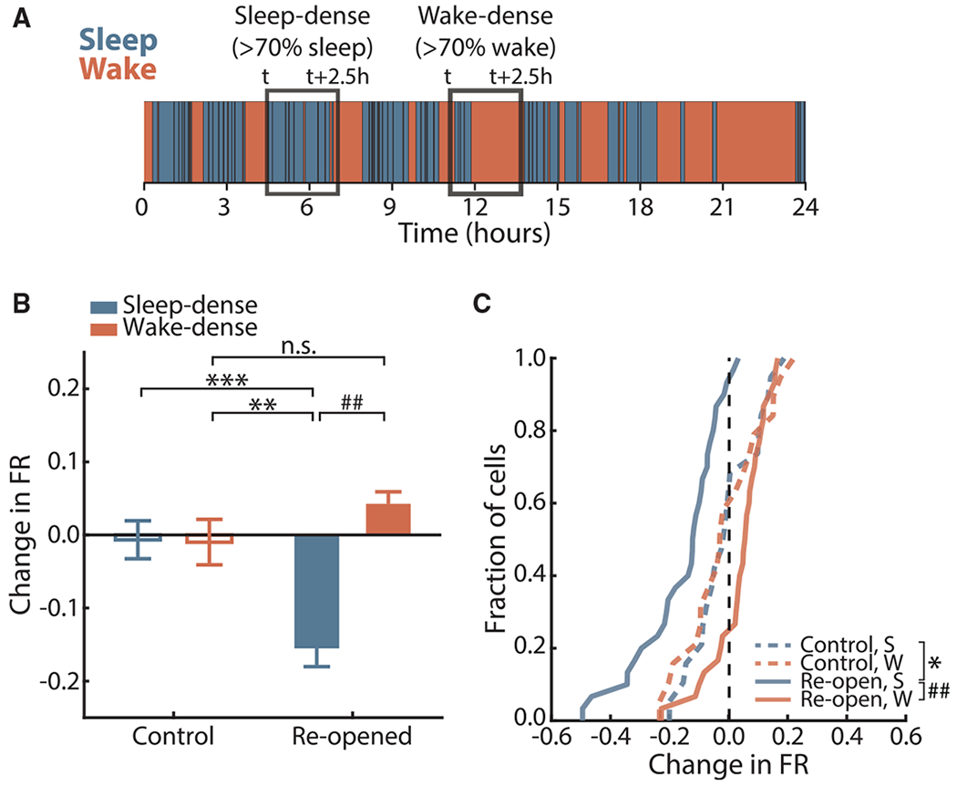
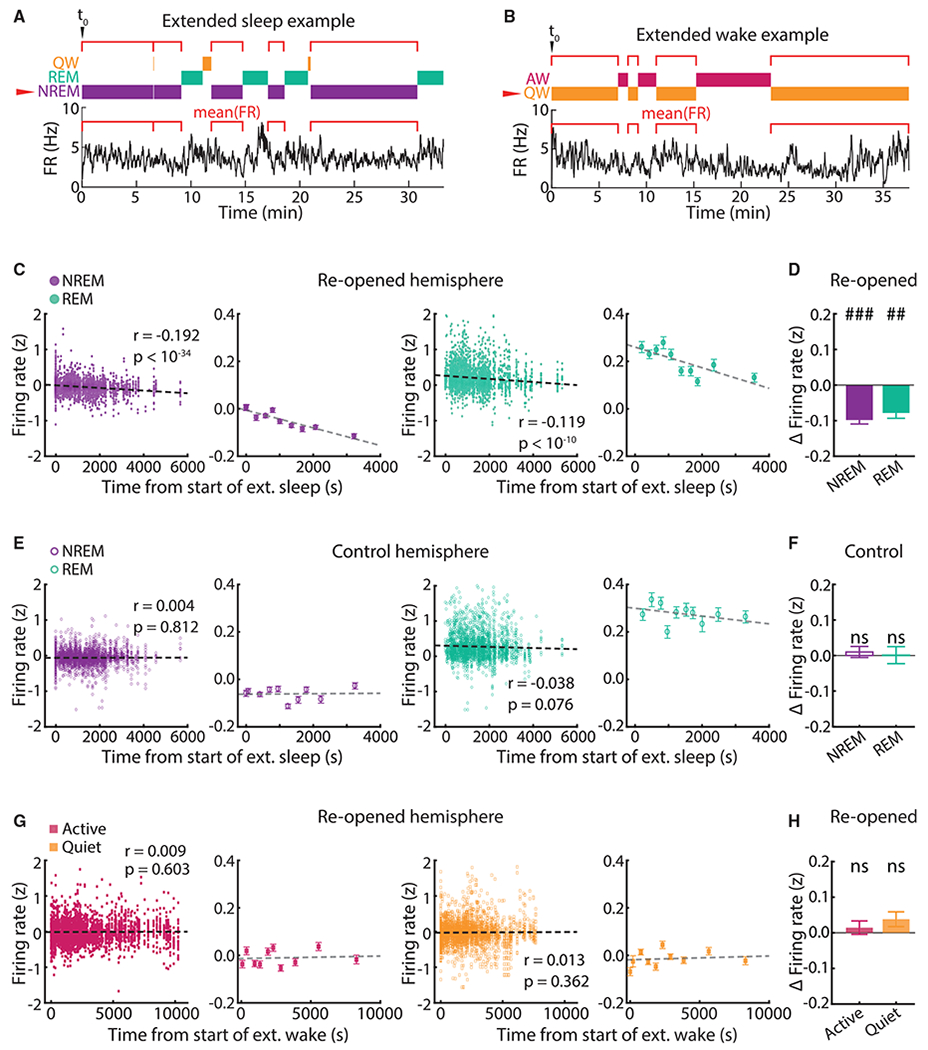
Homeostatic plasticity is hypothesized to bidirectionally regulate neuronal activity around a stable set point to compensate for learning-related plasticity, but to date only upward firing rate homeostasis (FRH) has been demonstrated in vivo. We combined chronic electrophysiology in freely behaving animals with an eye-reopening paradigm to enhance firing in primary visual cortex (V1) and found that neurons bidirectionally regulate firing rates around an individual set point. Downward FRH did not require N-methyl-D-aspartate receptor (NMDAR) signaling and was associated with homeostatic scaling down of synaptic strengths. Like upward FRH, downward FRH was gated by arousal state but in the opposite direction: it occurred during sleep, not during wake. In contrast, firing rate depression associated with Hebbian plasticity happened independently of sleep and wake. Thus, sleep and wake states temporally segregate upward and downward FRH, which might prevent interference or provide unopposed homeostatic compensation when it is needed most.

Keywords: Experience-dependent plasticity; Firing rate homeostasis; Homeostatic plasticity; Sleep; Synaptic scaling; Visual system.


Conflict of interest statement

Declaration of Interests G.G.T. is a member of the Neuron advisory board. The authors declare no competing interests.

Figures

Figure 1.
Figure 1.. Eye Reopening after MD Causes an Overshoot in Firing Followed by Homeostatic Recovery
(A) Example neurons recorded continuously from the control hemisphere for 5 days. Here and below, solid lines represent the firing rate of a single neuron, the dashed line indicates the mean firing rate (FR) of the neuron during the baseline (MD4) period, white and gray bars in the background indicate 12 h of light and dark, and the artifact from unplugging animals for ER surgery has been removed. (B) Example neurons recorded continuously from the reopened hemisphere for 5 days. (C and D) Average firing rate traces for all neurons recorded in control and reopened hemispheres, normalized to baseline (MD4) for each cell. (E) Comparison of individual neuronal firing rates between baseline (MD4) and early ER (left) or late ER (right). Each dot is the mean firing rate of a neuron averaged over the corresponding 12-h period. The yellow line indicates unity. (F) Percentage of change in firing rate from baseline at early ER and late ER for recorded neurons. Black lines indicate mean ± SEM. Control, n = 31; reopen, n = 36; Kruskal-Wallis test (p < 0.0001) with Tukey-Kramer post hoc; n.s. (not significant) p = 0.99, **p = 0.003, ***p = 0.002, ****p = 0.0004. (G) Mean firing rate of every cell at baseline (MD4), early ER, and late ER. Each dot represents the mean firing rate of a neuron over the corresponding 12-h period, and mean firing rates for the same neuron are connected via solid lines. Control, n = 31; reopen, n = 36; Wilcoxon sign-rank test with Bonferroni correction; ***p < 0.001.
Figure 2.
Figure 2.. ER-Induced Firing Rate Overshoot, but Not Downward Recovery, Is NMDAR-Dependent
(A and B) Experiment schematic: the NMDAR antagonist CPP (15 mg/kg) was injected subcutaneously once at the time of eye reopening (A) or twice after that at 24-h intervals (B). (C and D) Example firing rates of neurons recorded in each of the CPP experiments. (E) Change in firing rate from baseline to early ER to late ER for neurons recorded in the first CPP condition (one injection at time of ER). n = 15; Wilcoxon sign-rank test with Bonferroni correction; *p = 0.0251, **p = 0.0026. (F) Percentage of change in firing rate from baseline in the first CPP condition. One-sample t test compared with mean = 0; ER2, p = 0.723; ER4, p < 10−4. n = 15; two-sample t test; *p = 0.0293. (G) As in (E), but for the second CPP condition (two injections, 24 and 48 h after ER). n = 22; Wilcoxon sign-rank test with Bonferroni correction; *p = 0.0269, **p = 0.0014. (H) As in (F), but for the second CPP condition. One-sample t test compared with mean = 0; ER2, p = 0.003; ER4, p = 0.657. Two-sample t test; **p = 0.0058.
Figure 3.
Figure 3.. Downward Firing Rate Recovery Is Associated with Synaptic Scaling Down
(A) Schematic of experimental timeline. Slices were taken 24 h (ER2) or 72 h (ER4) after ER. (B) Example recordings of mEPSCs in L2/3 pyramidal neurons in V1. Average peak-aligned mEPSC waveforms for each condition are also shown. (C) Average mEPSC amplitude in each condition. Each dot represents one neuron. Black lines represent mean ± SEM. Kruskal-Wallis test (p < 0.001); ER2 control, n = 27; ER4 control, n = 17; ER2 reopen, n = 25; ER4 reopen, n = 22; **p < 0.01. (D) Cumulative distribution of mEPSC amplitudes. Kuiper test with Bonferroni correction; # p < 10−5, ## p < 10−6. (E) Cumulative distribution of mEPSC amplitudes. The black dotted line represents ER2 reopen distribution scaled according to the linear function f(x) = 0.877x + 0.318. Kuiper test; ## p < 10−6. (F) As in (E) but comparing the amplitude distribution at ER2 in control versus reopen conditions. The ER2 control distribution has been scaled according to the linear function f(x) = 0.999x + 0.692. Kuiper test; *p = 0.0377, ***p < 0.001.
Figure 4.
Figure 4.. Downward FRH Occurs during Sleep-Dense, but Not Wake-Dense, Periods
(A) Schematic of state-dense analysis. (B) Average change in firing rate over sleep-dense (S) or wake-dense (W) windows in control and reopened hemispheres. Bars represent mean ± SEM. Kruskal-Wallis test (p < 10−6) with Tukey-Kramer post hoc; control S, n = 19 neurons, 13 windows; control W, n = 19 neurons, 10 windows; reopen S, n = 36 neurons, 13 windows; reopen W, n = 36 neurons, 14 windows; **p = 0.0051, ***p = 0.0031, ## p < 10−7. (C) Cumulative distribution function of mean change in firing rate across S or W windows for all neurons. Vertical dashed line indicates no change. Two-sample Kolmogorov-Smirnov test with Bonferroni correction; control, n = 19; reopened, n = 36; *p =0.0380, ## p < 10−6.
Figure 5.
Figure 5.. During Downward FRH, Firing Rates Depress as a Function of Time Spent Asleep
(A) Schematic of extended sleep analysis for one neuron. Individual epochs of a state (NREM in this example) are found within an extended sleep episode, and the neuron’s mean firing rate is calculated in each one; these values are then plotted against the start time of that epoch, aligned to the start of the whole episode (t0). (B) As in (A), but showing an example extended wake episode. (C) Correlation between firing rate in NREM and REM and time from start of extended sleep in the reopened hemisphere. All data points are shown in the left panels; the right panels show data in 10 groups of equal size for ease of visualization (dots show mean ± SEM). Pearson r and associated p values were computed on the ungrouped data (NREM, n = 4,036 data points; REM, n = 3,161 data points). Firing rates were Z scored to the extended sleep episode. (D) Difference in firing rate between the last and the first NREM (left) or REM (right) epoch within an extended sleep episode, averaged across all episodes. Bars show mean ± SEM. One-sample t test compared with mean = 0; n = 74 episodes; ## p < 10−6, ### p < 10−17. (E and F) As in (C) and (D), but for NREM and REM in the control hemisphere (NREM, n = 2,933 data points; REM, n = 2,249 data points). In (F), one-sample t test compared with mean = 0; n = 47 episodes. (G and H) As in (C) and (D), but for active wake (AW, n = 4,156 data points) and quiet wake (QW, 5,131 data points) in the reopened hemisphere. In (H), one-sample t test compared with mean = 0; n = 70 episodes.
Figure 6.
Figure 6.. Sleep Deprivation (SD) during Downward FRH Slows the Restoration of Firing Rates
(A) Diagram of the SD paradigm and analysis. Yellow and gray rectangles represent 12 h of light and dark, respectively. Animals were sleep deprived during two 12-h periods during the light phase of ER2 and ER3. The 12-h bins corresponding to the dark phases were used to calculate average firing rates for individual neurons. The change in firing rate was then calculated across each SD period and for the last light period (no SD). (B) Change in firing rate for each recorded neuron in the SD condition (n = 22 neurons from 4 animals) and control condition (n = 36 neurons from 5 animals). Left, effect of SD on ER2 (change in firing rate between time points 1 and 2). Middle, effect of SD on ER3 (change in firing rate between time points 2 and 3). Right, effect of last light period (change in firing rate between time points 3 and 4, when no SD occurred). Yellow dashed lines indicate no change. **p = 0.0079, two-sample t test; # p = 0.0449, ## p = 0.0084, #### p < 10−5; one-sample t test versus mean of 0. (C) Average change in firing rate for each neuron across periods of sleep and wake for the control group, and periods of prolonged wake and recovery sleep for the SD group. Only data for the two 12-h SD periods (and corresponding times in the control dataset) were used. Data for both periods were pooled. The yellow dashed line indicates no change. *p = 0.0374, ***p = 0.0001, ****p < 10−4; one-way ANOVA (p < 10−4) with Tukey-Kramer post hoc; # p = 0.0247, #### p < 10−6; linear model comparing all group means to a mean of 0.
Figure 7.
Figure 7.. Downward FRH Occurs during Both NREM and REM Sleep
(A) Schematic for the analysis of NREM1-REM-NREM2 triplets. (B) Firing rate values are Z scored within each triplet and then averaged across all triplets for a given neuron. Each dot represents a neuron, and mean firing rate values for the same neuron are linked by horizontal lines. ****p < 0.0001; Wilcoxon sign-rank test. (C) Difference in mean firing rate values between the first 5 min of NREM2 and the last 5 min of NREM1. Black lines indicate mean ± SEM. The yellow dashed line indicates no change. ****p < 0.0001; one-sample t test versus mean of 0. (D) As in (A), but for REM1-NREM-REM2 triplets. (E) As in (B), but for REM1-NREM-REM2 triplets. *p = 0.05; Wilcoxon sign-rank test. (F) As in (C), but for REM1-NREM-REM2 triplets. *p = 0.0383; one-sample t test versus mean of 0.
Figure 8.
Figure 8.. Non-homeostatic Firing Rate Changes Happen Independently of Animals’ Behavioral State
(A) Two example neurons whose activity begins decreasing in the first light period after MD (early drop). Dashed lines indicate the baseline firing rate for each neuron. White/gray bars in the background indicate 12 h of light/dark. (B) As in (A), but for two neurons whose drop-in firing rate begins during the second light period after MD (late drop). (C) Average baseline-normalized firing rate of all early-drop neurons. The dashed line indicates the baseline firing rate. (D) As in (C), but for late-drop neurons. (E) Change in firing rate across sleep- or wake-dense epochs for all neurons in their respective 12-h drop period. Control S, n = 38 epochs; control W, n = 12 epochs; deprived S, n = 38 epochs; deprived W, n = 26 epochs. (F) Correlation between change in firing rate during drop and percentage of time spent asleep in the same period. Each data point represents the average change in firing rate across early-drop (circles) or late-drop (diamonds) neurons, and each color represents a different animal (n = 6 animals). Percentage of time asleep is calculated per animal in the early-drop or late-drop 12-h period. The black dashed line represents no change. The black solid line shows the linear fit to the data.

Comment in

  • Sleep calms firing rates.
    Whalley K. Whalley K. Nat Rev Neurosci. 2021 Feb;22(2):74-75. doi: 10.1038/s41583-020-00419-3. Nat Rev Neurosci. 2021. PMID: 33293691 No abstract available.
  • The Ups and Downs of Firing Rate Homeostasis.
    Jacobsen RI, Keck T. Jacobsen RI, et al. Neuron. 2021 Feb 3;109(3):401-403. doi: 10.1016/j.neuron.2020.12.024. Neuron. 2021. PMID: 33539775

References

    1. Abbott LF, and Nelson SB (2000). Synaptic plasticity: taming the beast. Nat. Neurosci 3 (Suppl), 1178–1183. - PubMed
    1. Abel T, Havekes R, Saletin JM, and Walker MP (2013). Sleep, plasticity and memory from molecules to whole-brain networks. Curr. Biol 23, R774–R788. - PMC - PubMed
    1. Aton SJ, Suresh A, Broussard C, and Frank MG (2014). Sleep promotes cortical response potentiation following visual experience. Sleep (Basel) 37, 1163–1170. - PMC - PubMed
    1. Barnes SJ, Sammons RP, Jacobsen RI, Mackie J, Keller GB, and Keck T (2015). Subnetwork-Specific Homeostatic Plasticity in Mouse Visual Cortex In Vivo. Neuron 86, 1290–1303. - PMC - PubMed
    1. Blackman MP, Djukic B, Nelson SB, and Turrigiano GG (2012).Acritical and cell-autonomous role for MeCP2 in synaptic scaling up. J. Neurosci 32, 13529–13536. - PMC - PubMed

Publication types

Substances