. 2020 Dec 3;107(6):1170-1177.
doi: 10.1016/j.ajhg.2020.11.001. Epub 2020 Nov 23.

Heterozygous Variants in KDM4B Lead to Global Developmental Delay and Neuroanatomical Defects

Affiliations

Heterozygous Variants in KDM4B Lead to Global Developmental Delay and Neuroanatomical Defects

Anna R Duncan et al. Am J Hum Genet. .

Abstract

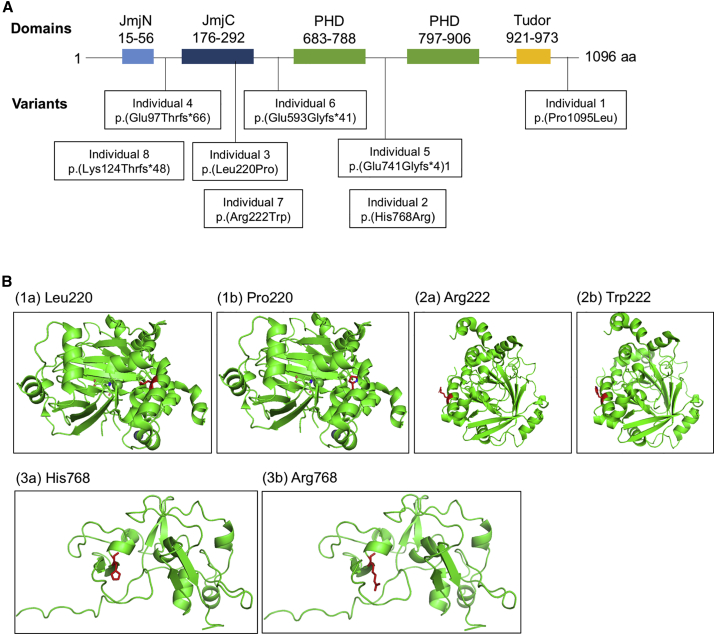
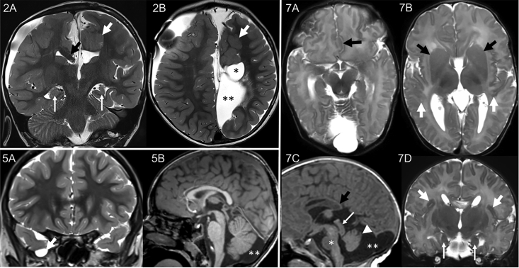
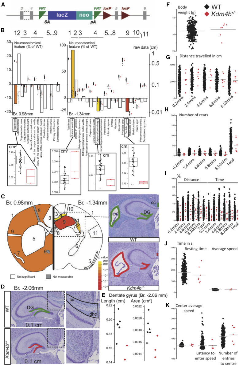
KDM4B is a lysine-specific demethylase with a preferential activity on H3K9 tri/di-methylation (H3K9me3/2)-modified histones. H3K9 tri/di-demethylation is an important epigenetic mechanism responsible for silencing of gene expression in animal development and cancer. However, the role of KDM4B on human development is still poorly characterized. Through international data sharing, we gathered a cohort of nine individuals with mono-allelic de novo or inherited variants in KDM4B. All individuals presented with dysmorphic features and global developmental delay (GDD) with language and motor skills most affected. Three individuals had a history of seizures, and four had anomalies on brain imaging ranging from agenesis of the corpus callosum with hydrocephalus to cystic formations, abnormal hippocampi, and polymicrogyria. In mice, lysine demethylase 4B is expressed during brain development with high levels in the hippocampus, a region important for learning and memory. To understand how KDM4B variants can lead to GDD in humans, we assessed the effect of KDM4B disruption on brain anatomy and behavior through an in vivo heterozygous mouse model (Kdm4b+/-), focusing on neuroanatomical changes. In mutant mice, the total brain volume was significantly reduced with decreased size of the hippocampal dentate gyrus, partial agenesis of the corpus callosum, and ventriculomegaly. This report demonstrates that variants in KDM4B are associated with GDD/ intellectual disability and neuroanatomical defects. Our findings suggest that KDM4B variation leads to a chromatinopathy, broadening the spectrum of this group of Mendelian disorders caused by alterations in epigenetic machinery.

Keywords: JMJD2B; KDM4B; agenesis of the corpus callosum; dysmorphic hippocampi; global developmental delay; heterozygous variant; intellectual disability; neurodevelopmental disorder.


Conflict of interest statement

M.J.G.S. and T.S.S. are employees of GeneDx. P.B.A. is on the Scientific Advisory Board of Illumina, Inc. and GeneDx. The other authors declare no competing interests.

Figures

Figure 1
Figure 1
KDM4B Variant Localization and Molecular Modeling (A) KDM4B with domains and patient individual variants noted. (B) Molecular modeling of the missense variants in individuals (I) 2, 3, and 7. (1a) shows the helix formed by the catalytic domain of KDM4B. Residue Leu220 is in red. (2b) shows the local destruction of the helical structure when Pro220 is substituted. Pro220 is shown in red. (2a) shows the residue Arg222 within the catalytic domain of KDM4B; Arg222 is highlighted in red. (2b) shows the substitution of Arg222 for Trp222 in I:7, which creates a bulky hydrophobic side chain most likely altering the folding and stability of the helix formation; Trp222 is shown in red. (3a) shows His768 and the zinc knuckle formation; His768 highlighted in red. (3b) shows the unraveling of the zinc knuckle with the substitution of Arg768 in I:2; Arg768 is shown in red.
Figure 2
Figure 2
MRI Findings in Individuals 2, 5, and 7 I:2 had many abnormalities, including agenesis of the corpus callosum with a right bundle of Probst (2A, black arrow), white matter cysts (2B, asterisk), interhemispheric cysts (2B, two asterisks), polymicrogyria (2A and 2B, white arrows), and incompletely rotated dysmorphic hippocampi with the left decreased in volume (2A, thin arrows). The following are not shown: absent left fornix with right fornix fused with the bundle of Probst, subcortical gray matter heterotopia, stenogyria of the right hemisphere (probably due to prior hydrocephalus), and decreased white matter volume. I:5 had a small right middle cranial fossa meningocele (5A, arrow) and mega cisterna magna (5B, two asterisks). I:7 also had many abnormalities, including partial agenesis of the corpus callosum (7C, black arrow), polymicrogyria (7B and 7D, arrows), dysmorphic hippocampi (7D, thin arrows), interdigitation of the frontal lobes (7A, arrow), fused caudate and lentiform nuclei with absent anterior limb internal capsule (7B, arrows), mega cisterna magna (7C, two asterisks), prominent tectum (7C, arrow), incompletely rotated small vermis (7C, arrowhead), and hypoplastic brainstem (7C, asterisk). The following are not shown: dysmorphic partially fused fornices, azygous ACA, small optic nerves, and small cerebellar hemispheres.
Figure 3
Figure 3
Mouse Studies Reveal a Role of KDM4B in the Anatomy of the Brain (A) Construction of the Kdm4btm1a(EUCOMM)Wtsi allele. (B) Histograms for three heterozygous Kdm4b mice showing percentage difference relative to 40 WTs. The overlapping dots show actual data points. To present areas and lengths on the same scale, the square root of areas was used. Inserts are full dot and box plots of WT (black) versus Kdm4b+/− mice (red) for each of the four significant parameters (total brain area, lateral ventricle area, corpus callosum height, dentate gyrus length). (C and D) Left: schematic representation of a section at Bregma +0.98 mm and Bregma −1.34 mm. Colored regions indicate the presence of at least one significant parameter within the brain region at the 0.05 level. White coloring indicates a p value higher than 0.05 and gray shows there is not enough data to calculate a p value. Right: illustrating example of WT and Kdm4b+/− brain images in coronal sections double-stained for Nissl and Luxol at Bregma −1.34 (C) and Bregma −2.06 mm (D). (E) Dot plot of the coronal section at Bregma −2.06 mm showing further characterization of the hippocampus. (F) Body weight of seven Kdm4b+/− and 771 baseline control mice. (G) Distance traveled in cm in the open-field arena shown in sessions of 2 min each. (H) Number of rears measured in the open-field arena. (I) Distance and time in the open-field arena expressed in percentages. (J) Resting time and average speed in the open-field test. (K) Average speed in the center of the open-field arena, latency to enter the center, and the number of entries to the center.

References

    1. Moeschler J.B., Shevell M., Committee on Genetics Comprehensive evaluation of the child with intellectual disability or global developmental delays. Pediatrics. 2014;134:e903–e918. - PMC - PubMed
    1. Centers for Disease Control and Prevention (CDC) Economic costs associated with mental retardation, cerebral palsy, hearing loss, and vision impairment--United States, 2003. MMWR Morb. Mortal. Wkly. Rep. 2004;53:57–59. - PubMed
    1. Dikow N., Moog U., Karch S., Sander A., Kilian S., Blank R., Reuner G. What do parents expect from a genetic diagnosis of their child with intellectual disability? J. Appl. Res. Intellect. Disabil. 2019;32:1129–1137. - PubMed
    1. Gilissen C., Hehir-Kwa J.Y., Thung D.T., van de Vorst M., van Bon B.W., Willemsen M.H., Kwint M., Janssen I.M., Hoischen A., Schenck A. Genome sequencing identifies major causes of severe intellectual disability. Nature. 2014;511:344–347. - PubMed
    1. Vissers L.E., Gilissen C., Veltman J.A. Genetic studies in intellectual disability and related disorders. Nat. Rev. Genet. 2016;17:9–18. - PubMed

Publication types

MeSH terms