. 2021 Apr:137:106823.
doi: 10.1016/j.vph.2020.106823. Epub 2020 Nov 21.

SARS-CoV-2 spike protein-mediated cell signaling in lung vascular cells

Affiliations

SARS-CoV-2 spike protein-mediated cell signaling in lung vascular cells

Yuichiro J Suzuki et al. Vascul Pharmacol. 2021 Apr.

Abstract

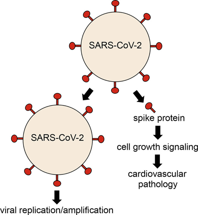
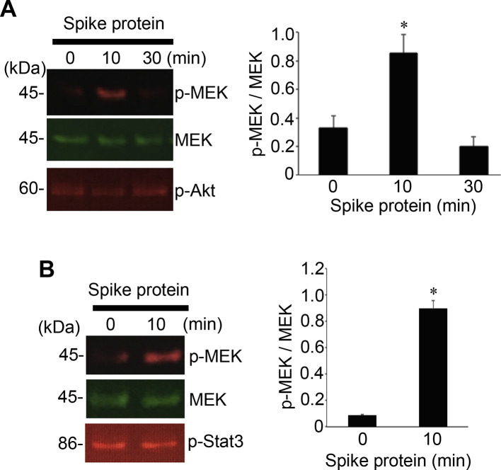
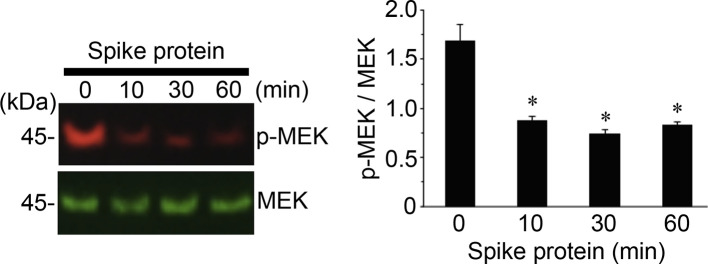
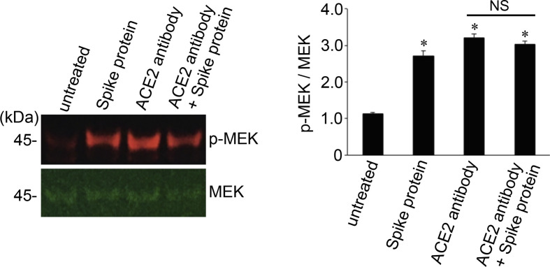
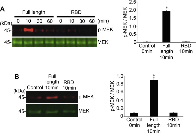
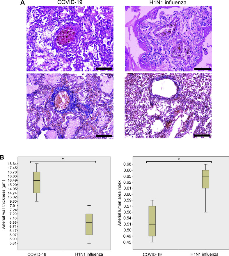
Currently, the world is suffering from the pandemic of coronavirus disease 2019 (COVID-19), caused by severe acute respiratory syndrome coronavirus 2 (SARS-CoV-2) that uses angiotensin-converting enzyme 2 (ACE2) as a receptor to enter the host cells. So far, 60 million people have been infected with SARS-CoV-2, and 1.4 million people have died because of COVID-19 worldwide, causing serious health, economical, and sociological problems. However, the mechanism of the effect of SARS-CoV-2 on human host cells has not been defined. The present study reports that the SARS-CoV-2 spike protein alone without the rest of the viral components is sufficient to elicit cell signaling in lung vascular cells. The treatment of human pulmonary artery smooth muscle cells or human pulmonary artery endothelial cells with recombinant SARS-CoV-2 spike protein S1 subunit (Val16 - Gln690) at 10 ng/ml (0.13 nM) caused an activation of MEK phosphorylation. The activation kinetics was transient with a peak at 10 min. The recombinant protein that contains only the ACE2 receptor-binding domain of the SARS-CoV-2 spike protein S1 subunit (Arg319 - Phe541), on the other hand, did not cause this activation. Consistent with the activation of cell growth signaling in lung vascular cells by the SARS-CoV-2 spike protein, pulmonary vascular walls were found to be thickened in COVID-19 patients. Thus, SARS-CoV-2 spike protein-mediated cell growth signaling may participate in adverse cardiovascular/pulmonary outcomes, and this mechanism may provide new therapeutic targets to combat COVID-19.

Keywords: COVID-19; Cell signaling; Coronavirus; SARS-CoV-2; Vascular.


Conflict of interest statement

None.

Figures

Unlabelled Image
Graphical abstract
Fig. 1
Fig. 1
The SARS-CoV-2 spike protein S1 promotes the MEK phosphorylation in human cells. (A) Human pulmonary artery smooth cells (N = 5) and (B) human pulmonary artery endothelial cells (N = 3) were treated with recombinant full length S1 subunit of the SARS-CoV-2 spike protein (Val16 - Gln690) at 10 ng/ml for the durations indicated. Cell lysates were prepared and subjected to Western blotting using antibodies against phosphorylated MEK (p-MEK), MEK protein, phosphorylated Akt (p-Akt), and phosphorylated Stat3 (p-Stat3). Bar graphs represent means ± SEM. *Significantly different from 0 min at p < 0.05.
Fig. 2
Fig. 2
In rat cells, the SARS-CoV-2 spike protein S1 does not phosphorylate, but rather dephosphorylates MEK. Rat pulmonary artery smooth muscle cells were treated with recombinant full length S1 subunit of the SARS-CoV-2 spike protein (Val16 - Gln690) at 10 ng/ml for the durations indicated. Cell lysates were prepared and subjected to Western blotting using antibodies against phosphorylated MEK (p-MEK) and MEK protein. The bar graph represents means ± SEM (N = 4). *Significantly different from 0 min at p < 0.05.
Fig. 3
Fig. 3
The SARS-CoV-2 spike protein S1 does not promote the phosphorylation of MEK in the presence of the neutralizing antibody against ACE2. Human pulmonary artery smooth muscle cells were pre-treated with the ACE2 antibody for 1 h and then treated with recombinant full length S1 subunit of the SARSCoV-2 spike protein (Val16 – Gln690) for 10 min. Cell lysates were prepared and subjected to Western blotting using antibodies against phosphorylated MEK (p-MEK) and MEK protein. The bar graph represents means ± SEM (N = 4).*Significantly different from untreated control at p < 0.05. NS denotes that the two values are not significantly different from each other at p < 0.05.
Fig. 4
Fig. 4
The RBD only containing SARS-CoV-2 spike protein S1 does not activate the MEK phosphorylation. (A) Human pulmonary artery smooth muscle cells and (B) human pulmonary artery endothelial cells were treated with recombinant full length S1 subunit (Val16 - Gln690) or the RBD region of S1 subunit (Arg319 - Phe541) at 100 ng/ml for the durations indicated. Cell lysates were prepared and subjected to Western blotting using antibodies against phosphorylated MEK (p-MEK) and the MEK protein. Bar graphs represent means ± SEM (N = 3). *Significantly different from 0 min control at p < 0.05.
Fig. 5
Fig. 5
Pulmonary arterial walls are thickened in COVID-19 patients. (A) Representative hematoxylin and eosin staining images of the pulmonary arteries of patients who died of ARDS due to COVID-19 and H1N1 influenza. In COVID-19 patients, but not in H1N1 influenza patients, thickening of the pulmonary arterial walls was observed. Scale bars indicate 100 μm. (B) Box plots represent quantifications of pulmonary arterial wall thicknesses and lumen areas in COVID-19 and H1N1 influenza patients using H&E stained slides. The number of patients used for these analyses was 10 for COVID-19 and 10 for H1N1. *The Mann-Whitney U Test indicated that the two values are significantly different at p = 0.001.

Update of

Similar articles

Cited by

References

    1. Su S., Wong G., Shi W., Liu J., Lai A.C.K., Zhou J., Liu W., Bi Y., Gao G.F. Epidemiology, genetic recombination, and pathogenesis of coronaviruses. Trends Microbiol. 2016;24:490–502. - PMC - PubMed
    1. Satija N., Lal S.K. The molecular biology of SARS coronavirus. Ann. N. Y. Acad. Sci. 2007;1102:26–38. - PMC - PubMed
    1. Wu F., Zhao S., Yu B., Chen Y.M., Wang W., Song Z.G., Hu Y., Tao Z.W., Tian J.H., Pei Y.Y., Yuan M.L., Zhang Y.L., Dai F.H., Liu Y., Wang Q.M., Zheng J.J., Xu L., Holmes E.C., Zhang Y.Z. A new coronavirus associated with human respiratory disease in China. Nature. 2020;579:265–269. - PMC - PubMed
    1. Huang C., Wang Y., Li X., Ren L., Zhao J., Hu Y., Zhang L., Fan G., Xu J., Gu X., Cheng Z., Yu T., Xia J., Wei Y., Wu W., Xie X., Yin W., Li H., Liu M., Xiao Y., Gao H., Guo L., Xie J., Wang G., Jiang R., Gao Z., Jin Q., Wang J., Cao B. Clinical features of patients infected with 2019 novel coronavirus in Wuhan, China. Lancet. 2020;395:497–506. - PMC - PubMed
    1. Yan R., Zhang Y., Li Y., Xia L., Guo Y., Zhou Q. Structural basis for the recognition of SARS-CoV-2 by full-length human ACE2. Science. 2020;367:1444–1448. - PMC - PubMed

Publication types

MeSH terms

Substances