

Deep learning-based classification of primary bone tumors on radiographs: A preliminary study
- PMID: 33232868
- PMCID: PMC7689511
- DOI: 10.1016/j.ebiom.2020.103121
Deep learning-based classification of primary bone tumors on radiographs: A preliminary study
Abstract
Background: To develop a deep learning model to classify primary bone tumors from preoperative radiographs and compare performance with radiologists.
Methods: A total of 1356 patients (2899 images) with histologically confirmed primary bone tumors and pre-operative radiographs were identified from five institutions' pathology databases. Manual cropping was performed by radiologists to label the lesions. Binary discriminatory capacity (benign versus not-benign and malignant versus not-malignant) and three-way classification (benign versus intermediate versus malignant) performance of our model were evaluated. The generalizability of our model was investigated on data from external test set. Final model performance was compared with interpretation from five radiologists of varying level of experience using the Permutations tests.
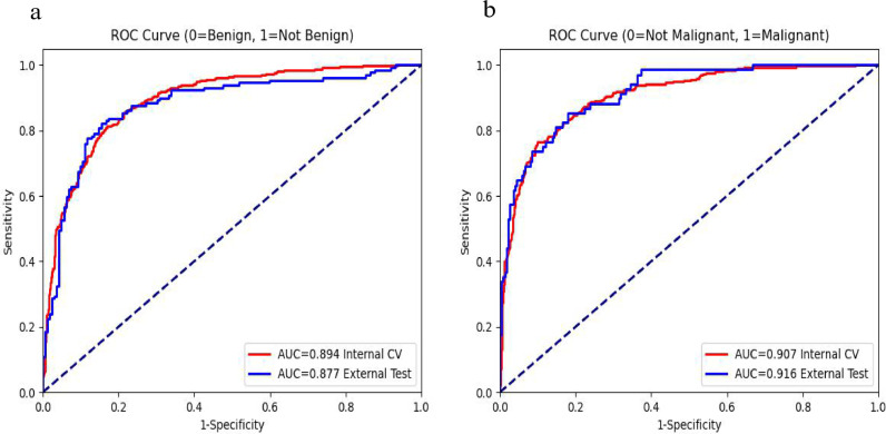
Findings: For benign vs. not benign, model achieved area under curve (AUC) of 0•894 and 0•877 on cross-validation and external testing, respectively. For malignant vs. not malignant, model achieved AUC of 0•907 and 0•916 on cross-validation and external testing, respectively. For three-way classification, model achieved 72•1% accuracy vs. 74•6% and 72•1% for the two subspecialists on cross-validation (p = 0•03 and p = 0•52, respectively). On external testing, model achieved 73•4% accuracy vs. 69•3%, 73•4%, 73•1%, 67•9%, and 63•4% for the two subspecialists and three junior radiologists (p = 0•14, p = 0•89, p = 0•93, p = 0•02, p < 0•01 for radiologists 1-5, respectively).
Interpretation: Deep learning can classify primary bone tumors using conventional radiographs in a multi-institutional dataset with similar accuracy compared to subspecialists, and better performance than junior radiologists.
Funding: The project described was supported by RSNA Research & Education Foundation, through grant number RSCH2004 to Harrison X. Bai.
Keywords: Convolutional neural network; Deep learning; Plain radiograph; Primary bone tumor.
Copyright © 2020 The Authors. Published by Elsevier B.V. All rights reserved.
Figures

Similar articles
-
A deep learning-machine learning fusion approach for the classification of benign, malignant, and intermediate bone tumors.Eur Radiol. 2022 Feb;32(2):1371-1383. doi: 10.1007/s00330-021-08195-z. Epub 2021 Aug 25. Eur Radiol. 2022. PMID: 34432121
-
Deep Learning for Classification of Bone Lesions on Routine MRI.EBioMedicine. 2021 Jun;68:103402. doi: 10.1016/j.ebiom.2021.103402. Epub 2021 Jun 5. EBioMedicine. 2021. PMID: 34098339 Free PMC article.
-
Multitask Deep Learning for Segmentation and Classification of Primary Bone Tumors on Radiographs.Radiology. 2021 Nov;301(2):398-406. doi: 10.1148/radiol.2021204531. Epub 2021 Sep 7. Radiology. 2021. PMID: 34491126
-
A radiograph-based deep learning model improves radiologists' performance for classification of histological types of primary bone tumors: A multicenter study.Eur J Radiol. 2024 Jul;176:111496. doi: 10.1016/j.ejrad.2024.111496. Epub 2024 May 7. Eur J Radiol. 2024. PMID: 38733705
-
A proposed "Radiological Evaluation Score for Bone Tumors" (REST): An objective system for assessment of a radiograph in patients with suspected bone tumor.Musculoskelet Surg. 2022 Dec;106(4):371-382. doi: 10.1007/s12306-021-00711-0. Epub 2021 May 12. Musculoskelet Surg. 2022. PMID: 33982208 Review.
Cited by
-
Detecting bone lesions in X-ray under diverse acquisition conditions.J Med Imaging (Bellingham). 2024 Mar;11(2):024502. doi: 10.1117/1.JMI.11.2.024502. Epub 2024 Mar 19. J Med Imaging (Bellingham). 2024. PMID: 38510544 Free PMC article.
-
AI in radiological imaging of soft-tissue and bone tumours: a systematic review evaluating against CLAIM and FUTURE-AI guidelines.EBioMedicine. 2025 Apr;114:105642. doi: 10.1016/j.ebiom.2025.105642. Epub 2025 Mar 20. EBioMedicine. 2025. PMID: 40118007 Free PMC article.
-
Artificial intelligence and machine learning applications for the imaging of bone and soft tissue tumors.Front Radiol. 2024 Sep 5;4:1332535. doi: 10.3389/fradi.2024.1332535. eCollection 2024. Front Radiol. 2024. PMID: 39301168 Free PMC article. Review.
-
A Radiograph Dataset for the Classification, Localization, and Segmentation of Primary Bone Tumors.Sci Data. 2025 Jan 16;12(1):88. doi: 10.1038/s41597-024-04311-y. Sci Data. 2025. PMID: 39820508 Free PMC article.
-
Applications of artificial intelligence in orthopaedic surgery.Front Med Technol. 2022 Dec 15;4:995526. doi: 10.3389/fmedt.2022.995526. eCollection 2022. Front Med Technol. 2022. PMID: 36590152 Free PMC article. Review.
References
-
- Ward E, DeSantis C, Robbins A, Kohler B, Jemal A. Childhood and adolescent cancer statistics, 2014. CA Cancer J Clin. 2014;64:83–103. - PubMed
-
- Siegel RL, Miller KD, Jemal A. Cancer statistics, 2018. CA Cancer J Clin. 2018;68:7–30. - PubMed
-
- Fletcher CDM. International Agency for research on cancer. WHO classification of tumours of soft tissue and bone. 4th ed. IARC Press; Lyon: 2013. World Health Organization; p. 468. p. p.
-
- Helms . Wolters Kluwer Health; Philadelphia: 2012. WEBCA. Fundamentals of diagnostic radiology; p. 1420.
Publication types
MeSH terms
LinkOut - more resources
Full Text Sources
Medical
