

Generation of an Oncolytic Herpes Simplex Viral Vector Completely Retargeted to the GDNF Receptor GFRα1 for Specific Infection of Breast Cancer Cells
- PMID: 33233403
- PMCID: PMC7700293
- DOI: 10.3390/ijms21228815
Generation of an Oncolytic Herpes Simplex Viral Vector Completely Retargeted to the GDNF Receptor GFRα1 for Specific Infection of Breast Cancer Cells
Abstract
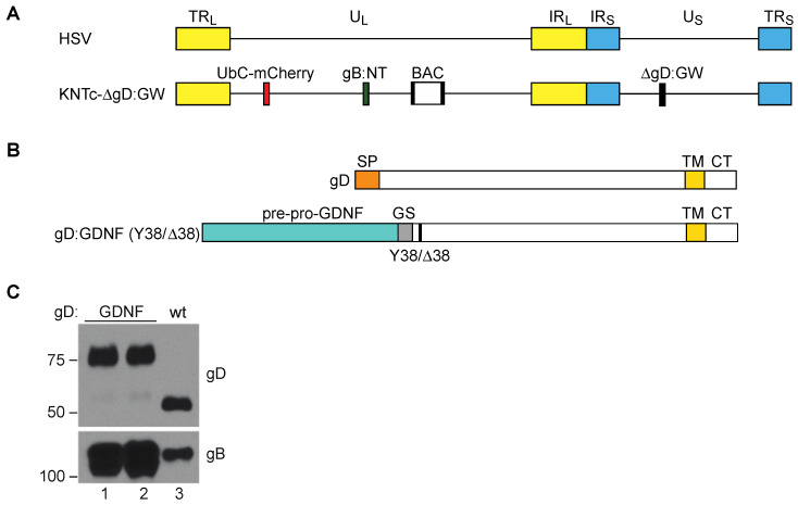
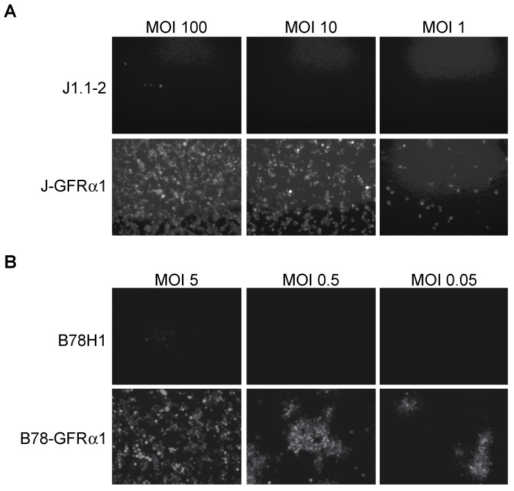
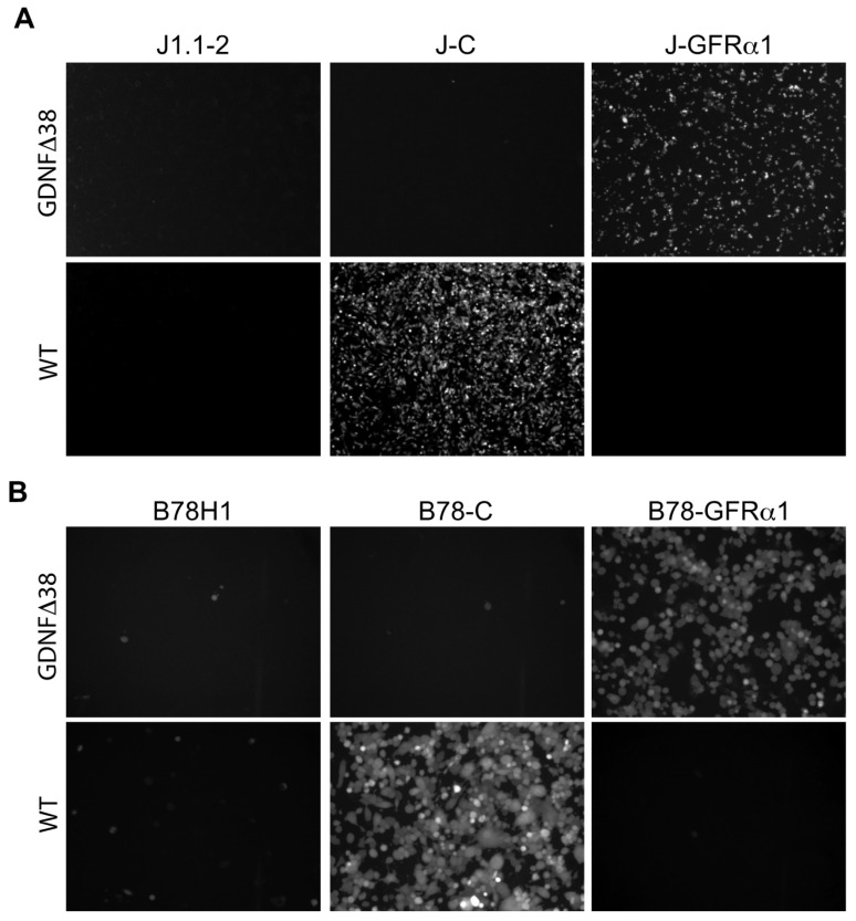
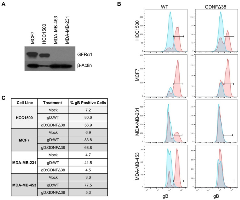
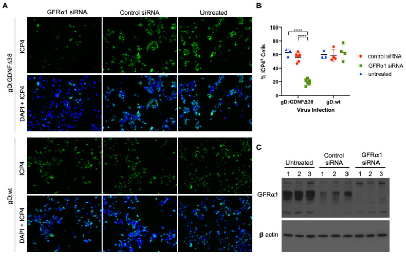
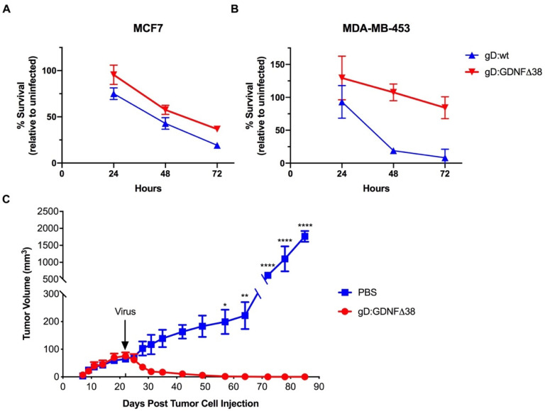
Oncolytic herpes simplex viruses (oHSV) are under development for the treatment of a variety of human cancers, including breast cancer, a leading cause of cancer mortality among women worldwide. Here we report the design of a fully retargeted oHSV for preferential infection of breast cancer cells through virus recognition of GFRα1, the cellular receptor for glial cell-derived neurotrophic factor (GDNF). GFRα1 displays a limited expression profile in normal adult tissue, but is upregulated in a subset of breast cancers. We generated a recombinant HSV expressing a completely retargeted glycoprotein D (gD), the viral attachment/entry protein, that incorporates pre-pro-GDNF in place of the signal peptide and HVEM binding domain of gD and contains a deletion of amino acid 38 to eliminate nectin-1 binding. We show that GFRα1 is necessary and sufficient for infection by the purified recombinant virus. Moreover, this virus enters and spreads in GFRα1-positive breast cancer cells in vitro and caused tumor regression upon intratumoral injection in vivo. Given the heterogeneity observed between and within individual breast cancers at the molecular level, these results expand our ability to deliver oHSV to specific tumors and suggest opportunities to enhance drug or viral treatments aimed at other receptors.
Keywords: breast cancer; herpes simplex virus; oncolytic.
Conflict of interest statement
J.C.G. is an inventor of intellectual property licensed to Coda Biotherapeutics, Inc. (San Francisco, CA). J.B.C. and J.C.G. are inventors of intellectual property licensed to Oncorus, Inc. (Cambridge, MA, USA). J.C.G. is a consultant and member of the Scientific Advisory Boards of Coda Biotherapeutics, Inc. and Oncorus, Inc. W.F.G. is a consultant of Oncorus, Inc.
Figures
Similar articles
-
A Strategy for Cultivation of Retargeted Oncolytic Herpes Simplex Viruses in Non-cancer Cells.J Virol. 2017 Apr 28;91(10):e00067-17. doi: 10.1128/JVI.00067-17. Print 2017 May 15. J Virol. 2017. PMID: 28250120 Free PMC article.
-
Antibody Screening System Using a Herpes Simplex Virus (HSV)-Based Probe To Identify a Novel Target for Receptor-Retargeted Oncolytic HSVs.J Virol. 2021 Apr 12;95(9):e01766-20. doi: 10.1128/JVI.01766-20. Print 2021 Apr 12. J Virol. 2021. PMID: 33627393 Free PMC article.
-
Restriction of Replication of Oncolytic Herpes Simplex Virus with a Deletion of γ34.5 in Glioblastoma Stem-Like Cells.J Virol. 2018 Jul 17;92(15):e00246-18. doi: 10.1128/JVI.00246-18. Print 2018 Aug 1. J Virol. 2018. PMID: 29793956 Free PMC article.
-
Oncolytic Herpes Simplex Virus Vectors Fully Retargeted to Tumor- Associated Antigens.Curr Cancer Drug Targets. 2018;18(2):162-170. doi: 10.2174/1568009617666170206105855. Curr Cancer Drug Targets. 2018. PMID: 28176649 Review.
-
Employing tumor hypoxia for oncolytic therapy in breast cancer.J Mammary Gland Biol Neoplasia. 2005 Oct;10(4):311-8. doi: 10.1007/s10911-006-9004-6. J Mammary Gland Biol Neoplasia. 2005. PMID: 16826462 Review.
Cited by
-
Generation of a Novel Mesothelin-Targeted Oncolytic Herpes Virus and Implemented Strategies for Manufacturing.Int J Mol Sci. 2021 Jan 6;22(2):477. doi: 10.3390/ijms22020477. Int J Mol Sci. 2021. PMID: 33418877 Free PMC article.
-
Next-generation replication-defective HSV vectors for delivery of large DNA payloads.Mol Ther. 2025 May 7;33(5):2205-2216. doi: 10.1016/j.ymthe.2025.03.055. Epub 2025 Apr 2. Mol Ther. 2025. PMID: 40181547 Review.
-
Viral and cellular insulators promote sustained HSV vector-mediated transgene expression in brain.Mol Ther. 2025 Apr 2;33(4):1420-1433. doi: 10.1016/j.ymthe.2025.02.039. Epub 2025 Feb 28. Mol Ther. 2025. PMID: 40022446 Free PMC article.
-
Immunotherapeutic Efficacy of Retargeted oHSVs Designed for Propagation in an Ad Hoc Cell Line.Cancers (Basel). 2021 Jan 12;13(2):266. doi: 10.3390/cancers13020266. Cancers (Basel). 2021. PMID: 33445744 Free PMC article. Review.
-
Novel mutations in UL24 and gH rescue efficient infection of an HSV vector retargeted to TrkA.Mol Ther Methods Clin Dev. 2023 Jul 3;30:208-220. doi: 10.1016/j.omtm.2023.06.012. eCollection 2023 Sep 14. Mol Ther Methods Clin Dev. 2023. PMID: 37519407 Free PMC article.
References
-
- Bernstein V., Ellard S.L., Dent S., Tu D., Mates M., Dhesy-Thind S.K., Panasci L., Gelmon K.A., Salim M., Song X., et al. A randomized phase II study of weekly paclitaxel with or without pelareorep in patients with metastatic breast cancer: Final analysis of Canadian Cancer Trials Group IND.213. Breast Cancer Res. Treat. 2018;167:485–493. doi: 10.1007/s10549-017-4538-4. - DOI - PubMed
-
- Hu J.C., Coffin R.S., Davis C.J., Graham N.J., Groves N., Guest P.J., Harrington K., James N.D., Love C.A., McNeish I., et al. A Phase I Study of OncoVEXGM-CSF, a Second-Generation Oncolytic Herpes Simplex Virus Expressing Granulocyte Macrophage Colony-Stimulating Factor. Clin. Cancer Res. 2006;12:6737–6747. doi: 10.1158/1078-0432.CCR-06-0759. - DOI - PubMed
-
- Kimata H., Imai T., Kikumori T., Teshigahara O., Nagasaka T., Goshima F., Nishiyama Y., Nakao A. Pilot Study of Oncolytic Viral Therapy Using Mutant Herpes Simplex Virus (HF10) Against Recurrent Metastatic Breast Cancer. Ann. Surg. Oncol. 2006;13:1078–1084. doi: 10.1245/ASO.2006.08.035. - DOI - PubMed
MeSH terms
Substances
Grants and funding
LinkOut - more resources
Full Text Sources
Medical
Research Materials
Miscellaneous
