. 2020 Nov 21;9(11):3753.
doi: 10.3390/jcm9113753.

A Revised Protocol for Culture of Airway Epithelial Cells as a Diagnostic Tool for Primary Ciliary Dyskinesia

Affiliations

A Revised Protocol for Culture of Airway Epithelial Cells as a Diagnostic Tool for Primary Ciliary Dyskinesia

Janice L Coles et al. J Clin Med. .

Abstract

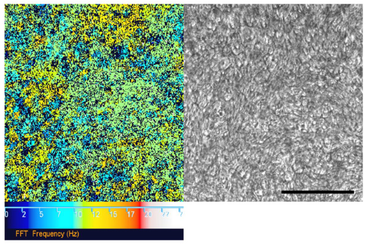
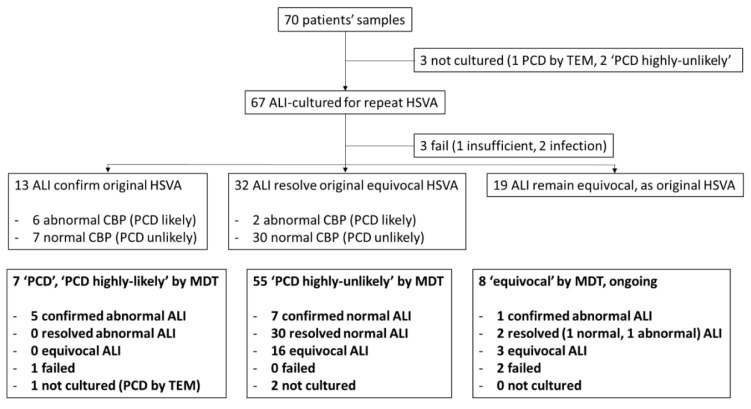
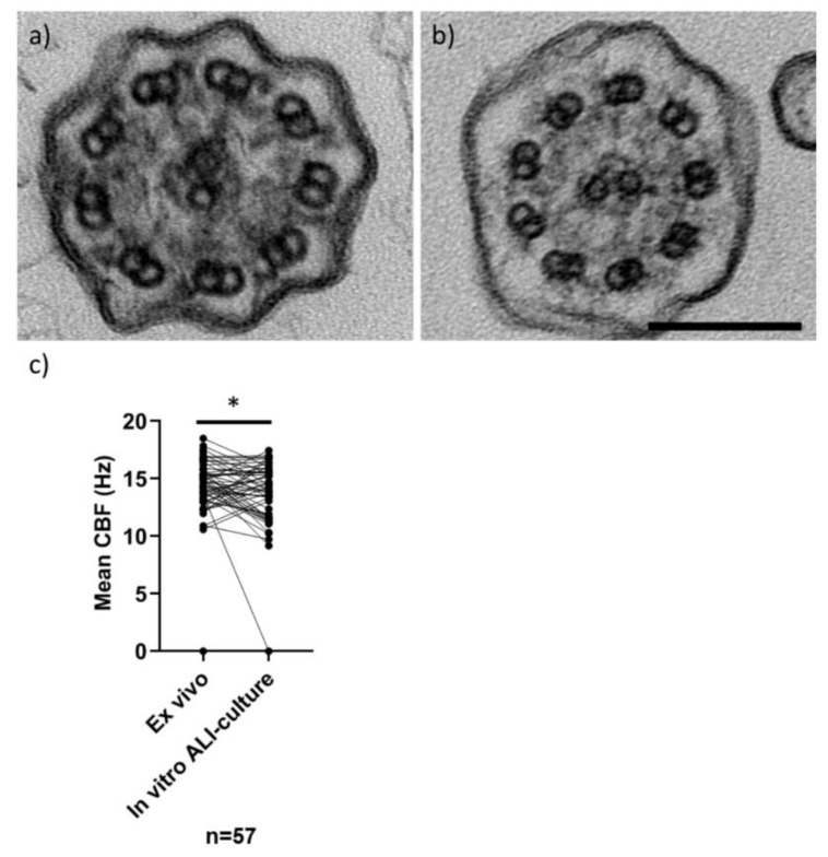
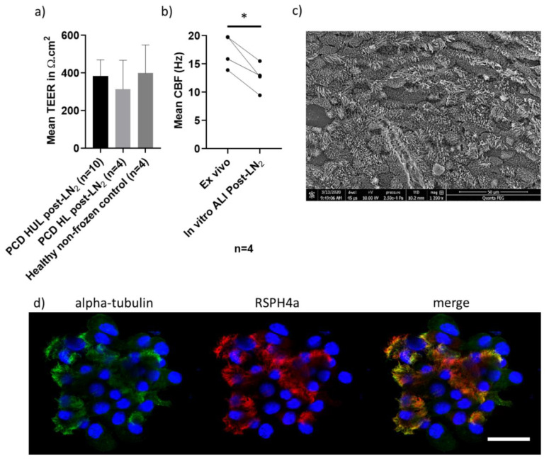
Air-liquid interface (ALI) culture of nasal epithelial cells is a valuable tool in the diagnosis and research of primary ciliary dyskinesia (PCD). Ex vivo samples often display secondary dyskinesia from cell damage during sampling, infection or inflammation confounding PCD diagnostic results. ALI culture enables regeneration of healthy cilia facilitating differentiation of primary from secondary ciliary dyskinesia. We describe a revised ALI culture method adopted from April 2018 across three collaborating PCD diagnostic sites, including current University Hospital Southampton COVID-19 risk mitigation measures, and present results. Two hundred and forty nasal epithelial cell samples were seeded for ALI culture and 199 (82.9%) were ciliated. Fifty-four of 83 (63.9%) ex vivo samples which were originally equivocal or insufficient provided diagnostic information following in vitro culture. Surplus basal epithelial cells from 181 nasal brushing samples were frozen in liquid nitrogen; 39 samples were ALI-cultured after cryostorage and all ciliated. The ciliary beat patterns of ex vivo samples (by high-speed video microscopy) were recapitulated, scanning electron microscopy demonstrated excellent ciliation, and cilia could be immuno-fluorescently labelled (anti-alpha-tubulin and anti-RSPH4a) in representative cases that were ALI-cultured after cryostorage. In summary, our ALI culture protocol provides high ciliation rates across three centres, minimising patient recall for repeat brushing biopsies and improving diagnostic certainty. Cryostorage of surplus diagnostic samples was successful, facilitating PCD research.

Keywords: ALI culture; PCD; bio-resource; diagnostics; primary nasal epithelium.


Conflict of interest statement

All authors certify that they have no conflicts of interest to declare.

Figures

Figure 1
Figure 1
Fast Fourier transform (FFT) analysis of ciliary movement using high-speed video microscopy analysis (HSVA) data. FFT analysis (left) “colour map” corresponding to ciliary movement detected by HSVA (using a 20× objective lens) on the surface of an air–liquid interface (ALI) culture (right). The colour scale (left to right) depicts increasing ciliary beat frequency (CBF) from 0 (black) to 25 Hz (white). Black pixels also represent a CBF measurement outside of the detection threshold (below 2 Hz or above 50 Hz). Normal mean CBF at 37 °C is 11–20 Hz. Scale bar represents 100 µm.
Figure 2
Figure 2
Flow diagram of diagnostic sample processing for ALI culture by repeat HSVA and MDT outcomes. Seventy patients’ ex vivo samples evaluated by HSVA (as part of the whole diagnostic process) and their ALI culture outcomes were followed. HSVA on ALI culture either confirmed the original HSVA finding, resolved an originally equivocal HSVA or remained equivocal despite culture. In bold, the ALI culture HSVA outcomes are shown by MDT outcome (“PCD”/“PCD highly-likely”; “PCD highly-unlikely” or “equivocal” pending follow up, repeat tests or further tests (genetics or additional IF for example).
Figure 3
Figure 3
TEM of cilia in transverse section and CBF (by HSVA) in nasal samples and after ALI culture. Representative TEM images (a,b) of in vitro ALI-cultured cilia in transverse section, showing a “9 + 2” microtubular arrangement. Cilia have normal ciliary ultrastructure in (a) a “PCD highly-unlikely” subject (with 3% microtubular defects, 18% inner and 4% outer dynein arm defects quantified from 102 cilia), and (b) a “PCD positive” subject (with 11% microtubular defects, 47% inner and 99% outer dynein arm absence quantified from 302 cilia). Scale bar represents 100 nm. Dot plot (c) demonstrates the mean CBFs (Hz) of 57 ex vivo nasal brushing samples compared to their matched in vitro ALI cultures (Wilcoxon paired test *p = 0.03). Data from ex vivo samples without a matched ALI sample were excluded (n = 13), which was due to 7 ex vivo samples with a variable ciliary beat pattern (CBP), 3 with failed ALI cultures and 3 that were not cultured. Normal CBF range of ex vivo samples at University Hospital Southampton (UHS) is 11–20 Hz.
Figure 4
Figure 4
Characteristics of in vitro ALI cultures (ciliated and differentiated at passage 2) derived from frozen liquid nitrogen storage. (a) There was no difference in the mean (±SD) trans-epithelial electrical resistance (TEER) (Ω.cm2) of n = 10 “PCD highly-unlikely” and n = 4 “PCD highly-likely” ALI cultures recovered from liquid nitrogen cryostorage (post-LN2) (t test), or compared to n = 4 healthy donor sample (non-frozen) controls (Mann–Whitney test) measured in triplicate per Transwell® insert at 4 weeks ALI culture, when cells were widely ciliated. (b) The mean CBF (Hz) of n = 4 matched PCD clinic samples differed before (ex vivo) and after liquid nitrogen storage (in vitro ALI culture) *p = 0.01 (paired t test). (c) A representative SEM image from a “PCD highly-unlikely” ALI culture showing typical ciliation at week 4 post-LN2. (d) Representative PCD diagnostic immunofluorescence [15] images from an SP8 laser scanning confocal microscope, showing a PCD clinic ALI culture after cryostorage with 4% paraformaldehyde fixation and immunofluorescence labelling with anti-alpha-tubulin (cilia marker-Alexa488 secondary antibody, green), anti-RSPH4a (radial spoke head protein-Alexa549 secondary antibody, red) and DAPI (nuclei DNA stain, blue). Scale 20 µm.

References

    1. Wallmeier J., Nielsen K.G., Kuehni C.E., Lucas J.S., Leigh M.W., Zariwala M.A., Omran H. Motile ciliopathies. Nat. Rev. Dis. Prim. 2020;6:1–29. doi: 10.1038/s41572-020-0209-6. - DOI - PubMed
    1. O’Callaghan C., Chetcuti P., Moya E. High prevalence of primary ciliary dyskinesia in a British Asian population. Arch. Dis. Child. 2010;95:51–52. doi: 10.1136/adc.2009.158493. - DOI - PubMed
    1. Lucas J.S., Davis S.D., Omran H., Shoemark A. Primary ciliary dyskinesia in the genomics age. Lancet Respir. Med. 2020;8:202–216. doi: 10.1016/S2213-2600(19)30374-1. - DOI - PubMed
    1. Behan L., Dimitrov B.D., Kuehni C.E., Hogg C., Carroll M., Evans H.J., Goutaki M., Harris A., Packham S., Walker W.T., et al. PICADAR: A diagnostic predictive tool for primary ciliary dyskinesia. Eur. Respir. J. 2016;47:1103–1112. doi: 10.1183/13993003.01551-2015. - DOI - PMC - PubMed
    1. Lucas J.S., Alanin M.C., Collins S., Harris A., Johansen H.K., Nielsen K.G., Papon J.F., Robinson P., Walker W.T. Clinical care of children with primary ciliary dyskinesia. Expert Rev. Respir. Med. 2017;11:779–790. doi: 10.1080/17476348.2017.1360770. - DOI - PubMed

LinkOut - more resources