

Three-Dimensional Biofabrication Models of Endometriosis and the Endometriotic Microenvironment
- PMID: 33233463
- PMCID: PMC7700676
- DOI: 10.3390/biomedicines8110525
Three-Dimensional Biofabrication Models of Endometriosis and the Endometriotic Microenvironment
Abstract
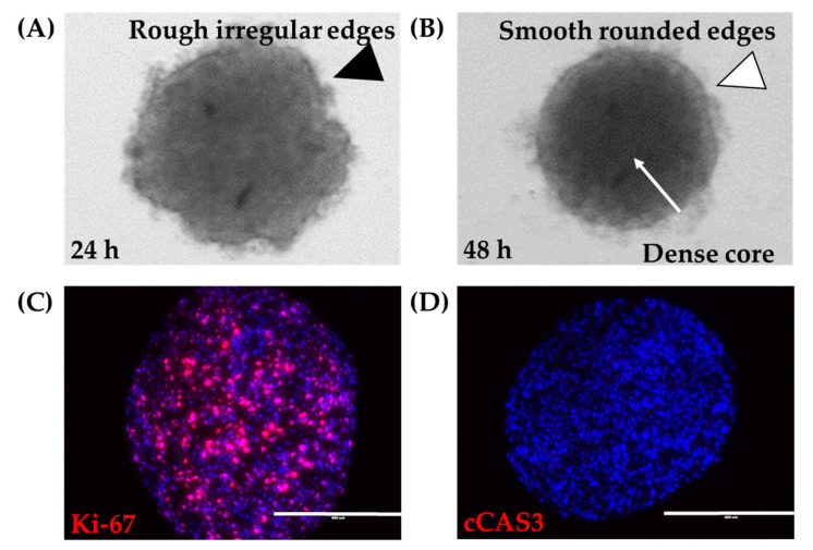
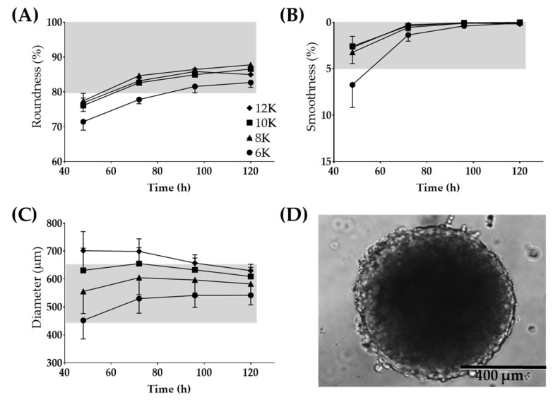
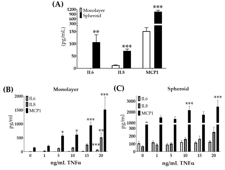
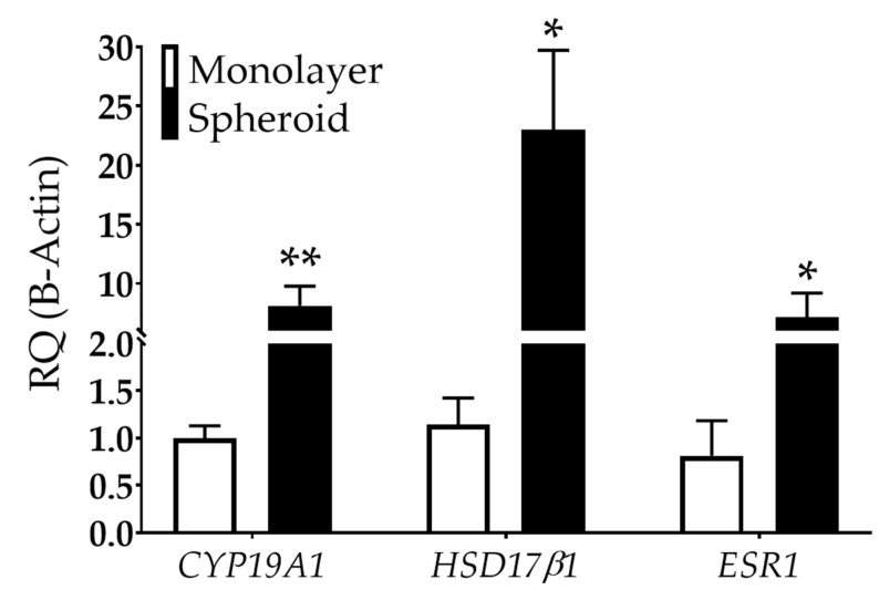
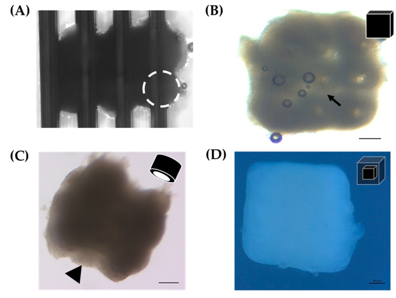
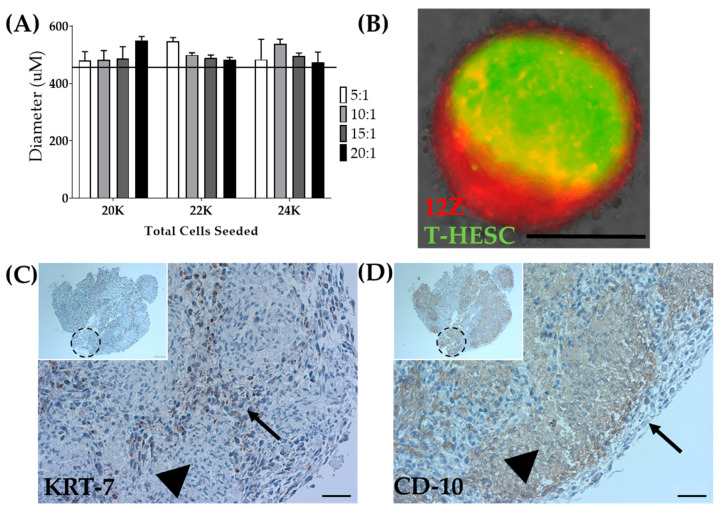
Endometriosis occurs when endometrial-like tissue grows outside the uterine cavity, leading to pelvic pain, infertility, and increased risk of ovarian cancer. The present study describes the optimization and characterization of cellular spheroids as building blocks for Kenzan scaffold-free method biofabrication and proof-of-concept models of endometriosis and the endometriotic microenvironment. The spheroid building blocks must be of a specific diameter (~500 μm), compact, round, and smooth to withstand Kenzan biofabrication. Under optimized spheroid conditions for biofabrication, the endometriotic epithelial-like cell line, 12Z, expressed high levels of estrogen-related genes and secreted high amounts of endometriotic inflammatory factors that were independent of TNFα stimulation. Heterotypic spheroids, composed of 12Z and T-HESC, an immortalized endometrial stromal cell line, self-assembled into a biologically relevant pattern, consisting of epithelial cells on the outside of the spheroids and stromal cells in the core. 12Z spheroids were biofabricated into large three-dimensional constructs alone, with HEYA8 spheroids, or as heterotypic spheroids with T-HESC. These three-dimensional biofabricated constructs containing multiple monotypic or heterotypic spheroids represent the first scaffold-free biofabricated in vitro models of endometriosis and the endometriotic microenvironment. These efficient and innovative models will allow us to study the complex interactions of multiple cell types within a biologically relevant microenvironment.
Keywords: 12Z; Kenzan; biofabrication; endometriosis; scaffold-free; spheroid.
Conflict of interest statement
The authors declare no conflict of interest. The funders had no role in the design of the study; in the collection, analyses, or interpretation of data; in the writing of the manuscript, or in the decision to publish the results.
Figures

Similar articles
-
Spheroids as a model for endometriotic lesions.JCI Insight. 2023 Jun 8;8(11):e160815. doi: 10.1172/jci.insight.160815. JCI Insight. 2023. PMID: 37104033 Free PMC article.
-
Co-operation between the AKT and ERK signaling pathways may support growth of deep endometriosis in a fibrotic microenvironment in vitro.Hum Reprod. 2015 Jul;30(7):1606-16. doi: 10.1093/humrep/dev108. Epub 2015 May 14. Hum Reprod. 2015. PMID: 25976656
-
iPSC-Derived Vascular Cell Spheroids as Building Blocks for Scaffold-Free Biofabrication.Biotechnol J. 2017 Dec;12(12). doi: 10.1002/biot.201700444. Epub 2017 Nov 14. Biotechnol J. 2017. PMID: 29030959
-
In vitro modeling of endometriosis and endometriotic microenvironment - Challenges and recent advances.Cell Signal. 2022 Sep;97:110375. doi: 10.1016/j.cellsig.2022.110375. Epub 2022 Jun 9. Cell Signal. 2022. PMID: 35690293 Review.
-
The Endometriotic Tumor Microenvironment in Ovarian Cancer.Cancers (Basel). 2018 Aug 7;10(8):261. doi: 10.3390/cancers10080261. Cancers (Basel). 2018. PMID: 30087267 Free PMC article. Review.
Cited by
-
Di-(2-ethylhexyl) Phthalate Triggers Proliferation, Migration, Stemness, and Epithelial-Mesenchymal Transition in Human Endometrial and Endometriotic Epithelial Cells via the Transforming Growth Factor-β/Smad Signaling Pathway.Int J Mol Sci. 2022 Apr 1;23(7):3938. doi: 10.3390/ijms23073938. Int J Mol Sci. 2022. PMID: 35409294 Free PMC article.
-
Three-dimensional culture models of human endometrium for studying trophoblast-endometrium interaction during implantation.Reprod Biol Endocrinol. 2022 Aug 13;20(1):120. doi: 10.1186/s12958-022-00973-8. Reprod Biol Endocrinol. 2022. PMID: 35964080 Free PMC article. Review.
-
Model Systems in Endometriosis Research: Translation, Translation, Translation!Front Reprod Health. 2022 Jan 11;3:809366. doi: 10.3389/frph.2021.809366. eCollection 2021. Front Reprod Health. 2022. PMID: 36304048 Free PMC article. No abstract available.
-
WERF Endometriosis Phenome and Biobanking Harmonisation Project for Experimental Models in Endometriosis Research (EPHect-EM-Organoids): endometrial organoids as an emerging technology for endometriosis research.Mol Hum Reprod. 2025 Jul 3;31(3):gaaf024. doi: 10.1093/molehr/gaaf024. Mol Hum Reprod. 2025. PMID: 40628401 Free PMC article. Review.
-
Scaffold-mediated liver regeneration: A comprehensive exploration of current advances.J Tissue Eng. 2024 Oct 13;15:20417314241286092. doi: 10.1177/20417314241286092. eCollection 2024 Jan-Dec. J Tissue Eng. 2024. PMID: 39411269 Free PMC article. Review.
References
Grants and funding
LinkOut - more resources
Full Text Sources
Research Materials
