Review
. 2020 Nov 20;6(4):302.
doi: 10.3390/jof6040302.

Dynamic Regulation of Peroxisomes and Mitochondria during Fungal Development

Affiliations
Review

Dynamic Regulation of Peroxisomes and Mitochondria during Fungal Development

Raful Navarro-Espíndola et al. J Fungi (Basel). .

Abstract

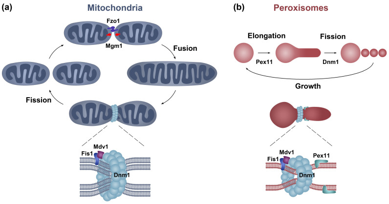
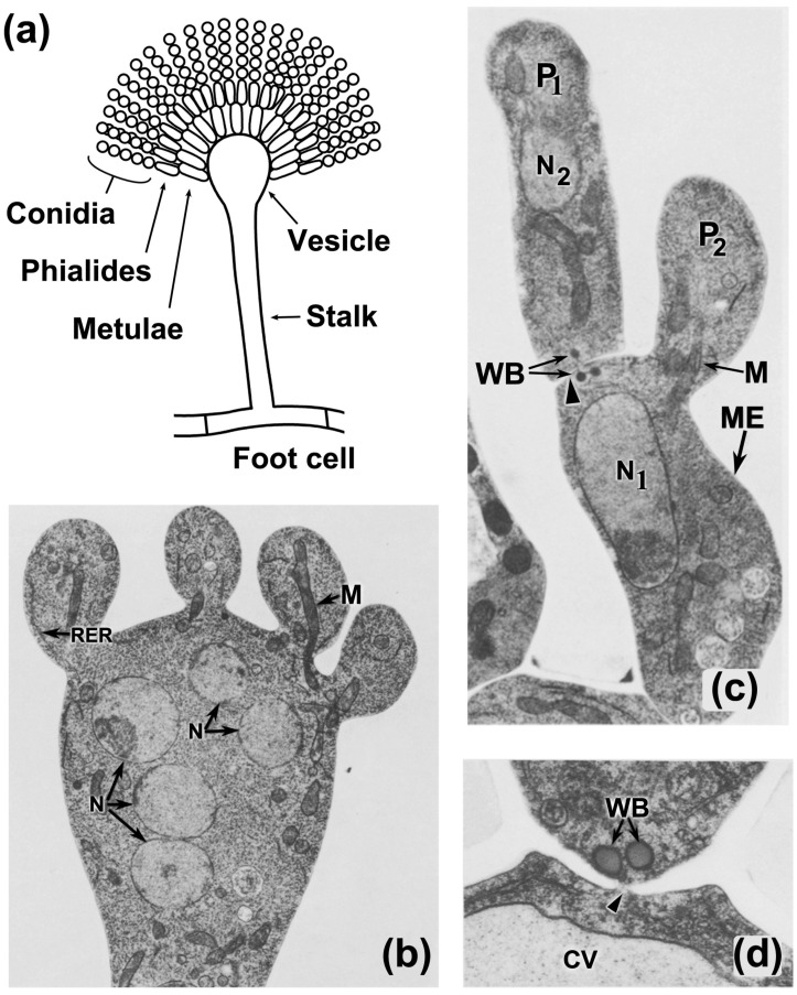
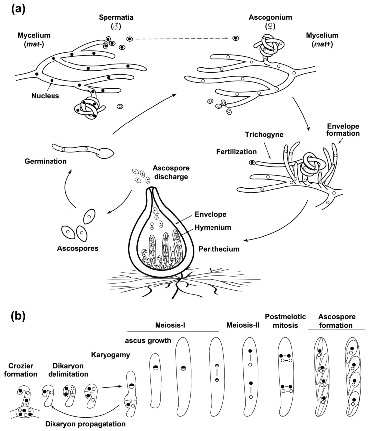
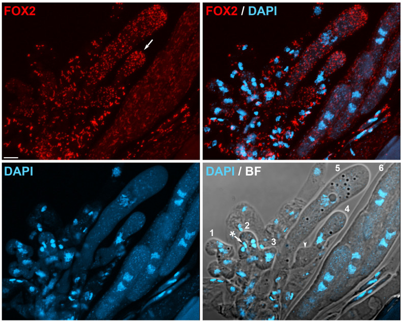
Peroxisomes and mitochondria are organelles that perform major functions in the cell and whose activity is very closely associated. In fungi, the function of these organelles is critical for many developmental processes. Recent studies have disclosed that, additionally, fungal development comprises a dynamic regulation of the activity of these organelles, which involves a developmental regulation of organelle assembly, as well as a dynamic modulation of the abundance, distribution, and morphology of these organelles. Furthermore, for many of these processes, the dynamics of peroxisomes and mitochondria are governed by common factors. Notably, intense research has revealed that the process that drives the division of mitochondria and peroxisomes contributes to several developmental processes-including the formation of asexual spores, the differentiation of infective structures by pathogenic fungi, and sexual development-and that these processes rely on selective removal of these organelles via autophagy. Furthermore, evidence has been obtained suggesting a coordinated regulation of organelle assembly and dynamics during development and supporting the existence of regulatory systems controlling fungal development in response to mitochondrial activity. Gathered information underscores an important role for mitochondrial and peroxisome dynamics in fungal development and suggests that this process involves the concerted activity of these organelles.

Keywords: cell differentiation; fungi; mitochondria; mitochondrial fission; mitophagy; organelle dynamics; peroxisome; peroxisome fission; pexophagy; sexual development.


Conflict of interest statement

The authors declare no conflict of interest.

Figures

Figure 1
Figure 1
Mitochondrial and peroxisome division in fungi. (a) The mitochondrial arrangement is determined by fusion and fission events that are mediated by dynamin proteins. Fusion is driven by the dynamins Fzo1 (mitofusin) and Mgm1 (Opa1) at the mitochondrial outer and inner membranes, respectively, while Dnm1 mediates fission. (b) Peroxisomes multiply by growth and division from preexisting organelles, in a process that also involves Dnm1. Dnm1 is recruited to mitochondria and peroxisomes by the membrane receptor Fis1 through interactions with the Mdv1 adapter (lower panels). Dnm1 then assembles into spirals around the organelles, which constrict and sever their membranes upon GTP hydrolysis. Peroxisome fission is preceded by the organelle elongation, which is promoted by the membrane elongation factor Pex11. This protein also serves as a Dnm1 activator.
Figure 2
Figure 2
Subcellular organization during Aspergillus nidulans conidiation. (a) Schematics of an A. nidulans conidiophore. Asexual spore (conidium) formation in A. nidulans takes place on specialized aerial conidiophores, which consist of a large specialized hypha—the stalk—that grows away from the substrate and swells at the tip to produce a conidiophore vesicle. This vesicle produces numerous primary sterigmata (metulae) by budding, which, in turn, differentiate secondary sporogenous sterigmata (phialides) at their apices. Conidia emerge by budding from the tip of these specialized cells. The stalk extends from a specialized foot cell, which anchors the conidiophore and connects it to the substratum mycelium. (b) Ultrastructural analyses of a conidiophore vesicle showing four young metulae. At this stage, mitochondria (M) and rough ER (RER) strands extend into metulae, while nuclei (N) remain positioned below metulae. (c) Metula (ME) has differentiated a phialide (P1) delimited by a septum and that is developing a second phialide (P2). Visible is a mitochondrion (M) extending into the developing phialide, and Woronin bodies (WB) bordering the septum (arrowhead) at the base of the first phialide. (d) Mature septum at the base of a metula. Note the Woronin bodies bordering the pore (arrowhead) of the septum; CV, conidiophore vesicle. (bd) Adapted from [79], with permission.
Figure 3
Figure 3
Sexual development of a model mycelial ascomycete. (a) The sexual life cycle of a heterothallic Sordariomycete. Mating in ascomycetes is controlled by a mating-type (MAT) locus, for which two alternative versions (idiomorphs) exist in heterothallic (self-sterile) species (denoted here as mat+ and mat-). Strains of either mating type differentiate both, female gametangia (ascogonia) and male gametes (spermatia), which cross-fertilize when bearing opposite mating-type (opposite mating-type nuclei are illustrated as dots with different shading). Fertilization is attained by a specialized hypha—the trichogyne—which is produced by the ascogonium and that exhibits tropic growth towards a pheromone-producing male gamete. The ascogonium recruits neighboring hyphae, which develop a protective envelope around the ascogonium, producing a protoperithecium. Upon fertilization, the protoperithecium develops into a perithecium, and the fertilized ascogonium develops the hymenium, the fertile tissue where karyogamy, meiosis, and meiotic spore (ascospore) formation are accomplished. Ultimately, haploid mature ascospores are expelled out of perithecia and produce a new mycelium upon germination. (b) Hymenium development. Inside perithecia, the nuclei of the opposite mating type present in the fertilized ascogonia migrate into ascogenous hyphae that differentiate from the ascogonial cells. These ascogenous hyphae then develop into specialized hook-shaped cells, referred to as croziers, in which dikaryotic compartmentalization takes place. This process results from the synchronized mitoses (lines linking the dots represent spindles) of the two crozier leading nuclei, which are positioned in the crook part of the crozier and that differ in their mating type, and results in the formation of three cells separated by septa: an upper binucleated cell flanked by two uninucleate cells. The upper dikaryotic cell suffers karyogamy and enters meiosis at the same time that differentiates into an ascus (the meiocyte), whereas the two flanking uninucleate cells fuse to produce a new dikaryotic crozier, perpetuating the dikaryotic stage. In the example provided, which illustrates the development of P. anserina, asci then elongate from about 5 to more than 150 μm during the first meiotic prophase. After meiosis, a mitotic division results in the formation of eight nuclei, which are enclosed by pairs into four ascospores. Finally, ascospores grow and maturate inside the original ascus (for review, [126]).
Figure 4
Figure 4
Peroxisome arrangement in representative stages of P. anserina sexual development. The numbering indicates successive developmental stages: (1) Young binucleate croziers. (2) Dikaryotic cell formation (note the lateral cell nucleus—asterisk—migrating into the basal cell to produce a new dikaryotic cell). (3) Karyogamy. (4–5) Asci at successive stages of meiotic prophase-I (arrow indicates peroxisomes concentrated at the ascus apex, arrowhead points to the reminiscent initial crozier cell). (6) Early ascospores. Peroxisomes were labeled with FOX2-mCherry. Nuclei and mitochondrial DNA (mtDNA) were stained by DAPI. BF, bright field. Scale bar, 5 μm. A section of this micrograph was previously reported in [102].
Figure 5
Figure 5
Contribution of the fission machinery to peroxisome dynamics during P. anserina sexual development. Examples of the effect of abrogating the fission machinery during sexual development: (a) Croziers. Visible are a dikaryotic tetra-nucleate crozier (right, arrow points to the dikaryotic cell, which lacks peroxisomes) and a crozier following karyogamy (left). (b) First meiotic prophase ascus. (c) Early ascospores. Four binucleate ascospores inside an ascus are visible; note the asymmetric distribution of peroxisomes and the cluster of elongated peroxisomes (arrow) that failed to be incorporated into ascospores. (d) Growing ascospores. Visible are two normal ascospores that have differentiated a large head and a slender tail, and a small aberrant ascospore (arrow, note the very reduced number of peroxisomes of this ascospore). Arrowheads in (c,d) point to mtDNA clusters. Cells were issued from homozygous sexual crosses of ∆dnm1 or ∆fis1 mutants, which exhibit a very similar phenotype. Please refer to Figure 4 to appreciate the morphology of wild-type peroxisomes at the equivalent stages. Peroxisomes were labeled with FOX2-GFP. Nuclei and mtDNA were stained by DAPI. BF, bright field. Scale bar, 5 μm.

Similar articles

Cited by

References

    1. Fransen M., Lismont C., Walton P. The Peroxisome-Mitochondria Connection: How and Why? Int. J. Mol. Sci. 2017;18:1126. doi: 10.3390/ijms18061126. - DOI - PMC - PubMed
    1. Hicks L., Fahimi H.D. Peroxisomes (microbodies) in the myocardium of rodents and primates. A comparative Ultrastructural cytochemical study. Cell Tissue Res. 1977;175:467–481. doi: 10.1007/BF00222413. - DOI - PubMed
    1. Frederick S.E., Newcomb E.H. Microbody-like organelles in leaf cells. Science. 1969;163:1353–1355. doi: 10.1126/science.163.3873.1353. - DOI - PubMed
    1. Shai N., Yifrach E., van Roermund C.W.T., Cohen N., Bibi C., IJlst L., Cavellini L., Meurisse J., Schuster R., Zada L., et al. Systematic mapping of contact sites reveals tethers and a function for the peroxisome-mitochondria contact. Nat. Commun. 2018;9:1761. doi: 10.1038/s41467-018-03957-8. - DOI - PMC - PubMed
    1. Mattiazzi Usaj M., Brloznik M., Kaferle P., Zitnik M., Wolinski H., Leitner F., Kohlwein S.D., Zupan B., Petrovic U. Genome-Wide Localization Study of Yeast Pex11 Identifies Peroxisome-Mitochondria Interactions through the ERMES Complex. J. Mol. Biol. 2015;427:2072–2087. doi: 10.1016/j.jmb.2015.03.004. - DOI - PMC - PubMed

LinkOut - more resources