. 2020 Nov 20;10(11):882.
doi: 10.3390/brainsci10110882.

ColorViz, a New and Rapid Tool for Assessing Collateral Circulation during Stroke

Affiliations

ColorViz, a New and Rapid Tool for Assessing Collateral Circulation during Stroke

Tommaso Verdolotti et al. Brain Sci. .

Abstract

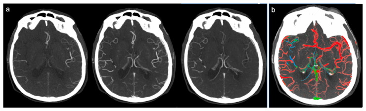
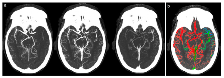
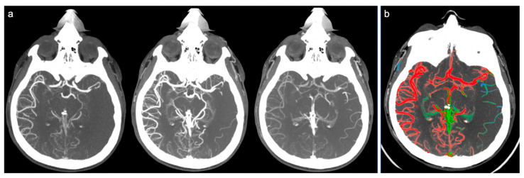
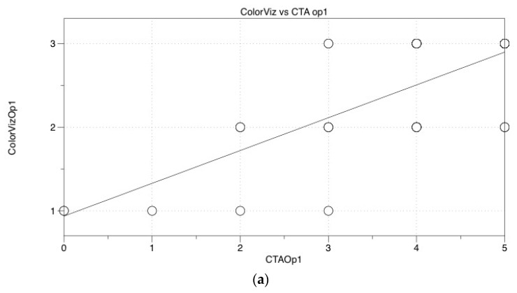
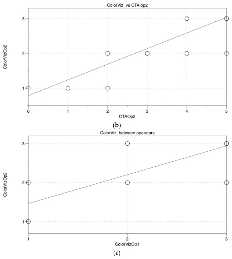
Prognosis of patients with acute ischemic stroke is strictly related to the patency and prominence of the collateral leptomeningeal pathways distal to the arterial occlusion. The gold standard for assessment of collateral circulation is conventional angiography, but it is invasive and used in selected cases. To date, the most reliable technique is multiphase CTA; currently, the available classifications of collateral circles are often complex, time-consuming, and require a trained observer. The purpose of our work is to establish the effectiveness of a new semi-automatic post-processing software (ColorViz FastStroke, GE Healthcare, Milwaukee, Wisconsin) in evaluation of collateral circulation compared to the six-point classifications of multiphase CTA already validated in literature. We selected 86 patients with anterior ischemic stroke symptoms who underwent multiphasic CTA in our emergency department. Two radiologists separately evaluated the collateral leptomeningeal vessels, analyzing respectively, the multiphase CTA (using the six-point scale and its trichotomized form) and ColorViz (using a three-point scale). Then the results were matched. We found a good correlation between the two different analyses; the main advantage of ColorViz is that, while maintaining fast diagnostic times, it allows a simpler and more immediate evaluation of collateral circulation, especially for less experienced radiologists.

Keywords: Collateral grading scales; Color Map; acute ischemic stroke; leptomeningeal collaterals; multiphase CT angiography.


Conflict of interest statement

The authors declare no conflict of interests.

Figures

Figure 1
Figure 1
Example of good collateral circulation at ColorViz. (a) mCTA shows a right-sided M1 segment occlusion with good pial artery filling (1 phase delay); (b) ColorViz map shows predominant red vessels in the affected territory downstream from the occlusion (score of 3).
Figure 2
Figure 2
Example of intermediate collateral circulation at ColorViz. (a) mCTA shows a left-sided M1 segment occlusion with intermediate pial artery filling; (b) ColorViz map shows predominant green vessels in the affected territory downstream from the occlusion (score of 2).
Figure 3
Figure 3
Example of poor collateral circulation at ColorViz. (a) mCTA shows a left-sided M1 segment occlusion with poor pial artery filling; (b) ColorViz map shows reduced extension of collateral circles vessels in the affected territory downstream from the occlusion (score of 1).
Figure 4
Figure 4
Statistical Graphs. (a,b), Graphs showing good accordance for both operators when comparing ColorViz to mCTA (r = 0.735, p < 0.001 and r = 0.839, p < 0.001, respectively); (c) Graph showing good accordance between operators when comparing ColorViz scores (r = 0.890, p < 0.001).
Figure 4
Figure 4
Statistical Graphs. (a,b), Graphs showing good accordance for both operators when comparing ColorViz to mCTA (r = 0.735, p < 0.001 and r = 0.839, p < 0.001, respectively); (c) Graph showing good accordance between operators when comparing ColorViz scores (r = 0.890, p < 0.001).

References

    1. Li G.-Z., Ding J., Leng X., Pu Y.-H., Huang L.-A., Xu A.-D., Wong K.S.L., Wang X., Wang Y. Guidelines for evaluation and management of cerebral collateral circulation in ischaemic stroke 2017. Stroke Vasc. Neurol. 2018;3:117–130. doi: 10.1136/svn-2017-000135. - DOI - PMC - PubMed
    1. Smith A., Hill C.R. Imaging assessment of acute ischaemic stroke: A review of radiological methods. Br. J. Radiol. 2017;91:20170573. doi: 10.1259/bjr.20170573. - DOI - PMC - PubMed
    1. Potter C.A., Vagal A., Goyal M., Nunez D.B., Leslie-Mazwi T.M., Lev M.H. CT for Treatment Selection in Acute Ischemic Stroke: A Code Stroke Primer. RadioGraphics. 2019;39:1717–1738. doi: 10.1148/rg.2019190142. - DOI - PubMed
    1. Powers W.J., Rabinstein A.A., Ackerson T., Adeoye O.M., Bambakidis N.C., Becker K., Biller J., Brown M., Demaerschalk B.M., Hoh B., et al. Guidelines for the Early Management of Patients WITH Acute Ischemic Stroke: 2019 Update to the 2018 Guidelines for the Early Management of Acute Ischemic Stroke: A Guideline for Healthcare Professionals From the American Heart Association/American Stroke Association. Stroke. 2019;50:e344–e418. doi: 10.1161/str.0000000000000211. - DOI - PubMed
    1. Menon B.K., D’Esterre C.D., Qazi E.M., Almekhlafi M., Hahn L., Demchuk A.M., Goyal M. Multiphase CT Angiography: A New Tool for the Imaging Triage of Patients with Acute Ischemic Stroke. Radiology. 2015;275:510–520. doi: 10.1148/radiol.15142256. - DOI - PubMed

LinkOut - more resources