Review
. 2020 Nov 20;21(22):8790.
doi: 10.3390/ijms21228790.

The Immune Functions of Keratinocytes in Skin Wound Healing

Affiliations
Review

The Immune Functions of Keratinocytes in Skin Wound Healing

Minna Piipponen et al. Int J Mol Sci. .

Abstract

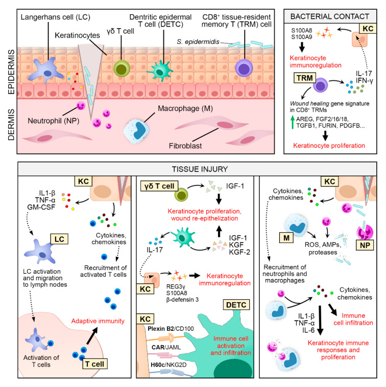
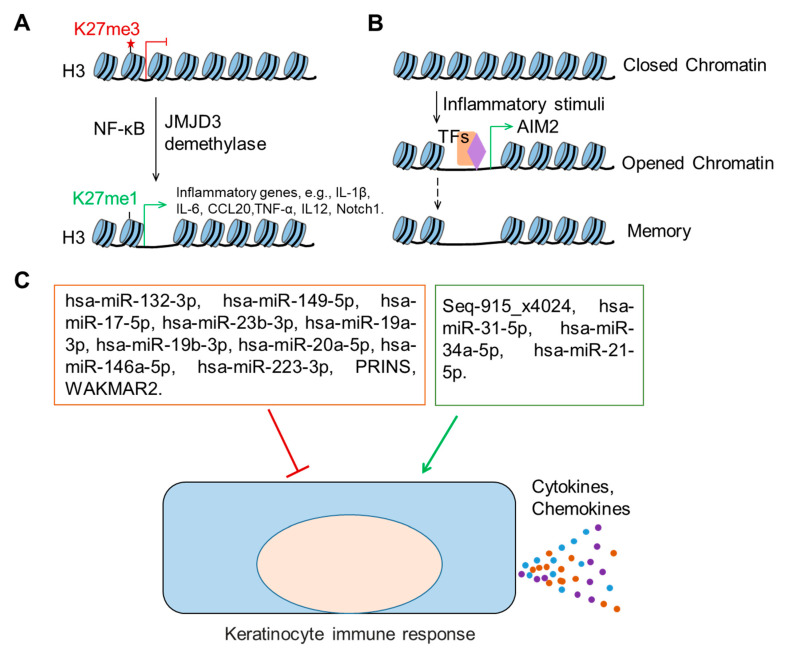
As the most dominant cell type in the skin, keratinocytes play critical roles in wound repair not only as structural cells but also exerting important immune functions. This review focuses on the communications between keratinocytes and immune cells in wound healing, which are mediated by various cytokines, chemokines, and extracellular vesicles. Keratinocytes can also directly interact with T cells via antigen presentation. Moreover, keratinocytes produce antimicrobial peptides that can directly kill the invading pathogens and contribute to wound repair in many aspects. We also reviewed the epigenetic mechanisms known to regulate keratinocyte immune functions, including histone modifications, non-protein-coding RNAs (e.g., microRNAs, and long noncoding RNAs), and chromatin dynamics. Lastly, we summarized the current evidence on the dysregulated immune functions of keratinocytes in chronic nonhealing wounds. Based on their crucial immune functions in skin wound healing, we propose that keratinocytes significantly contribute to the pathogenesis of chronic wound inflammation. We hope this review will trigger an interest in investigating the immune roles of keratinocytes in chronic wound pathology, which may open up new avenues for developing innovative wound treatments.

Keywords: antimicrobial peptide; chronic wounds; cytokine; epigenetic regulation; immune function; inflammation; keratinocyte; long noncoding RNA; microRNA; wound healing.


Conflict of interest statement

The authors declare no conflict of interest.

Figures

Figure 1
Figure 1
Toll-like receptors (TLR)-mediated signaling pathway in keratinocytes. TLR1, 2, 4, 5, and 6 are present on the cell membrane, while TLR3 and 9 reside in keratinocytes’ intracellular compartments. TLR2 recognizes a broad range of pathogen-associated molecular patterns, including triacyl lipopeptide (in combination with TLR1), diacyl lipopeptide (in combination with TLR6), peptidoglycan (PGN), lipoteichoic acid (LTA), and Zymosan. TLR5 recognizes flagellin, while TLR4 recognizes lipopolysaccharides (LPS), which is aided by two accessory proteins cluster of differentiation (CD14) and myeloid differentiation factor 2 (MD2). TLR3 and TLR9 reside on the endosomal membrane and recognize double-stranded RNA (dsRNA) and unmethylated CpG DNA, respectively. Upon stimulation, these TLRs activate the nuclear factor kappa-light-chain-enhancer of activated B cells (NF-kB) signaling pathway to induce the expression of inflammatory cytokines. TLR3 and TLR4 can also activate interferon regulatory factor 3 (IRF3), while TLR9 activates interferon regulatory factor 7 (IRF7) to induce the expression of type 1 interferons (IFNs).
Figure 2
Figure 2
Keratinocyte‒immune cell crosstalk in skin wound healing. Keratinocyte (KC)-secreted pro-inflammatory cytokines and chemokines contribute to the recruitment of neutrophils (NP) and macrophages (M) to the site of injury. NP and M remove cellular debris and destroy invading pathogens by phagocytosis and the release of reactive oxygen species (ROS), proteases, and antimicrobial proteins (AMPs). They further amplify the inflammatory response by secreting various pro-inflammatory cytokines and chemokines to recruit more immune cells. Some of these secreted factors, such as interleukin 1 beta (IL-1β), tumor necrosis factor (TNF-α), and interleukin 6 (IL-6) stimulate keratinocyte proliferation and immune responses. After tissue injury, Langerhans cells (LC) react to KC-derived factors and migrate to the draining lymph node to activate T cells, triggering an adaptive immune response. KC-derived cytokines and chemokines serve as a chemoattractant to recruit activated T cells to the wound. Epidermal γδ T cells (human) and dendritic epidermal T cells (DETCs, mouse) respond rapidly to tissue injury and produce growth factors to activate KC proliferation and wound re-epithelization. Vice versa, ligands expressed on the KC cell membrane, i.e., Plexin B2, coxsackievirus and adenovirus receptor (CAR), and H60c are detected by DETCs, triggering immune cell activation and infiltration. DETC-produced interleukin 17 (IL-17) also activates KC to produce antimicrobial peptides (AMPs). Even in the absence of tissue injury, bacterial contact with skin commensal Staphylococcus epidermidis induces CD8+ tissue-resident memory T (TRM) cells to produce IL-17 and Interferon gamma (IFN-γ), which in turn activate KC to produce AMPs. S. epidermidis-activated TRM cells upregulate the expression of KC mitogens, such as amphiregulin (AREG), fibroblast growth factor 2 (FGF2), and transforming growth factor beta 1 (TGFB1), to promote keratinocyte proliferation. In the later phase of wound healing, the engulfment of apoptotic NP by M is essential for M’s conversion into an anti-inflammatory phenotype and the production of several key factors for wound healing, including TGFB1, vascular endothelial growth factor (VEGF), and platelet-derived growth factor (PDGF).
Figure 3
Figure 3
Epigenetic regulation of keratinocyte immune functions in wound healing. (A), The H3K27me3 demethylase Jumonji domain containing-3 (JMJD3), cooperating with the nuclear factor kappa-light-chain-enhancer of activated B cells (NF-κB), activates the expression of several inflammatory genes, including interleukin 1 beta (IL-1β), interleukin 6 (IL-6), chemokine (C-C motif) ligand 20 (CCL20), tumor necrosis factor (TNF-α), interleukin 12 (IL12), and Notch1 that is a crucial transcription factor in skin development and homeostasis. (B), Inflammatory stimuli trigger the chromatin opening in epithelial stem cells. After the resolution of inflammation, the chromatin accessibility is maintained for several critical inflammatory response genes, such as absent in melanoma 2 (AIM2), allowing the rapid transcription of AIM2 upon secondary stimulus. (C), Noncoding RNAs regulate keratinocyte immune response. The noncoding RNAs in the red frame negatively regulate cytokine or chemokine secretion in keratinocytes, while the ones in the green frame positively regulate them.

References

    1. Landen N.X., Li D., Stahle M. Transition from inflammation to proliferation: A critical step during wound. Cell. Mol. Life Sci. 2016;73:3861–3885. doi: 10.1007/s00018-016-2268-0. - DOI - PMC - PubMed
    1. Pastar I., Stojadinovic O., Yin N.C., Ramirez H., Nusbaum A.G., Sawaya A., Patel S.B., Khalid L., Isseroff R.R., Tomic-Canic M. Epithelialization in Wound Healing: A Comprehensive Review. Adv. Wound Care. 2014;3:445–464. doi: 10.1089/wound.2013.0473. - DOI - PMC - PubMed
    1. Souren J.M., Ponec M., van Wijk R. Contraction of collagen by human fibroblasts and keratinocytes. In Vitro Cell. Dev. Biol. 1989;25:1039–1045. doi: 10.1007/BF02624138. - DOI - PubMed
    1. Isaac C., Paggiaro A.O., Aldunate J.L.C.B., Herson M.R., Altran S.C., Mônica Beatriz M., Ferreira M.C. Role of keratinocytes in wound contraction: An impact assessment using a model of collagen matrix populated with fibroblasts. Rev. Bras. Cir. Plást. 2011;26:402–406. doi: 10.1590/S1983-51752011000300007. - DOI
    1. Krausgruber T., Fortelny N., Fife-Gernedl V., Senekowitsch M., Schuster L.C., Lercher A., Nemc A., Schmidl C., Rendeiro A.F., Bergthaler A., et al. Structural cells are key regulators of organ-specific immune responses. Nature. 2020;583:296–302. doi: 10.1038/s41586-020-2424-4. - DOI - PMC - PubMed