. 2020 Nov 20;21(22):8799.
doi: 10.3390/ijms21228799.

Rbm38 Reduces the Transcription Elongation Defect of the SMEK2 Gene Caused by Splicing Deficiency

Affiliations

Rbm38 Reduces the Transcription Elongation Defect of the SMEK2 Gene Caused by Splicing Deficiency

Shintaro Muraoka et al. Int J Mol Sci. .

Abstract

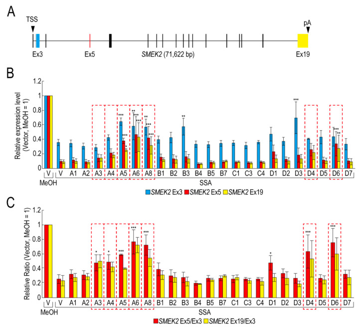
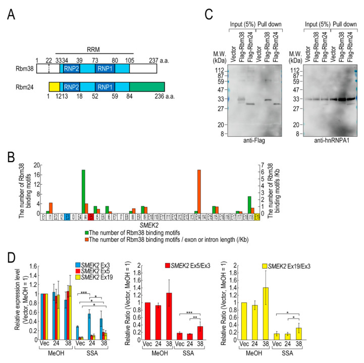
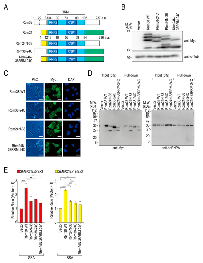
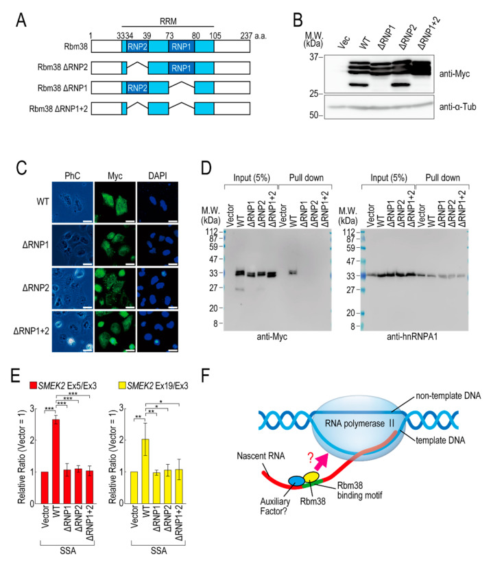
Pre-mRNA splicing is an essential mechanism for ensuring integrity of the transcriptome in eukaryotes. Therefore, splicing deficiency might cause a decrease in functional proteins and the production of nonfunctional, aberrant proteins. To prevent the production of such aberrant proteins, eukaryotic cells have several mRNA quality control mechanisms. In addition to the known mechanisms, we previously found that transcription elongation is attenuated to prevent the accumulation of pre-mRNA under splicing-deficient conditions. However, the detailed molecular mechanism behind the defect in transcription elongation remains unknown. Here, we showed that the RNA binding protein Rbm38 reduced the transcription elongation defect of the SMEK2 gene caused by splicing deficiency. This reduction was shown to require the N- and C-terminal regions of Rbm38, along with an important role being played by the RNA-recognition motif of Rbm38. These findings advance our understanding of the molecular mechanism of the transcription elongation defect caused by splicing deficiency.

Keywords: Rbm38; pre-mRNA splicing; spliceostatin A; transcription elongation.


Conflict of interest statement

The authors declare no conflict of interest.

Figures

Figure 1
Figure 1
RNA binding proteins (RBPs) reduce the transcription elongation defect caused by splicing deficiency. (A) Schematic representation of the SMEK2 gene. Black arrowheads indicate the transcription start site and the polyA site. Cyan, red, and yellow rectangles are Ex3, Ex5, and Ex19, respectively. (B,C) HeLa cells were transfected with a vector (V) or the indicated RBP in our library, and then cultured for 48 h after transfection. The cells were treated with MeOH or spliceostatin A (SSA) (10 ng/mL) for 1 h, and then treated with 200 µM 5-EU for an additional 2 h to label nascent RNA. The labeled RNA was analysed by quantitative RT-PCR. Relative expression levels of SMEK2 Ex3, Ex5, and Ex19 were plotted (B). Ratios of the expression levels of Ex5 or Ex19 relative to that of Ex3 (i.e., Ex5/Ex3 and Ex19/Ex3, respectively) were plotted (C). Error bars indicate S.D. (n = 3). Statistical significance was investigated by one-way ANOVA and Dunnett’s test (*: p < 0.05; **: p < 0.01; ***: p < 0.001). Red dotted line rectangles indicate RBPs that suppressed the transcription elongation defect.
Figure 2
Figure 2
Rbm38, but not Rbm24, suppresses the transcription elongation defect caused by splicing deficiency. (A) A schematic of the structure of the indicated RNA binding proteins. (B) The distribution of Rbm38 binding motifs in the SMEK2 gene. The number of Rbm38 binding sites listed in Table S2 was counted. (C) Flag-Rbm38 and Flag-Rbm24 were expressed in HEK293T cells and the biotinylated substrate RNA was prepared by in vitro transcription from an SMEK2 plasmid containing intron 4. The substrate RNA was purified using streptavidin beads and co-precipitated proteins were analysed by Western blotting. (D) HeLa cells were transfected with a vector (Vec), and Flag-Rbm24 (24) and Flag-Rbm38 (38) plasmids. The transfected cells were treated with SSA and 5-EU, and the labeled RNA was analysed as in Figure 1. Error bars indicate S.D. (n = 3). Statistical significance was investigated by one-way ANOVA and Tukey’s test (*: p < 0.05; **: p < 0.01; ***: p < 0.001).
Figure 3
Figure 3
The N- and C-terminal regions of Rbm38 are important for reducing the transcription elongation defect. (A) A schematic of the structure of chimeric RNA binding proteins. (B,C) HeLa cells were transfected with a vector or chimeric plasmids, and then cultured for 48 h after transfection. Protein samples were prepared and analysed by Western blotting (B). The subcellular localization of chimeric proteins was analysed by immunofluorescence microscopy (C). Bar = 20 µm. (D) A biotinylated RNA pull-down assay was performed using chimeric proteins as in Figure 2C. (E) HeLa cells were transfected with a vector or chimeric constructs, and then treated with SSA and 5-EU. The labeled RNA was analysed as in Figure 1. Error bars indicate S.D. (n = 3). Statistical significance was investigated by one-way ANOVA and Tukey’s test (*: p < 0.05; **: p < 0.01; ***: p < 0.001).
Figure 4
Figure 4
RNA binding motif of Rbm38 is important for suppressing the transcription elongation defect. (A) A schematic of the structure of the deleted RNA binding proteins. (B,C) HeLa cells were transfected with a vector or RNP deletion mutants, and then cultured for 48 h. Protein samples were prepared and analysed by Western blotting (B). The subcellular localization of the RNP deletion mutants was analysed by immunofluorescence microscopy (C). Bar = 20 µm. (D) A biotinylated RNA pull-down assay was performed using RNP deletion mutants as in Figure 2C. (E) HeLa cells were transfected with a vector or RNP deletion mutants, and then treated with SSA and 5-EU. The labeled RNA was analysed as in Figure 1. Error bars indicate S.D. (n = 3). Statistical significance was investigated by one-way ANOVA and Tukey’s test (*: p < 0.05; **: p < 0.01; ***: p < 0.001). (F) A schematic model of the mechanism by which Rbm38 suppresses the transcription defect caused by splicing deficiency. The N- and C-terminal domains and RBD/RRM of Rbm38 are required for the suppression. However, it is still unknown whether the RNA binding ability of Rbm38 is required for the suppression. Furthermore, an auxiliary factor might be involved in the suppression.

References

    1. Grzybowska E.A. Human intronless genes: Functional groups, associated diseases, evolution, and mRNA processing in absence of splicing. Biochem. Biophys. Res. Commun. 2012;424:1–6. doi: 10.1016/j.bbrc.2012.06.092. - DOI - PubMed
    1. Papasaikas P., Valcárcel J. The Spliceosome: The Ultimate RNA Chaperone and Sculptor. Trends Biochem. Sci. 2016;41:33–45. doi: 10.1016/j.tibs.2015.11.003. - DOI - PubMed
    1. Wahl M.C., Will C.L., Lührmann R. The Spliceosome: Design Principles of a Dynamic RNP Machine. Cell. 2009;136:701–718. doi: 10.1016/j.cell.2009.02.009. - DOI - PubMed
    1. Kaida D., Motoyoshi H., Tashiro E., Nojima T., Hagiwara M., Ishigami K., Watanabe H., Kitahara T., Yoshida T., Nakajima H., et al. Spliceostatin A targets SF3b and inhibits both splicing and nuclear retention of pre-mRNA. Nat. Chem. Biol. 2007;3:576–583. doi: 10.1038/nchembio.2007.18. - DOI - PubMed
    1. Satoh T., Kaida D. Upregulation of p27 cyclin-dependent kinase inhibitor and a C-terminus truncated form of p27 contributes to G1 phase arrest. Sci. Rep. 2016;6:27829. doi: 10.1038/srep27829. - DOI - PMC - PubMed

LinkOut - more resources