Review
. 2020 Nov 20;12(11):1334.
doi: 10.3390/v12111334.

Interplay between Hepatitis D Virus and the Interferon Response

Affiliations
Review

Interplay between Hepatitis D Virus and the Interferon Response

Zhenfeng Zhang et al. Viruses. .

Abstract

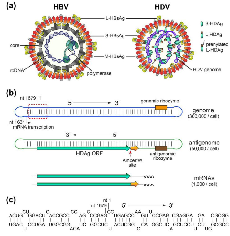
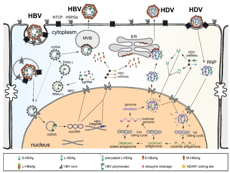
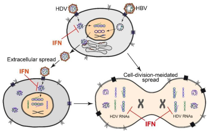
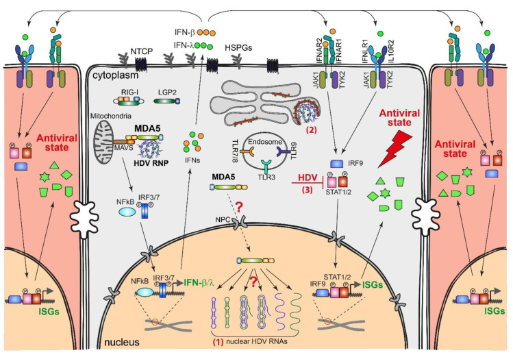
Chronic hepatitis D (CHD) is the most severe form of viral hepatitis, with rapid progression of liver-related diseases and high rates of development of hepatocellular carcinoma. The causative agent, hepatitis D virus (HDV), contains a small (approximately 1.7 kb) highly self-pairing single-strand circular RNA genome that assembles with the HDV antigen to form a ribonucleoprotein (RNP) complex. HDV depends on hepatitis B virus (HBV) envelope proteins for envelopment and de novo hepatocyte entry; however, its intracellular RNA replication is autonomous. In addition, HDV can amplify HBV independently through cell division. Cellular innate immune responses, mainly interferon (IFN) response, are crucial for controlling invading viruses, while viruses counteract these responses to favor their propagation. In contrast to HBV, HDV activates profound IFN response through the melanoma differentiation antigen 5 (MDA5) pathway. This cellular response efficiently suppresses cell-division-mediated HDV spread and, to some extent, early stages of HDV de novo infection, but only marginally impairs RNA replication in resting hepatocytes. In this review, we summarize the current knowledge on HDV structure, replication, and persistence and subsequently focus on the interplay between HDV and IFN response, including IFN activation, sensing, antiviral effects, and viral countermeasures. Finally, we discuss crosstalk with HBV.

Keywords: Hepcludex; Myrcludex B; cell-division-mediated spread; countermeasures; de novo infection; hepatitis B virus; hepatitis D virus; interferon response; pattern recognition receptors; persistence.


Conflict of interest statement

Z.Z. declares no conflicts of interest. S.U. is a co-applicant and co-inventor of patents protecting Hepcludex/bulevirtide. S.U. is a consultant for Gilead Sciences, Myr-GmbH, Galapagos NV, and Humabs BioMed SA. The funders had no role in the design of the study; in the collection, analyses, or interpretation of data; in the writing of the manuscript, or in the decision to publish the results.

Figures

Figure 1
Figure 1
Hepatitis D virus (HDV) and Hepatitis B virus (HBV) virions, and HDV RNAs. (a) Schematic representation of HDV and HBV virions. Both viruses share the same envelope containing three HBV envelope proteins: large- (L-), medium- (M-), and small- (S-) HBsAg. HDV (right panel) has a ribonucleoprotein (RNP) complex inside. The RNP consists of the HDV genome and two isoforms of hepatitis D antigen (HDAg), L- and S-HDAg. A portion of L-HDAg is prenylated, which is needed for its association with S-HBsAg [35]. On the other hand, HBV (left panel) has a nucleocapsid inside the envelope. The nucleocapsid consists of an HBV core protein shell and relaxed circular HBV DNA (rcDNA), with the latter associated with HBV polymerase. (b) HDV genome, antigenome and mRNAs. The HDV genome is a single-strand, negative-sense, circular RNA. It forms an unbranched rod-like structure due to its high degree of intramolecular base-pairing. The HDV antigenome is complementary to the genome and is predicted to form a similar structure to the genome. Two mRNAs encoding either S-HDAg or L-HDAg are transcribed using the genome as a template. Ribozymes, the ADAR1 editing (Amber/W) site, mRNA transcription starting site, and HDAg open reading frame (ORF) are indicated. Arrows indicate the 5′ to 3′ direction. (c) Structure of a representative region of the genome (red dash line box in (b)) consisting of short stems and bulges.
Figure 2
Figure 2
HDV and HBV life cycles. Right half: HDV life cycle. HDV virions first attach to heparan sulfate proteoglycans (HSPGs) [36,37,38,39] and then to the viral receptor sodium tautocholate co-transporting peptide (NTCP) [9,10]. After membrane fusion, the ribonucleoprotein (RNP) is released into the cytoplasm and further transported to the nucleus where RNA replication occurs [43,44]. The genome serves as the template for the first rolling circle amplification. The resulting antigenome multimers are cleaved in cis by the intrinsic ribozyme and ligated into circular antigenome monomers [35,45]. After a second rolling cycle using the antigenome as the template, HDV genome multimers are synthesized and self-cleaved to produce circular HDV genome monomers. The HDV antigenome might be edited by cellular adenosine deaminases acting on RNA 1 (ADAR1), yielding an extended HDAg ORF that produces L-HDAg [46]. These genomes, with or without ADAR1 editing, are used as the template for mRNA transcription. The mRNAs are translated into S-HDAg and L-HDAg. A portion of the L-HDAg molecules are prenylated for envelope acquirement [35]. S-HDAg and L-HDAg are transported into the nucleus to regulate virus replication or bind to the genome to form RNP, which is exported to the cytoplasm. Through the interaction between L-HDAg and S-HBsAg, RNP acquires an envelope and is released through the endoplasmic reticulum (ER)–Golgi secretory pathway. Left half: HBV life cycle. After binding to HSPG and NTCP, HBV is internalized through endocytosis [47]. The fusion of the HBV envelope with the endosome membrane releases the nucleocapsid, which is further transported to the nuclear pore complex (NPC) where rcDNA is imported into the nucleus. The rcDNA is processed into covalently closed circular DNA (cccDNA). This cccDNA serves as the template for HBV mRNAs and pregenomic RNA (pgRNA), with the latter captured in the HBV capsid and reverse-transcribed to the DNA of the progeny virus via HBV polymerase. The progeny HBV is considered to be secreted through a multivesicular body (MVB) [48]. Notably, HBV DNA might be integrated into cellular chromosomes [49,50]. These integrates can produce HBV envelope proteins that support HDV packaging [50,51,52,53].
Figure 3
Figure 3
HDV spreading pathways and the targets of the interferon (IFN) response. Left: de novo infection-mediated extracellular spreading pathway. HBV/HDV co-infection produces progeny HDV that infect neighboring intact hepatocytes. The IFN response inhibits early stages of HDV de novo infection but does not significantly impair HDV RNA replication in the nucleus. Right: cell-division-mediated HDV spread. HDV survives cell division and efficiently establishes replication in both daughter cells. The IFN response causes efficient degradation of HDV RNA during cell division and/or prevents the re-establishment of replication in daughter cells.
Figure 4
Figure 4
HDV-induced IFN response and possible HDV countermeasures. HDV RNA in the cytoplasm; likely, the RNP complex is recognized by the pattern recognition receptor (PRR) MDA5 [106]. This recognition activates the mitochondrial antiviral signaling protein (MAVS) on the mitochondria and downstream transcription factors, likely IFN regulatory factor (IRF) 3/7 and nuclear factor-κB (NFκB). The activated transcription factors are translocated into the nucleus and initiate the transcription of IFN-β/λ. Secreted IFN-β/λ binds to their receptors (IFNAR1/IFNAR2 for IFN-α/β and IFNLR1/IL10R2 for IFN-λ) on the infected cell or neighboring cells, which further activates Janus kinases (JAK) 1/2, tyrosine kinase (TYK) 2, and transcription factors signal transducer and activator of transcription (STAT) 1/2 and IRF9. STAT1/2 and IRF9 are translocated into the nucleus and activate hundreds of IFN-stimulated genes (ISGs), which directly inhibit HDV replication and protect the uninfected cells against subsequent infection. It is unknown whether MDA5 can also be transported to the nucleus and capture nuclear HDV replication intermediates. HDV may counteract the IFN response through different strategies: (1) HDV may replicate in a “safe” compartment, the nucleus, to avoid exposure of the replication intermediates to PRRs; (2) HDV genomic RNA in the cytoplasm may fold into RNP with HDAg and bud into an HBV envelope to avoid being recognized by the PRRs; and (3) HDV may directly inhibit STAT1/2 activation.

References

    1. Farci P., Niro G.A. Clinical features of hepatitis D. Semin. Liver Dis. 2012;32:228–236. doi: 10.1055/s-0032-1323628. - DOI - PubMed
    1. Botelho-Souza L.F., Vasconcelos M.P.A., Dos Santos A.O., Salcedo J.M.V., Vieira D.S. Hepatitis delta: Virological and clinical aspects. Virol. J. 2017;14:177. doi: 10.1186/s12985-017-0845-y. - DOI - PMC - PubMed
    1. Negro F. Hepatitis D virus coinfection and superinfection. Cold Spring Harb. Perspect. Med. 2014;4:a021550. doi: 10.1101/cshperspect.a021550. - DOI - PMC - PubMed
    1. Stockdale A.J., Kreuels B., Henrion M.Y.R., Giorgi E., Kyomuhangi I., de Martel C., Hutin Y., Geretti A.M. The global prevalence of hepatitis D virus infection: Systematic review and meta-analysis. J. Hepatol. 2020;73:523–532. doi: 10.1016/j.jhep.2020.04.008. - DOI - PMC - PubMed
    1. Chen H.Y., Shen D.T., Ji D.Z., Han P.C., Zhang W.M., Ma J.F., Chen W.S., Goyal H., Pan S., Xu H.G. Prevalence and burden of hepatitis D virus infection in the global population: A systematic review and meta-analysis. Gut. 2019;68:512–521. doi: 10.1136/gutjnl-2018-316601. - DOI - PubMed

Publication types

LinkOut - more resources