. 2021 Jan 20;41(3):502-512.
doi: 10.1523/JNEUROSCI.2015-20.2020. Epub 2020 Nov 24.

The Cell-Autonomous Clock of VIP Receptor VPAC2 Cells Regulates Period and Coherence of Circadian Behavior

Affiliations

The Cell-Autonomous Clock of VIP Receptor VPAC2 Cells Regulates Period and Coherence of Circadian Behavior

Ryan Hamnett et al. J Neurosci. .

Abstract

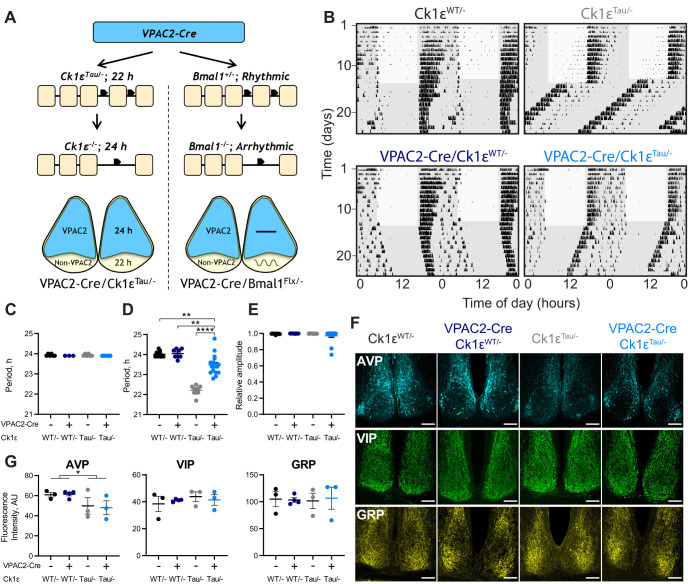
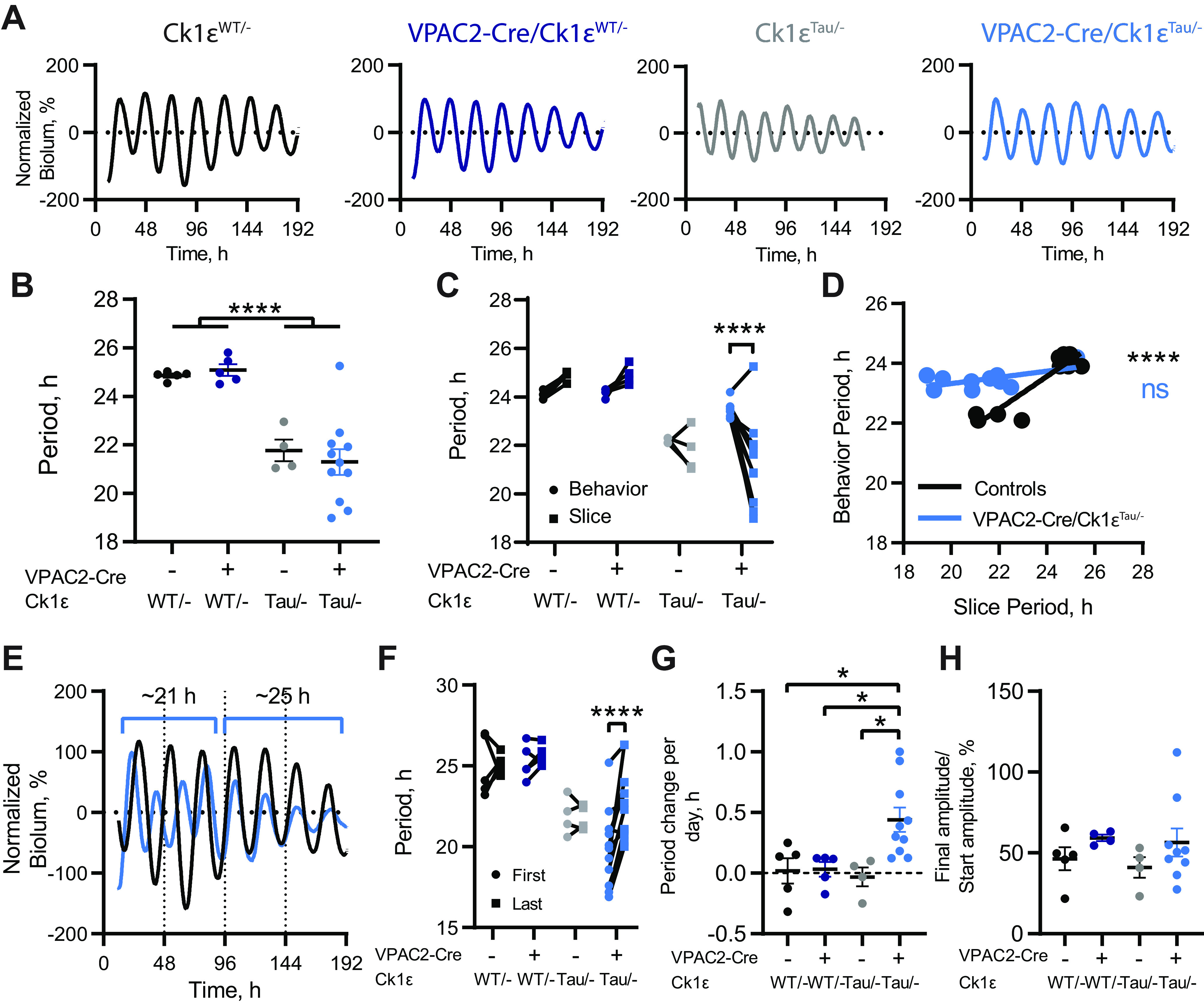
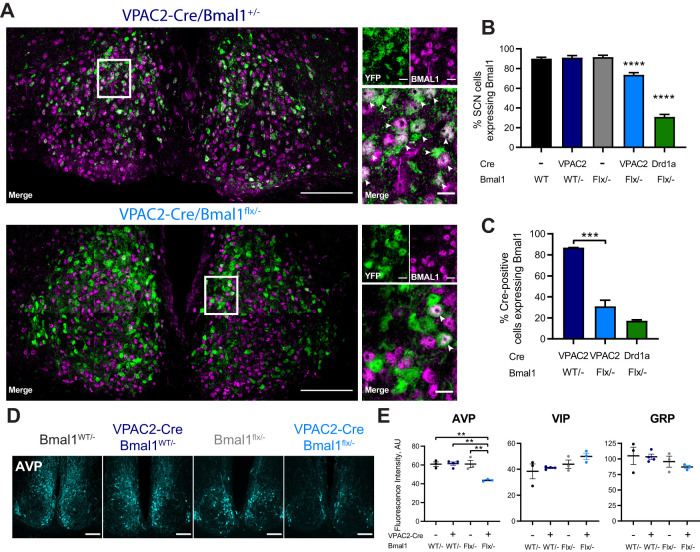
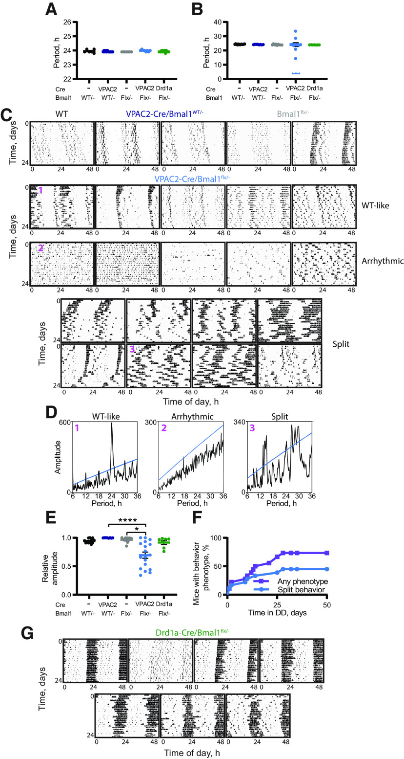
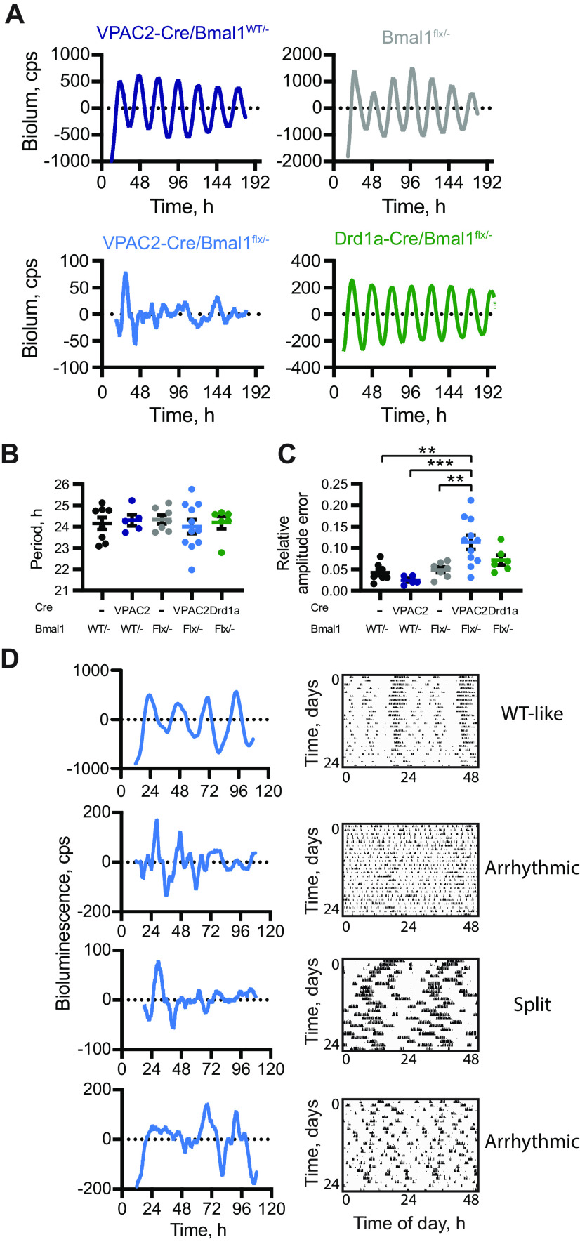
Circadian (approximately daily) rhythms pervade mammalian behavior. They are generated by cell-autonomous, transcriptional/translational feedback loops (TTFLs), active in all tissues. This distributed clock network is coordinated by the principal circadian pacemaker, the hypothalamic suprachiasmatic nucleus (SCN). Its robust and accurate time-keeping arises from circuit-level interactions that bind its individual cellular clocks into a coherent time-keeper. Cells that express the neuropeptide vasoactive intestinal peptide (VIP) mediate retinal entrainment of the SCN; and in the absence of VIP, or its cognate receptor VPAC2, circadian behavior is compromised because SCN cells cannot synchronize. The contributions to pace-making of other cell types, including VPAC2-expressing target cells of VIP, are, however, not understood. We therefore used intersectional genetics to manipulate the cell-autonomous TTFLs of VPAC2-expressing cells. Measuring circadian behavioral and SCN rhythmicity in these temporally chimeric male mice thus enabled us to determine the contribution of VPAC2-expressing cells (∼35% of SCN cells) to SCN time-keeping. Lengthening of the intrinsic TTFL period of VPAC2 cells by deletion of the CK1εTau allele concomitantly lengthened the period of circadian behavioral rhythms. It also increased the variability of the circadian period of bioluminescent TTFL rhythms in SCN slices recorded ex vivo Abrogation of circadian competence in VPAC2 cells by deletion of Bmal1 severely disrupted circadian behavioral rhythms and compromised TTFL time-keeping in the corresponding SCN slices. Thus, VPAC2-expressing cells are a distinct, functionally powerful subset of the SCN circuit, contributing to computation of ensemble period and maintenance of circadian robustness. These findings extend our understanding of SCN circuit topology.

Keywords: Bmal1; bioluminescence; casein kinase; circadian rhythm; neuropeptide; suprachiasmatic nucleus.


Conflict of interest statement

The authors declare no competing financial interests.

Figures

Figure 1.
Figure 1.
Intersectional genetics reveals that VPAC2-expressing cells determine the circadian period of mouse wheel-running behavior. A, Schematic view of the intersectional approach, whereby mice carrying Cre recombinase as a transgene controlled by the VPAC2 promoter were crossed with either Ck1εTau/– or Bmal1flx/– mice to create a chimeric SCN. The result is for VPAC2 cells to lack either the Tau allele or Bmal1, reverting them to a 24 h period as Ck1ε−/− or rendering them arrhythmic as Bmal1−/−, respectively. B, Representative double-plotted actograms of wheel-running activity of VPAC2-Cre/Ck1εTau/– mice and their respective controls exposed to a 12:12 light/dim red light (LD) cycle followed by continuous dim red light (DD) conditions. Gray shading represents dim red light. C, Periods (mean ± SEM) observed in 12:12 LD: n = 6 (Ck1εWT/–), n = 3 (VPAC2-Cre/Ck1εWT/–), n = 6 (Ck1εTau/–), and n = 7 (VPAC2-Cre/Ck1εTau/–). D, Periods (mean ± SEM) observed in DD (n = 9, n = 8, n = 9, n = 17) of VPAC2-Cre/Ck1εTau/– mice and their respective controls. E, RA scores (mean ± SEM) from 10 d of wheel-running activity under DD (n values as in D). Kruskal–Wallis test. F, Representative images of immunohistochemical staining of SCN of VPAC2-Cre/Ck1εTau/– mice and their respective controls for AVP-ir (top), VIP-ir (middle), and GRP-ir (bottom). Scale bars, 100 µm. G, Fluorescence intensities (mean ± SEM) for immunohistochemical staining of AVP-ir (left), VIP-ir (middle), and GRP-ir (right) in SCN sections of VPAC2-Cre/Ck1εTau/– mice with their respective controls (n = 3 per genotype). *p <0.05; **p <0.01; ****p <0.0001; two-way ANOVAs (main effects of Cre and Tau allele) with Tukey's post hoc test.
Figure 2.
Figure 2.
Circadian periods of bioluminescent rhythms of SCN slices from VPAC2-Cre/Ck1εTau/– mice do not reflect behavioral period. A, Representative baseline-corrected PER2::LUCIFERASE bioluminescence rhythms from Ck1εWT/–, VPAC2-Cre/Ck1εWT/–, Ck1εTau/–, and VPAC2-Cre/Ck1εTau/– SCN dissected following wheel-running recordings. B, Periods (mean ± SEM) of the first 4 bioluminescent cycles of adult SCN slices as in A. C, Comparison of circadian periods of free-running behavior and slice bioluminescence rhythms (measured from the first 4 cycles) from individual mice. D, Scatter plot of slice periods (measured from the first 4 cycles) versus behavioral periods. VPAC2-Cre/Ck1εTau/– mice show no significant correlation (p =0.07, Pearson's correlation), but grouped control slices do (****p <0.0001, Pearson's correlation). Lines indicate linear regression: VPAC2-Cre/Ck1εTau/–, r2 = 0.36, Y = 0.1037*X + 21.25; Controls: r2 = 0.85, Y = 0.5458*X + 10.47. E, Representative baseline-corrected PER2::LUCIFERASE bioluminescence rhythms from Ck1εWT/– and VPAC2-Cre/Ck1εTau/– SCN slices displaying no phase alignment initially, followed by period lengthening in the VPAC2-Cre/Ck1εTau/– slice and resultant phase alignment. F, Comparison of periods between the first two bioluminescent cycles (First) and final two cycles (Last) within each SCN slice. G, Period change per day (mean ± SEM) in SCN slices. H, Bioluminescence amplitude (mean ± SEM) in the last 3 cycles as a percentage of the amplitude in the first 3 cycles. B, F-H, n = 5 (Ck1εWT/–), n = 5 (VPAC2-Cre/Ck1εWT/–), n = 4 (Ck1εTau/–), and n = 11 (VPAC2-Cre/Ck1εTau/–). C, D, n = 5 (Ck1εWT/–), n = 4 (VPAC2-Cre/Ck1εWT/–), n = 4 (Ck1εTau/–), and n = 10 (VPAC2-Cre/Ck1εTau/–). *p <0.05; ****p <0.0001; two-way ANOVAs with Tukey's post hoc test.
Figure 3.
Figure 3.
Targeted deletion of BMAL1 from VPAC2-Cre-expressing SCN cells. A, Representative 63× tiled confocal micrographs of Cre recombinase activity, as reported by a genetically encoded EYFP reporter (green), and BMAL1 immunohistochemistry (magenta) in VPAC2-Cre/Bmal1WT/– and VPAC2-Cre/Bmal1flx/– SCN sections. White rectangles represent locations of magnified images. White arrowheads indicate colocalization between EYFP and BMAL1-ir. Scale bars: stitched images, 100 µm; magnified images, 10 µm. B, Percentage of SCN cells (marked by DAPI; mean ± SEM) expressing BMAL1-ir across genotypes, including Drd1a-Cre/Bmal1flx/–. C, Percentage of Cre-positive cells (marked by EYFP; mean ± SEM) expressing BMAL1-ir across genotypes. B, C, One-way ANOVA with Tukey's post hoc test. D, Representative images of AVP-ir in SCN of VPAC2-Cre/Bmal1flx/– mice and controls. Scale bars, 100 µm. E, Fluorescence intensities (mean ± SEM) for immunohistochemical staining of AVP (left), VIP (middle), and GRP (right) in SCN sections of VPAC2-Cre/Bmal1flx/– mice and controls. B, C, n = 7 (Bmal1WT/–), n = 7 (Bmal1flx/–), n = 2 (VPAC2-Cre/Bmal1WT/–), n = 8 (VPAC2-Cre/Bmal1flx/–), and n = 3 (Drd1a-Cre/Bmal1flx/–). E, n = 3 per group. **p <0.01; ***p <0.001; ****p <0.0001; two-way ANOVA with Tukey's post hoc test.
Figure 4.
Figure 4.
Deletion of BMAL1 from VPAC2-Cre-expressing cells compromises circadian behavior. A, B, Circadian periods (mean ± SEM) of VPAC2-Cre/Bmal1flx/– mice and controls under (A) 12:12 LD and (B) DD. For 6 VPAC2-Cre/Bmal1flx/– mice, an explicit single period could not be determined because of their disturbed behavior; and so, these are added as stars as a nominal 3 h, and were excluded from statistical analysis. Two-way ANOVA (excluding Drd1a-Cre/Bmal1flx/– mice); one-way ANOVA, including all groups for Drd1a-Cre/Bmal1flx/– comparison. C, Representative double-plotted actograms of final 24 d of wheel-running activity of control mice (1 Bmal1WT/–, 2 VPAC2-Cre/Bmal1WT/–, and 2 Bmal1flx/–) and of all VPAC2-Cre/Bmal1flx/– mice. VPAC2-Cre/Bmal1flx/– actograms are divided into three groups (WT-like, Arrhythmic, and Split) and then ranked in descending order of RA score under DD (final 14 d of recording). D, χ2 periodograms for 3 VPAC2-Cre/Bmal1flx/– as indicated in C: D1, has a single significant peak at ∼24 h; D2, has no significant period; D3, displays multiple significant periods. E, RA scores (mean ± SEM) from the last 14 d of wheel-running activity under DD. Kruskal–Wallis test with Dunn's multiple comparisons. n = 12 (Bmal1WT/–), n = 8 (Bmal1flx/–), n = 8 (VPAC2-Cre/Bmal1WT/–), n = 18 (VPAC2-Cre/Bmal1flx/–), and n = 7 (Drd1a-Cre/Bmal1flx/–). F, Percentage of VPAC2-Cre/Bmal1flx/– mice displaying behavioral phenotypes (purple, squares) or specifically split behavior (blue, circles) over time. G, Double-plotted actograms of wheel-running activity of all Drd1a-Cre/Bmal1flx/– mice. *p <0.05. ****p <0.0001.
Figure 5.
Figure 5.
Deletion of BMAL1 from VPAC2-Cre-expressing cells compromises SCN molecular pace-making. A, Representative, baseline-corrected PER2::LUCIFERASE bioluminescence traces from control, VPAC2-Cre/Bmal1flx/–, and Drd1a-Cre/Bmal1flx/– SCN dissected in dim red light following recording of wheel-running rhythms in DD. B, Period (mean ± SEM) of the first 4 bioluminescent cycles recorded from adult SCN slices; genotypes as in A. n = 8 (Bmal1WT/–), n = 7 (Bmal1flx/–), n = 5 (VPAC2-Cre/Bmal1WT/–), n = 11 (VPAC2-Cre/Bmal1flx/–), and n = 6 (Drd1a-Cre/Bmal1flx/–). C, RAE scores (mean ± SEM) for bioluminescent recordings from SCN slices. n values same as in B. D, Representative PER2::LUCIFERASE bioluminescence rhythms from VPAC2-Cre/Bmal1flx/– SCN slices alongside respective actograms from corresponding mice. “WT-like,” “Arrhythmic,” or “Split” beside the actograms indicates the phenotypic category. **p <0.01; ***p <0.001; one-way ANOVA, with Tukey's post hoc test.

Similar articles

Cited by

References

    1. Abrahamson EE, Moore RY (2001) Suprachiasmatic nucleus in the mouse: retinal innervation, intrinsic organization and efferent projections. Brain Res 916:172–191. 10.1016/s0006-8993(01)02890-6 - DOI - PubMed
    1. Antle MC, Silver R (2005) Orchestrating time: arrangements of the brain circadian clock. Trends Neurosci 28:145–151. 10.1016/j.tins.2005.01.003 - DOI - PubMed
    1. Aton SJ, Colwell CS, Harmar AJ, Waschek J, Herzog ED (2005) Vasoactive intestinal polypeptide mediates circadian rhythmicity and synchrony in mammalian clock neurons. Nat Neurosci 8:476–483. 10.1038/nn1419 - DOI - PMC - PubMed
    1. Brancaccio M, Patton AP, Chesham JE, Maywood ES, Hastings MH (2017) Astrocytes control circadian timekeeping in the suprachiasmatic nucleus via glutamatergic signaling. Neuron 93:1420–1435.e5. 10.1016/j.neuron.2017.02.030 - DOI - PMC - PubMed
    1. Brown TM, Hughes AT, Piggins HD (2005) Gastrin-releasing peptide promotes suprachiasmatic nuclei cellular rhythmicity in the absence of vasoactive intestinal polypeptide-VPAC2 receptor signaling. J Neurosci 25:11155–11164. 10.1523/JNEUROSCI.3821-05.2005 - DOI - PMC - PubMed

Publication types

MeSH terms

Substances

LinkOut - more resources