. 2020 Nov 24;13(659):eaba9902.
doi: 10.1126/scisignal.aba9902.

Kallikrein 13 serves as a priming protease during infection by the human coronavirus HKU1

Affiliations

Kallikrein 13 serves as a priming protease during infection by the human coronavirus HKU1

Aleksandra Milewska et al. Sci Signal. .

Abstract

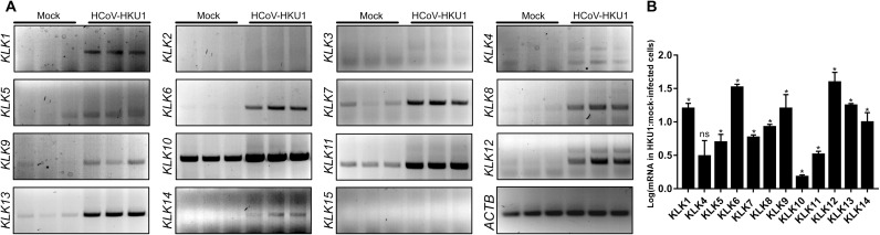
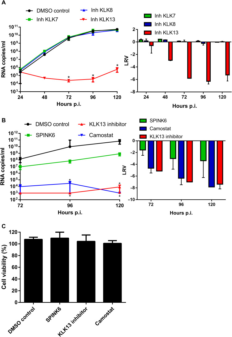
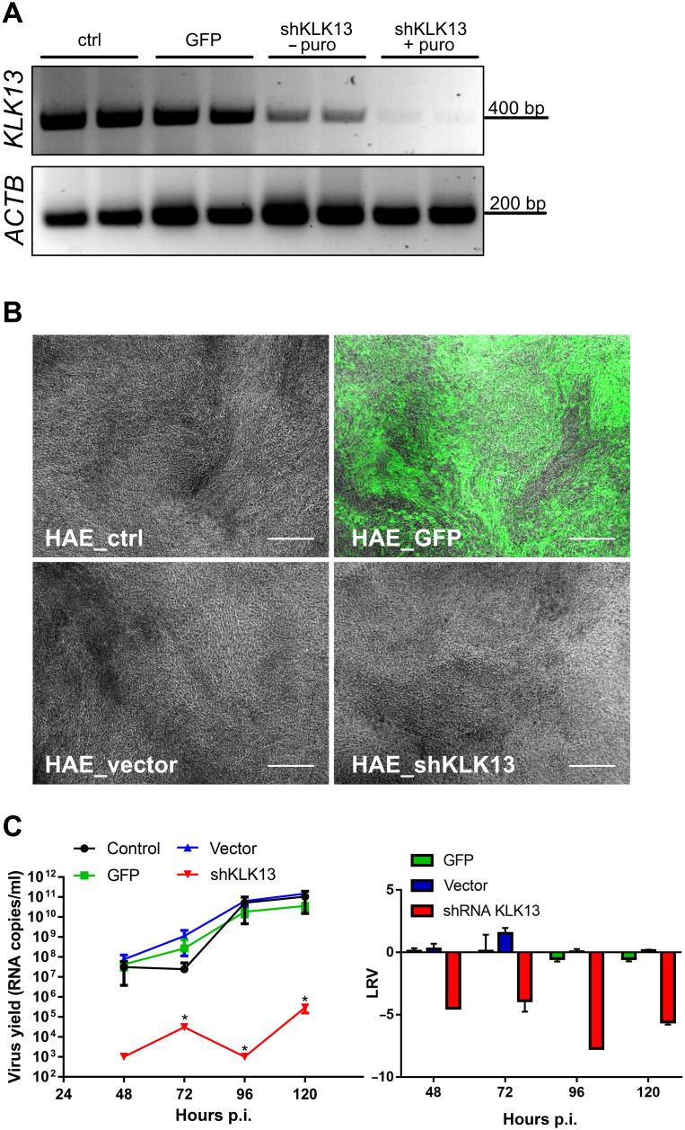
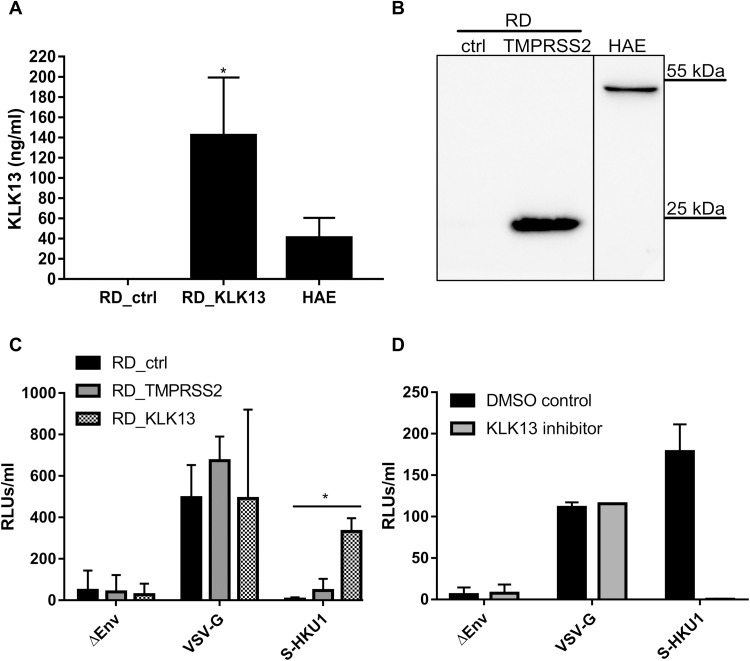
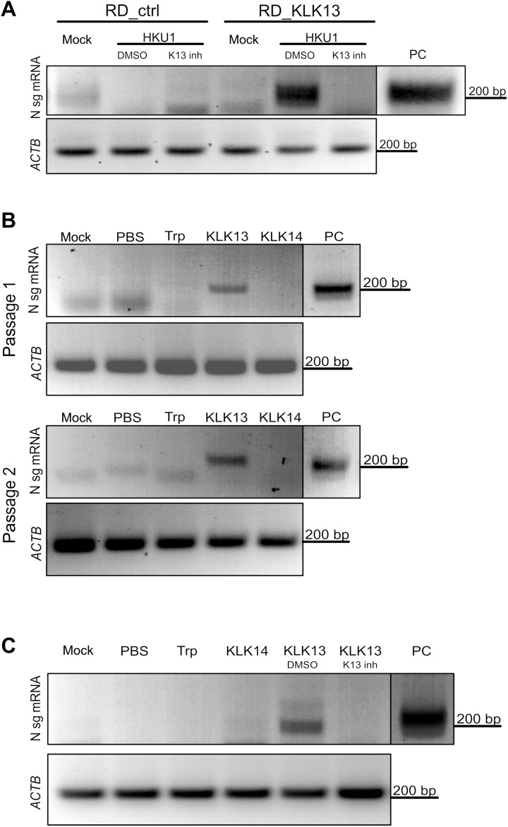
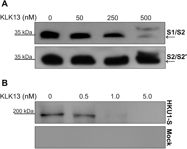
Human coronavirus HKU1 (HCoV-HKU1) is associated with respiratory disease and is prevalent worldwide, but an in vitro model for viral replication is lacking. An interaction between the coronaviral spike (S) protein and its receptor is the primary determinant of tissue and host specificity; however, viral entry is a complex process requiring the concerted action of multiple cellular elements. Here, we found that the protease kallikrein 13 (KLK13) was required for the infection of human respiratory epithelial cells and was sufficient to mediate the entry of HCoV-HKU1 into nonpermissive RD cells. We also demonstrated the cleavage of the HCoV-HKU1 S protein by KLK13 in the S1/S2 region, suggesting that KLK13 is the priming enzyme for this virus. Together, these data suggest that protease distribution and specificity determine the tissue and cell specificity of the virus and may also regulate interspecies transmission.


Figures

Fig. 1
Fig. 1. HCoV-HKU1 infection of HAE cultures induces the expression of several KLKs.
(A and B) HAE cultures were left uninfected (mock) or were infected with HCoV-HKU1 (106 RNA copies/ml) for 2 hours at 32°C and cultured for 5 days. Cellular RNA was then isolated, treated with DNase, and subjected to reverse transcription, and the mRNAs for the indicated KLKs were amplified using specific primers. The analysis was performed twice using cells obtained from different donors, each time in triplicate. (A) The indicated amplified PCR products were resolved and detected in 1.5% (w/v) agarose gel in 1× TAE buffer. (B) The relative abundance of the indicated KLK mRNAs normalized to that of ACTB was assessed semiquantitively by densitometric analysis. Data are presented as a log change of signal specific for the indicated KLK mRNA in HCoV-HKU1–infected cells compared to that in the mock-infected cells. The experiments were performed twice with cells from different donors, each time with two biological replicates. For comparisons by Student’s t test, *P < 0.05; ns, not significant.
Fig. 2
Fig. 2. HCoV-HKU1 infection is dependent on KLK13 activity.
(A and B) HAE cultures were inoculated with HCoV-HKU1 (106 RNA copies/ml) for 2 hours at 32°C in the presence of the indicated KLK inhibitors (each at 10 μM, table S1) or DMSO (A) or else in the presence of SPINK6 (10 μg/ml), 10 μM KLK13 inhibitor, 100 μM camostat, or DMSO. Statistical significance was assessed with the Kruskal-Wallis test. *P < 0.05. (B) To analyze viral replication kinetics, each day post-infection (p.i.), 100 μl of PBS containing a given inhibitor was applied to the apical surface of the HAE cultures and collected after 10 min of incubation at 32°C. Replication of HCoV-HKU1 was evaluated by RT-qPCR analysis, and the data are presented as RNA copy numbers/ml (left) and as the log removal value (LRV) compared to the untreated sample (right). The assays were performed twice, each time in triplicate (N = 3), and average values with standard errors are presented. (C) Assessment of the cytotoxicity of inhibitors in the HAE cultures. Cell viability was assessed with the XTT assay on mock-treated cells at 120 hours p.i. Data on the y axis represent the percentage values obtained for the untreated reference samples. The assays were performed in triplicate (N = 3), and average values with standard errors are presented.
Fig. 3
Fig. 3. HCoV-HKU1 does not replicate in HAE cultures deficient in KLK13.
(A to C) Primary human epithelial cells were transduced with lentiviral vectors expressing GFP (HAE_GFP), empty pLKO.1-TRC vector (HAE_vector), or shRNA specific for KLK13 mRNA (HAE_shKLK13) and differentiated to form HAE cultures. As a control, HAE cultures differentiated from untransduced cells were used (HAE_ctrl). (A) KLK13 mRNA was evaluated before (− puro) and after puromycin (+ puro) selection of positively transduced cells. ACTB mRNA was used as an internal control. (B) Microscopic examination of all HAE cultures after 4 weeks of culture in ALI at 37°C. The images show the fully differentiated, genetically engineered HAE cultures in which KLK13 was silenced and GFP was overexpressed. Scale bars, 200 μm. (C) All HAE cultures were inoculated with HCoV-HKU1 (106 RNA copies/ml) for 2 hours at 32°C and cultured for 5 days. Each day p.i., 100 μl of PBS was applied to the apical surface of the HAE cell cultures and collected after 10 min of incubation at 32°C. Replication of HCoV-HKU1 was evaluated by RT-qPCR analysis. The data are presented as RNA copy number/ml (left) and as log removal value (LRV) compared to the untreated sample (right). The assays were performed twice, each time in triplicate (N = 3), and average values with standard errors are presented. Statistical significance was assessed with the Kruskal-Wallis test. *P < 0.05.
Fig. 4
Fig. 4. RD cells expressing KLK13 are permissive for HCoV-HKU1 pseudoviruses.
(A) RD cells were transduced with lentiviral vectors expressing the KLK13 gene (KLK13) or with empty vector (ctrl). The abundance of KLK13 secreted by RD cells and HAE cultures into the cell culture medium was determined with a KLK13-specific ELISA. The assays were performed twice, each time in triplicate (N = 3). Statistical significance was assessed with the Mann-Whitney test. *P < 0.05. (B) RD cells were transduced with lentiviral vectors expressing the TMPRSS2 gene (TMPRSS2) or empty vector (ctrl). After blasticidin selection, the cells were lysed and proteins were resolved by SDS-PAGE and analyzed by Western blotting. TMPRSS2 was detected in RD cell lysates (50 μg of protein per lane) and HAE cultures lysate (25 μg of protein per lane) using a specific antibody. The vertical black line indicates that the lanes are not contiguous. Blots are representative of three experiments. (C) RD control cells (RD_ctrl), TMPRSS2-expressing RD cells (RD_TMPRSS2), and KLK13-expressing RD cells (RD_KLK13) were transduced with HIV pseudoviruses decorated with VSV-G protein (VSV-G) or S-HKU1 glycoprotein (S-HKU1) or with control viruses without the fusion protein (ΔEnv). After 72 hours at 37°C, pseudovirus entry was measured by measurement of the luminescence signal in the cell lysates [relative light units (RLUs)/ml of lysate sample]. The assays were performed twice, each time in triplicate (N = 3). Data are means ± SEM. Statistical significance was assessed with the Kruskal-Wallis test. *P < 0.05. (D) HAE cultures were inoculated with HIV pseudoviruses expressing the VSV-G control protein or S-HKU1 or with control viruses without the fusion protein (ΔEnv) in the presence of KLK13 inhibitor (10 μM) or DMSO. After 72 hours at 37°C, the entry of the pseudoviruses was determined by measuring the luminescence signal in the cell lysates. The assays were performed in duplicate (N = 2). Data are means ± SEM.
Fig. 5
Fig. 5. HCoV-HKU1 replicates in RD cells expressing KLK13.
(A) Control (RD_ctrl) and KLK13-expressing RD cells (RD_KLK13) were inoculated with HCoV-HKU1 (106 RNA copies/ml) or were left uninfected (mock) in the presence of 10 μM KLK 13 inhibitor (K13 inh) or with DMSO as a control. After 7 days of culture at 32°C, total RNA was isolated and reverse-transcribed, and subgenomic mRNA for the viral N protein was detected by seminested PCR analysis. ACTB was used as an internal control. PC, positive control from virus-infected HAE cultures. (B) HCoV-HKU1 was incubated with 200 nM trypsin (Trp), KLK13, KLK14, or PBS for 2 hours at 32°C and further applied onto the RD cells. Top: After 7 days at 32°C, total RNA was isolated and reverse-transcribed, and subgenomic mRNA for the viral N protein was detected by seminested PCR analysis (passage 1). Bottom: In addition, 1 ml of the cell culture medium was harvested and applied to freshly seeded RD cells with medium supplemented with fresh enzymes. After 7 days at 32°C, subgenomic mRNA for the N protein was detected by seminested PCR (passage 2). ACTB was used as an internal control. (C) HCoV-HKU1 was incubated with 200 nM trypsin (Trp), KLK14, KLK13, KLK13 in the presence of the KLK13 inhibitor (K13 inh), DMSO (DMSO), or PBS for 2 hours at 32°C and further applied onto RD cells. Subgenomic mRNA for N protein was detected by seminested PCR. ACTB was used as an internal control. The vertical black lines indicate that the lanes are not contiguous.
Fig. 6
Fig. 6. KLK13 cleaves the HCoV-HKU1 S protein between the S1 and S2 domains.
(A) Fifteen nanograms of CleavEx proteins harboring the S1/S2 or S2′ sites was incubated at 37°C for 3 hours with the indicated concentrations of purified KLK13 protein. The samples were then denatured at 95°C, and the proteins were resolved by SDS-PAGE and analyzed by Western blotting with antibodies specific for His-tagged proteins. Blots are representative of three experiments. (B) The full-length HKU1-S protein or control sample was incubated at 37°C for 3 hours with the indicated concentrations of purified KLK13 protein. The samples were then denatured at 95°C, and the proteins were resolved by SDS-PAGE and analyzed by Western blotting with HRP-conjugated antibody against the His tag to detect the full-length protein with the N-terminal His tag (~200 kD) and the S1 after KLK13 cleavage (~80 kD). Blots are representative of three experiments.

Similar articles

Cited by

References

    1. B. N. Fields, D. M. Knipe, P. M. Howley, Fields Virology (Wolters Kluwer Health/Lippincott Williams & Wilkins, ed. 6, 2013).
    1. Peiris J. S., Yuen K. Y., Osterhaus A. D. M. E., Stöhr K., The severe acute respiratory syndrome. N. Engl. J. Med. 349, 2431–2441 (2003). - PubMed
    1. de Groot R. J., Baker S. C., Baric R. S., Brown C. S., Drosten C., Enjuanes L., Fouchier R. A. M., Galiano M., Gorbalenya A. E., Memish Z. A., Perlman S., Poon L. L. M., Snijder E. J., Stephens G. M., Woo P. C. Y., Zaki A. M., Zambon M., Ziebuhr J., Middle East respiratory syndrome coronavirus (MERS-CoV): Announcement of the Coronavirus Study Group. J. Virol. 87, 7790–7792 (2013). - PMC - PubMed
    1. Zaki A. M., van Boheemen S., Bestebroer T. M., Osterhaus A. D. M. E., Fouchier R. A., Isolation of a novel coronavirus from a man with pneumonia in Saudi Arabia. N. Engl. J. Med. 367, 1814–1820 (2012). - PubMed
    1. van der Hoek L., Pyrc K., Jebbink M. F., Vermeulen-Oost W., Berkhout R. J. M., Wolthers K. C., Wertheim-van Dillen P. M. E., Kaandorp J., Spaargaren J., Berkhout B., Identification of a new human coronavirus. Nat. Med. 10, 368–373 (2004). - PMC - PubMed

Publication types

MeSH terms

LinkOut - more resources