. 2020 Dec 21;9(12):bio052969.
doi: 10.1242/bio.052969.

Harmine enhances the activity of the HIV-1 latency-reversing agents ingenol A and SAHA

Affiliations

Harmine enhances the activity of the HIV-1 latency-reversing agents ingenol A and SAHA

Jared P Taylor et al. Biol Open. .

Abstract

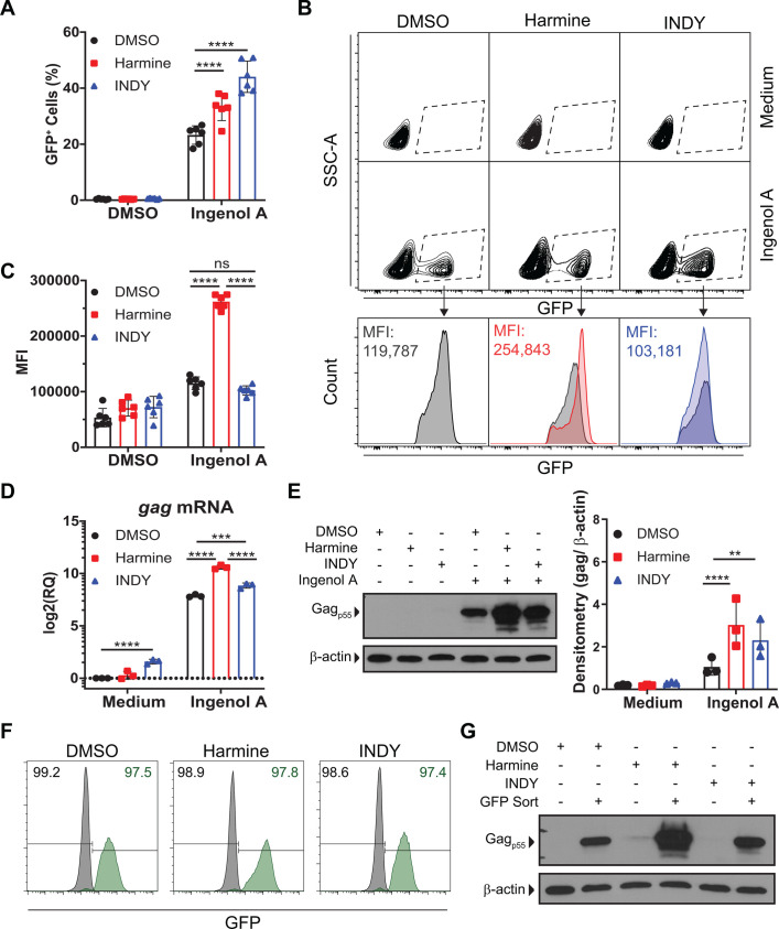
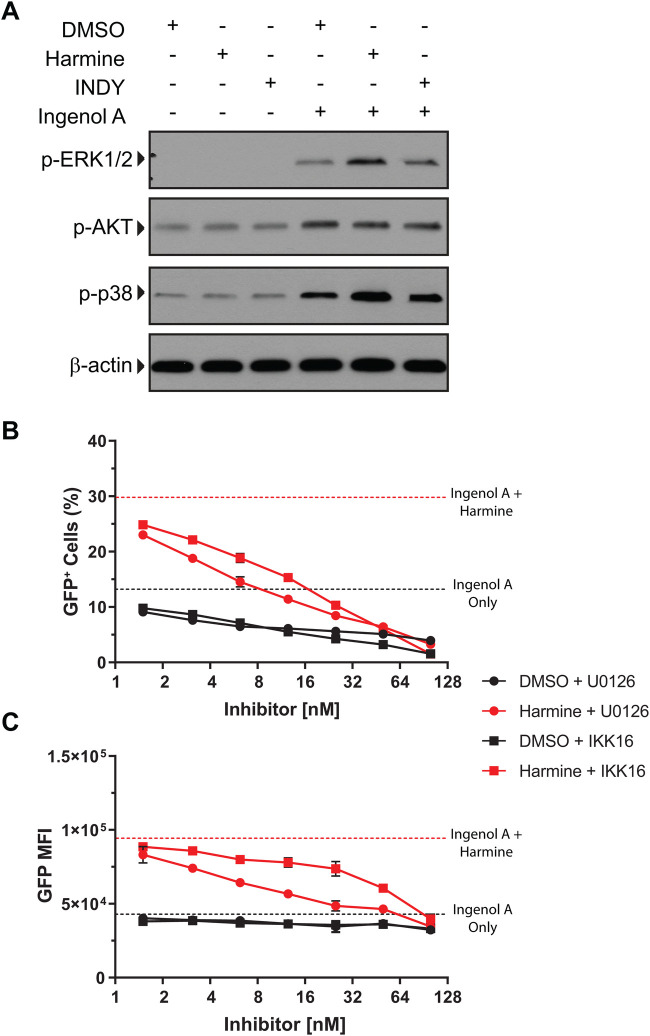
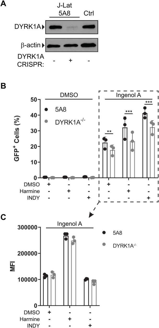
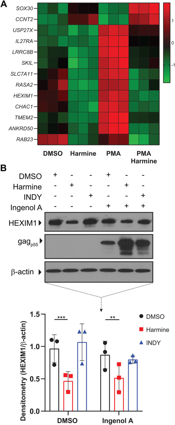
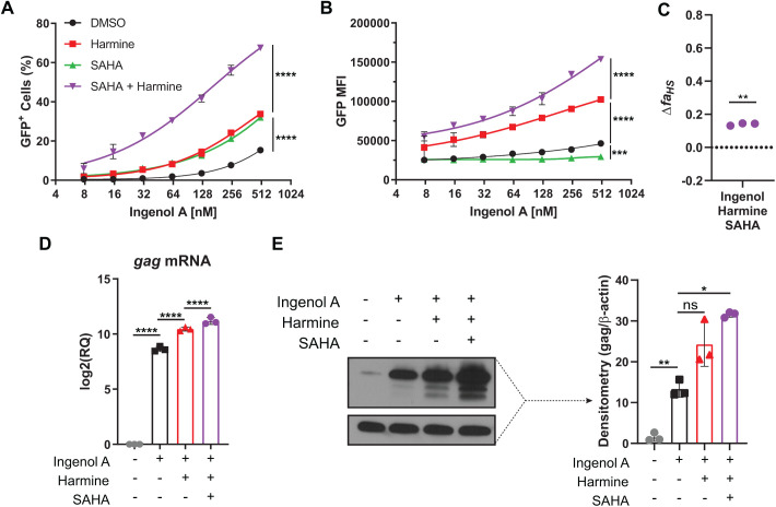
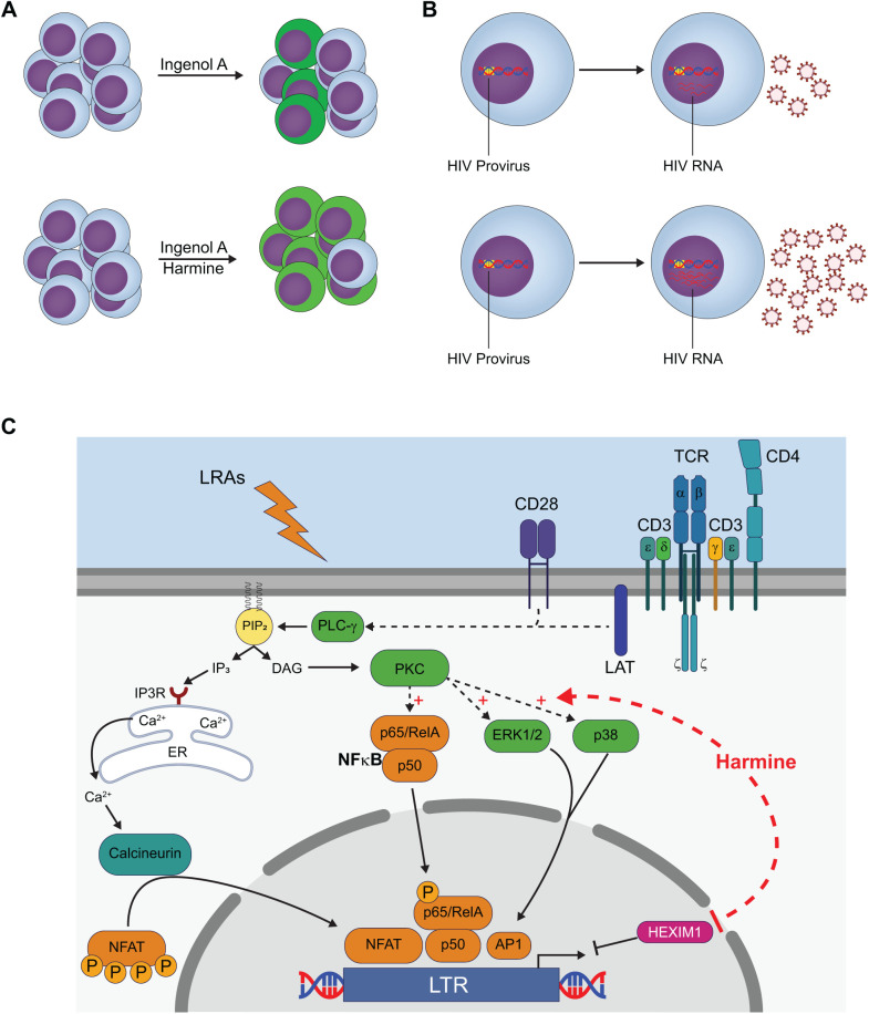
Infection with human immunodeficiency virus 1 (HIV-1) remains incurable because long-lived, latently-infected cells persist during prolonged antiretroviral therapy. Attempts to pharmacologically reactivate and purge the latent reservoir with latency reactivating agents (LRAs) such as protein kinase C (PKC) agonists (e.g. ingenol A) or histone deacetylase (HDAC) inhibitors (e.g. SAHA) have shown promising but incomplete efficacy. Using the J-Lat T cell model of HIV latency, we found that the plant-derived compound harmine enhanced the efficacy of existing PKC agonist LRAs in reactivating latently-infected cells. Treatment with harmine increased not only the number of reactivated cells but also increased HIV transcription and protein expression on a per-cell basis. Importantly, we observed a synergistic effect when harmine was used in combination with ingenol A and the HDAC inhibitor SAHA. An investigation into the mechanism revealed that harmine, when used with LRAs, increased the activity of NFκB, MAPK p38, and ERK1/2. Harmine treatment also resulted in reduced expression of HEXIM1, a negative regulator of transcriptional elongation. Thus, harmine enhanced the effects of LRAs by increasing the availability of transcription factors needed for HIV reactivation and promoting transcriptional elongation. Combination therapies with harmine and LRAs could benefit patients by achieving deeper reactivation of the latent pool of HIV provirus.

Keywords: HIV; Harmine; Latency.


Conflict of interest statement

Competing interestsThe authors declare no competing or financial interests.

Figures

Fig. 1.
Fig. 1.
DYRK1A inhibitors enhance the efficacy of PKC agonist ingenol A. (A) The percentage of GFP+ J-Lat 5A8 cells after reactivation with ingenol A in the presence of inhibitors (n=6), (B) representative flow cytometry plots of the mean fluorescence intensity of GFP+ cells, and (C) summary mean fluorescence intensity data (n=6). (D) gag mRNA expression determined by RT-qPCR. (E) Gag protein expression determined by western blot with representative blot and densitometry (n=3). GFP+ and GFP cells were sorted after reactivation with ingenol A. (F) Post-sort purity measured by flow cytometry. The percentage of GFP population (black) and the GFP+ population (green). (G) Gag protein expression of post-sort GFP+ cells measured by western blot. Error bars represent standard deviation. Statistical analysis was performed by two-way ANOVA and corrected for multiple comparisons by Tukey's test. **P<0.01; ***P<0.001; ****P<0.0001; ns, not significant.
Fig. 2.
Fig. 2.
Harmine's boosting effect is through NFκB, not NFAT. (A) Ionomycin or ingenol A were titrated on J-Lat 5A8 luciferase reporter cells. The dashed lines indicate the luciferase activity with no ionomycin or ingenol A treatment. (B) NFAT or NFκB luciferase reporter cells reactivated by ionomycin or ingenol A in the presence of inhibitors. Dashed lines represent luciferase activity with no ionomycin or ingenol A treatment. (C) J-Lat 5A8 cells were stained with Calcium Sensor Dye eFluor 514 (2 µM) and treated with DMSO, harmine, or INDY for 30 min. Geometric mean fluorescence was measured by flow cytometry for 240 s. Ionomycin (250 nM) or ingenol A (50 nM) was added after 75 s (arrows). Plots show mean geometric fluorescence intensity over time and the change in area under the curve (n=3). (D) J-Lat 5A8 cells were pretreated with DMSO or an IκB kinase inhibitor, IKK 16, at 1 µM (+) or 10 µM (++) followed by ingenol A treatment (31.25 nM) for 18 h (n=3). Error bars represent standard deviation. Statistical analysis was performed by one-way-ANOVA corrected for multiple comparisons with Dunnett's test (D) and one-way-ANOVA of the area under the curve corrected for multiple comparisons by Tukey's test (C). *P<0.05; ***P<0.001; ****P<0.0001; ns, not significant; RLU, relative light units.
Fig. 3.
Fig. 3.
Harmine boosts phospho-ERK1/2 and phospho-p38 levels after ingenol A stimulation. J-Lat 5A8 cells were reactivated with ingenol A (31.25 nM) in the presence of inhibitors. (A) Whole cell lysates were analyzed by western blot for phospho-ERK1/2, phospho-AKT, and phospho-p38. (B) J-Lat 5A8 cells were pretreated with DMSO, a MEK inhibitor (U0126), or an IκB kinase inhibitor (IKK 16) for 30 min followed by overnight treatment with ingenol A (100 nM). GFP expression was assessed by flow cytometry. The percentage and (C) mean fluorescence intensity of the GFP+ cells are shown. Dashed lines represent treatment with ingenol A alone.
Fig. 4.
Fig. 4.
Harmine boosts independently of DYRK1A. DYRK1A was knocked out with CRISPR/Cas9 in J-Lat 5A8 cells. (A) Western blot of J-Lat 5A8 cells after treatment with CRISPR compared to Jurkat cells (Ctrl). The CRISPR knockout cells were reactivated with ingenol A (31.25 nM) in the presence of inhibitors. (B) The percentage of GFP+ cells and the (C) mean fluorescence intensity of GFP+ cells was measured by flow cytometry. Error bars represent standard deviation. Statistical analysis was performed by two-way ANOVA corrected for multiple comparisons with Bonferroni's test. **p<0.01; ***p<0.001.
Fig. 5.
Fig. 5.
Harmine downregulates HEXIM1 expression. J-Lat 5A8 cells were treated pretreated with DMSO, harmine, or INDY for 30 min followed by PMA stimulation for 2 hours and analyzed by whole transcriptome microarray. (A) Hierarchical clustering of transcripts that were upregulated or downregulated at least twofold between PMA and harmine+PMA treatments. (B) Representative western blot analysis of HEXIM1 expression and densitometry (n=3). Error bars represent standard deviation. Statistical analysis was performed by two-way ANOVA corrected for multiple comparisons with Tukey's test. **P<0.01; ***P<0.001.
Fig. 6.
Fig. 6.
SAHA and harmine act synergistically to boost ingenol A activation of J-Lat cells. J-Lat 5A8 cells were pretreated with DMSO, harmine, SAHA, or SAHA+harmine for 30 min followed by ingenol A (31.25 nM) stimulation for 18 h. (A) The percentage of GFP+ cells and (B) the mean fluorescence intensity (n=3). (C) Synergy calculated using the Bliss Independnce Model. Expression of gag mRNA and gag protein were measured by (D) qPCR and (E) western blot, respectively (n=3). Statistical analysis was performed by calculating the area under the curve and performing a two-way ANOVA corrected for multiple comparisons with Tukey's test (A,B) or by one-way ANOVA analysis corrected for multiple comparisons with Tukey's test (D,E). Statistical analysis for (C) was performed by a ratio paired t-test comparing the predicted affected fraction faHS,P to the observed affected fraction faHS,O. *P<0.05; **P<0.01; ****P<0.0001; ns, not significant.
Fig. 7.
Fig. 7.
Harmine treatment results in increased HIV reactivation by PKC-agonists. (A) Combinatorial treatment of ingenol A and harmine results in increased frequency of GFP+ cells and increased MFI in GFP+ cells in J-Lat model. (B) Combinatorial treatment of ingenol A and harmine results in increased LTR activity and expression of HIV RNA. (C) Harmine treatment in combination with PKC agonists results in increased availability of transcription factor NFκB, increased MAPK p38 and ERK1/2 activity, and decreased HEXIM1 expression.

Similar articles

Cited by

References

    1. Adayev T., Wegiel J. and Hwang Y.-W. (2011). Harmine is an ATP-competitive inhibitor for dual-specificity tyrosine phosphorylation-regulated kinase 1A (Dyrk1A). Arch. Biochem. Biophys. 507, 212-218. 10.1016/j.abb.2010.12.024 - DOI - PMC - PubMed
    1. Archin N. M., Espeseth A., Parker D., Cheema M., Hazuda D. and Margolis D. M. (2009a). Expression of latent HIV induced by the potent HDAC inhibitor suberoylanilide hydroxamic acid. AIDS Res. Hum. Retroviruses 25, 207-212. 10.1089/aid.2008.0191 - DOI - PMC - PubMed
    1. Archin N. M., Keedy K. S., Espeseth A., Dang H., Hazuda D. J. and Margolis D. M. (2009b). Expression of latent human immunodeficiency type 1 is induced by novel and selective histone deacetylase inhibitors. AIDS 23, 1799-1806. 10.1097/QAD.0b013e32832ec1dc - DOI - PMC - PubMed
    1. Archin N. M., Liberty A. L., Kashuba A. D., Choudhary S. K., Kuruc J. D., Crooks A. M., Parker D. C., Anderson E. M., Kearney M. F., Strain M. C. et al. (2012). Administration of vorinostat disrupts HIV-1 latency in patients on antiretroviral therapy. Nature 487, 482-485. 10.1038/nature11286 - DOI - PMC - PubMed
    1. Arron J. R., Winslow M. M., Polleri A., Chang C. P., Wu H., Gao X., Neilson J. R., Chen L., Heit J. J., Kim S. K. et al. (2006). NFAT dysregulation by increased dosage of DSCR1 and DYRK1A on chromosome 21. Nature 441, 595-600. 10.1038/nature04678 - DOI - PubMed

Publication types

MeSH terms