Review
. 2020 Nov 24;8(1):e914.
doi: 10.1212/NXI.0000000000000914. Print 2021 Jan.

Is MS affecting the CNS only? Lessons from clinic to myelin pathophysiology

Affiliations
Review

Is MS affecting the CNS only? Lessons from clinic to myelin pathophysiology

Ellen Oudejans et al. Neurol Neuroimmunol Neuroinflamm. .

Abstract

MS is regarded as a disease of the CNS where a combination of demyelination, inflammation, and axonal degeneration results in neurologic disability. However, various studies have also shown that the peripheral nervous system (PNS) can be involved in MS, expanding the consequences of this disorder outside the brain and spinal cord, and providing food for thought to the still unanswered questions about MS origin and treatment. Here, we review the emerging concept of PNS involvement in MS by looking at it from a clinical, molecular, and biochemical point of view. Clinical, pathologic, electrophysiologic, and imaging studies give evidence that the PNS is functionally affected during MS and suggest that the disease might be part of a spectrum of demyelinating disorders instead of being a distinct entity. At the molecular level, similarities between the anatomic structure of the myelin and its interaction with axons in CNS and PNS are evident. In addition, a number of biochemical alterations that affect the myelin during MS can be assumed to be shared between CNS and PNS. Involvement of the PNS as a relevant disease target in MS pathology may have consequences for reaching the diagnosis and for therapeutic approaches of patients with MS. Hence, future MS studies should pay attention to the involvement of the PNS, i.e., its myelin, in MS pathogenesis, which could advance MS research.


Figures

Figure 1
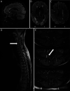
Figure 1. MRI observations in the CNS and PNS of a patient with MS
MRI scans of a patient who was diagnosed with MS based on clinical presentation in combination with the presence of CNS lesions suggestive of demyelination with dissemination in space and time. The diagnosis was confirmed by the presence of unique oligoclonal bands in the spinal fluid, in the absence of any other inflammatory signs that are atypical for MS such as a severe pleiocytosis. In addition, we excluded a diagnosis of neurosarcoidosis, systemic inflammatory condition, or central nervous infection. At 18 months after the diagnosis of MS, the patient developed severe radicular pain in the trajectory of L4 on the right side, with an absent patellar tendon reflex. Subsequent MRI and laboratory investigations systematically ruled out neurosarcoidosis, infection of the CNS, or a systemic inflammatory condition. (A–C) FLAIR images of multiple confluent lesions periventricular, juxtacortical, and in the corpus callosum with a Dawson finger aspect. (D) Focal hyperintensity (arrow) on the T2-PD-weighted image of the spinal cord at the level of C4. There was also a smaller lesion (not depicted) at the level of Th8-Th9. A follow-up scan 1 year after these images showed a new, small, focal lesion at the level of C2-C3. (E) At 6 months after the images shown in (A–D), 3 axial T1-weighted images after contrast enhancement on the level of the exit of root L4 of the spinal cord were made. We observed isolated intradural contrast enhancement of the nerve root L4 with some postganglionic nerve root enhancement (arrow). There was neither spinal disc protrusion nor nerve root compression. No leptomeningeal enhancement was seen. The patient with MS gave permission to present the imaging data as shown in this figure.
Figure 2
Figure 2. The periaxonal region of a myelinated axon in the CNS is similar to the PNS
(A) Overview of the myelinated axonal domains in the CNS and PNS. The upper half shows an axon that is myelinated by a Schwann cell, including the basal lamina, microvilli, Schmidt-Lanterman incisures, sodium (Na+) channels and potassium (K+) channels, and myelin proteins that are highly abundant in the PNS. The lower half represents an axon that is myelinated by an oligodendrocyte, including the process from a perinodal astrocyte/oligodendrocyte progenitor cells (OPCs), Na+ channels and K+ channels, and myelin proteins that are highly abundant in the CNS. (B) NFasc155 and NFasc186 are required to ensure the integrity of the clustered Na+ and K+ channels in the CNS and PNS. Paranodal NFasc155 binds to axolemmal Caspr and Contactin to form the paranodal complex and ensure paranodal integrity. Axolemmal NFasc186 ensures nodal integrity by clustering Na+ channels at the node of Ranvier. (C) The periaxonal region is suggested to function as a synapse in the CNS and PNS. The upper half represents a myelinated axon in the PNS. On arrival of the action potential, the voltage-gated K+ channel opens, resulting in a potassium efflux into the periaxonal region. Potassium is taken up by the myelin sheaths and eventually exits the myelin via nodal abaxonal voltage-gated K+ channels. The lower half represents a myelinated axon in the CNS. On arrival of the action potential, the voltage-gated periaxonal calcium (Ca2+) channel initiates subsequent calcium release from the axoplasmic reticulum. This results in the release of glutamate into the periaxonal region, which in turn binds to myelinic AMPA receptors (AMPARs) and NMDA receptors (NMDARs) to stimulate Ca2+ release in the myelin.58,e18 CLDN11 = claudin 11; CNP = cyclic nucleotide phosphodiesterase; FASN = fatty acid synthase; MAG = myelin-associated glycoprotein; MOG = myelin oligodendrocyte glycoprotein; P0 = myelin protein 0; PLP = proteolipid protein; SIRT2 = sirtuin 2; 4.1 G = band 4.1-like protein G.
Figure 3
Figure 3. The axo-myelinic synapse in the CNS might be involved in the pathogenesis of MS
It is thought that oligodendrocytes produce lactate that is transported to the axonal mitochondria for the production of ATP. If the oligodendrocyte is unable to transport lactate, this would result in a reduction of axonal ATP. This in turn results in the pathologic depolarization of the axon. As a consequence, voltage-gated calcium (Ca2+) channels become activated and cause an increased release of Ca2+ from the axoplasmic reticulum and a subsequent increase of glutamate release into the periaxonal region. Glutamate activates the myelinic NMDA receptor (NMDAR), resulting in the activation of Ca2+-dependent peptidyl arginine deiminases (PADs). PADs will citrullinate myelin basic protein (MBP), which hinders the function of MBPs and might lead to the breakdown of myelin.

References

    1. Koch-Henriksen N, Sørensen PS. The changing demographic pattern of multiple sclerosis epidemiology. Lancet Neurol 2010;9:520–532. - PubMed
    1. Nave K-A, Werner HB. Myelination of the nervous system: mechanisms and functions. Ann Rev Cell Dev Biol 2014;30:503–533. - PubMed
    1. Compston A, Coles A. Multiple sclerosis. Lancet 2002;359:1221–1231. - PubMed
    1. Lassmann H, Budka H, Schnaberth G. Inflammatory demyelinating polyradiculitis in a patient with multiple sclerosis. Arch Neurol 1981;38:99–102. - PubMed
    1. Drake ME. Peripheral neuropathy in multiple sclerosis. J Natl Med Assoc 1987;79:672–673. - PMC - PubMed