. 2020 Nov 24;11(1):5792.
doi: 10.1038/s41467-020-19566-3.

Biomineral armor in leaf-cutter ants

Affiliations

Biomineral armor in leaf-cutter ants

Hongjie Li et al. Nat Commun. .

Abstract

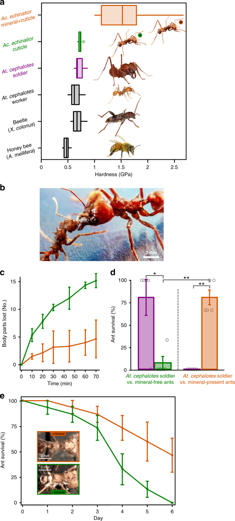
Although calcareous anatomical structures have evolved in diverse animal groups, such structures have been unknown in insects. Here, we report the discovery of high-magnesium calcite [CaMg(CO3)2] armor overlaying the exoskeletons of major workers of the leaf-cutter ant Acromyrmex echinatior. Live-rearing and in vitro synthesis experiments indicate that the biomineral layer accumulates rapidly as ant workers mature, that the layer is continuously distributed, covering nearly the entire integument, and that the ant epicuticle catalyzes biomineral nucleation and growth. In situ nanoindentation demonstrates that the biomineral layer significantly hardens the exoskeleton. Increased survival of ant workers with biomineralized exoskeletons during aggressive encounters with other ants and reduced infection by entomopathogenic fungi demonstrate the protective role of the biomineral layer. The discovery of biogenic high-magnesium calcite in the relatively well-studied leaf-cutting ants suggests that calcareous biominerals enriched in magnesium may be more common in metazoans than previously recognized.


Conflict of interest statement

The authors declare no competing interests.

Figures

Fig. 1
Fig. 1. Morphological and structural characterization of minerals on the cuticle of Ac. echinatior.
a Ac. echinatior ant with a whitish cuticular coating (Photo T.R.S.). b SEM image of ant cuticle with crystalline coating. c Backscattered electron (BSE) image of a polished cuticular cross-section of an ant. This layer is brighter than the cuticle in backscattered electron (BSE) mode scanning electron microscopy (SEM), indicating that it consists of heavier elements and is continuous, covering nearly the entire surface. d BSE image close-up of a polished cuticular cross-section of an ant.
Fig. 2
Fig. 2. Chemical characterization of minerals on the cuticle of Ac. echinatior.
a In situ XRD analysis identifying the cuticular crystalline layer as high-Mg calcite. b–g XANES spectroscopy and mapping with PEEM of a cuticular cross-section. b Average of PEEM images acquired across the C K-edge, showing crystalline layer tightly attached to the cuticle. Three distinct component spectra were identified in the regions labeled cuticle, epicuticle, and mineral, from the most internal part of the ant to the outer surface. c Normalized component spectra extracted from the corresponding labeled regions. Characteristic peaks are marked, including the 285.2 eV (C=C), 288.2 eV (C=O) and 290.3 eV (carbonate) peaks. d Component map where each pixel is colored according to the chemical components it contains. Black pixels are masked areas containing epoxy or gaps. Faint carbonate components within the cuticle and epicuticle were emphasized by enhancing the blue channel 5×, thus this is a semi-quantitative map. A fully quantitative RGB component map is presented in Supplementary Fig. 21. Individual maps of each component are presented in Supplementary Fig. 22, clearly showing an increasing gradient of carbonates towards the surface in the cuticle. e O K-edge spectra extracted from the mineral crystals correspondingly colored in the Polarization-dependent Imaging Contrast (PIC) maps in f and g. f Magnified PIC maps for the regions represented by boxes in the complete PIC map in g. g PIC map quantitatively displaying the orientations of the mineral crystals’ c-axes in colors. This map was acquired from the same area shown in b and d at precisely the same magnification. These are interspersed high- and low-Mg calcite, and heterogenous at the nanoscale. Biomineral crystals do not show preferred orientations but are randomly oriented. High-magnesium calcite in carbon spectra is identified by the carbonate peak at 290.3 eV, which occurs in all carbonates, amorphous, or crystalline. The O spectra in d clearly indicate crystallinity, and their line shape indicates a mixture of high-magnesium calcite and low Mg-bearing calcite.
Fig. 3
Fig. 3. Mineral precipitation on the cuticle in both in vitro cuticle synthetic studies and ant-rearing experiments.
a Scheme of in vitro mineralization experiment using Acromyrmex echinatior leaf-cutting ant cuticles as templates for biomineralization (Photo C.M.C.). b, c Pre- and post-incubation SEM images showing the original, uncoated cuticle (b) and the cuticle covered by a layer of precipitated carbonate (c) after incubation in Mg2+/Ca2+/Cl/Na+/HCO3 solution for 7 days at 19 °C. d, XRD patterns of, from top to bottom, an uncoated ant cuticle, a cuticle after incubation in Mg2+/Ca2+/Cl/Na+/HCO3 solution, a platinum-coated cuticle incubated in Mg2+/Ca2+/Cl/Na+/HCO3 solution, and a cuticle after KOH protein hydrolysis incubated in Mg2+/Ca2+/Cl/Na+/HCO3 solution. H: high-magnesium-calcite, A: aragonite, Pt: platinum. e XRD patterns of cuticles of ants representing different developmental stages, ranging from (from bottom to top), a newly formed pupa to an older worker, after incubation in Mg2+/Ca2+/Cl/Na+/HCO3 solution. f Environmental scanning electron micrographs (eSEM) of ant epicuticles taken over a 10-day time series, from immediately after eclosion from pupa to adult (left), to 10 days post-eclosion (right), showing the formation of the biomineral layer over time (Photo H.L.). g Estimated magnesium concentration of the biomineral layer during 30 days of ant development based on the XRD d(104) value according to Graf and Goldsmith, showing the rapid integration of magnesium from days 6 to 8 and the continued presence of high-magnesium content for up to 30 days (n = 2 per treatment and the corresponding standard error are shown).
Fig. 4
Fig. 4. Mechanical protection afforded by the epicuticular mineral layer.
a Quantitative nano-mechanical properties of insect cuticles, including honeybee (Apis mellifera), beetle (Xylotrechus colonus), leaf-cutting ants [Atta cephalotes worker, Atta cephalotes soldier (purple), and Acromyrmex echinatior worker without biomineral (green, with green minus circle beside the ant image)] and Ac. echinatior ant worker with biomineral epicuticular layer (orange, with orange plus circle beside the ant image), measured by an in situ nanoindenters with a cube-corner probe (n = 12, 15, 13, 15, 12, and 13 for insect measured above, respectively; center, median; box, upper and lower quantiles; whisker, 1.5× interquartile range; points, outlier). Atta ants images, Xylotrechus beetle image, and Apis bee image provided with permission from the copyright holder, Alexander L. Wild, Jon Rapp, and Don Farrall, respectively. bd Aggressive interaction between three Ac. echinatior workers (with/without biomineral, respectively) and Atta cephalotes soldier (Photo C.M.C.). b Ac. echinatior worker (left) aggressively interacts with Atta cephalotes soldier (right). c In aggressive encounters with Atta cephalotes soldiers, Ac. echinatior workers with biomineral armor (orange) lose substantially fewer body parts (i.e., legs, antennae, abdomen, and head) compared to Ac. echinatior worker without biomineral (green). d Survivorship of Ac. echinatior workers without (green) and with (orange) biomineral armor in aggressive encounters with Atta cephalotes soldiers (purple). Asterisks indicate significant differences via a two-sample t-test (*P < 0.05, **P < 0.001; P-value = 0.0184, 0.0001, and 0.0006 from left to right, respectively; n = 5 per treatment and the corresponding standard error are shown.). e Survivorship curves of Ac. echinatior worker with (orange) and without (green) an epicuticular biomineral layer exposed to the entomopathogenic fungus Metarhizium. The inset images show more substantial fungal growth and emergence from biomineral-free workers (Photo H.L.). c, e n = 3 per treatment, and the corresponding standard error are shown.

References

    1. Knoll AH. Biomineralization and evolutionary history. Rev. Mineral. Geochem. 2003;54:329–356. doi: 10.2113/0540329. - DOI
    1. Eder M, Amini S, Fratzl P. Biological composites-complex structures for functional diversity. Science. 2018;362:543–547. doi: 10.1126/science.aat8297. - DOI - PubMed
    1. Sperling EA, et al. Oxygen, ecology, and the Cambrian radiation of animals. Proc. Natl Acad. Sci. USA. 2013;110:13446–13451. doi: 10.1073/pnas.1312778110. - DOI - PMC - PubMed
    1. Gilbert PUPA, et al. Biomineralization by particle attachment in early animals. Proc. Natl Acad. Sci. USA. 2019;116:17659–17665. doi: 10.1073/pnas.1902273116. - DOI - PMC - PubMed
    1. Gilbert PUPA, Abrecht M, Frazer BH. The organic-mineral interface in biominerals. Mol. Geomicrobiol. 2018;59:157–185.

Publication types