. 2020 Nov 24;10(1):20460.
doi: 10.1038/s41598-020-77452-w.

Addressing the challenges of E-cigarette safety profiling by assessment of pulmonary toxicological response in bronchial and alveolar mucosa models

Affiliations

Addressing the challenges of E-cigarette safety profiling by assessment of pulmonary toxicological response in bronchial and alveolar mucosa models

Koustav Ganguly et al. Sci Rep. .

Abstract

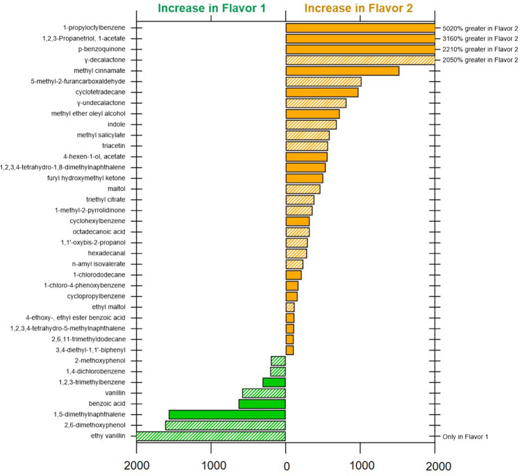
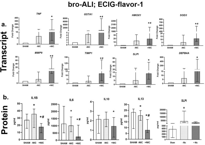
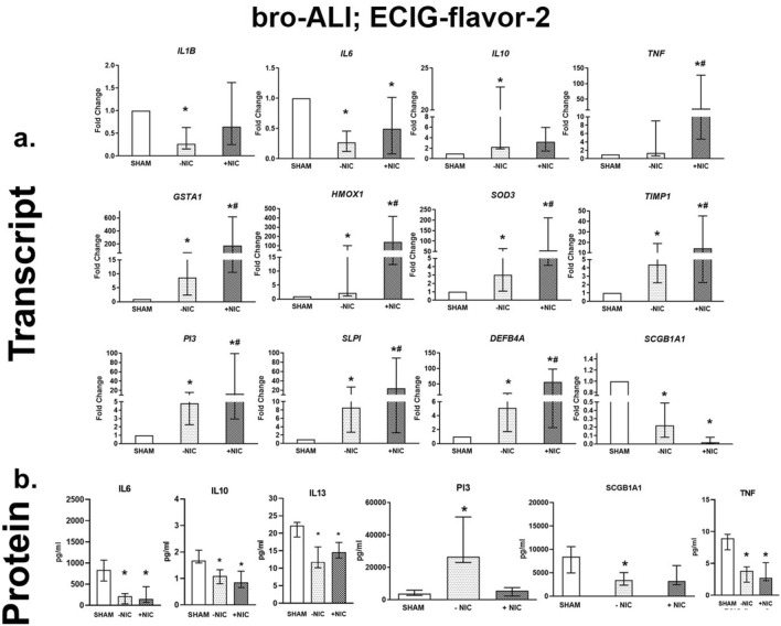
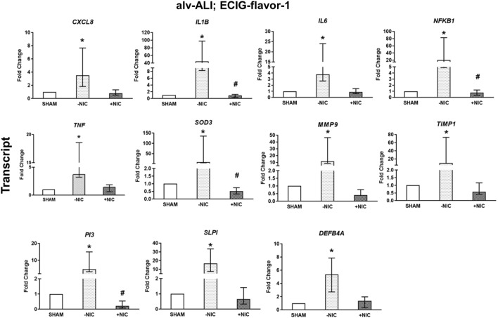
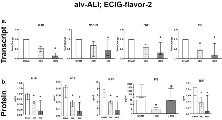
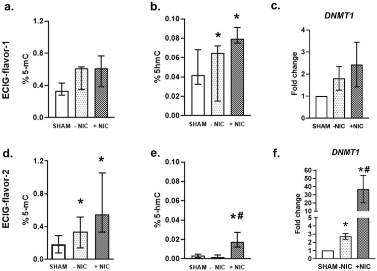
Limited toxicity data on electronic cigarette (ECIG) impede evidence-based policy recommendations. We compared two popular mixed fruit flavored ECIG-liquids with and without nicotine aerosolized at 40 W (E-smoke) with respect to particle number concentrations, chemical composition, and response on physiologically relevant human bronchial and alveolar lung mucosa models cultured at air-liquid interface. E-smoke was characterized by significantly increased particle number concentrations with increased wattage (25, 40, and 55 W) and nicotine presence. The chemical composition of E-smoke differed across the two tested flavors in terms of cytotoxic compounds including p-benzoquinone, nicotyrine, and flavoring agents (for example vanillin, ethyl vanillin). Significant differences in the expression of markers for pro-inflammation, oxidative stress, tissue injury/repair, alarm anti-protease, anti-microbial defense, epithelial barrier function, and epigenetic modification were observed between the flavors, nicotine content, and/ or lung models (bronchial or alveolar). Our findings indicate that ECIG toxicity is influenced by combination of multiple factors including flavor, nicotine content, vaping regime, and the region of respiratory tree (bronchial or alveolar). Toxic chemicals and flavoring agents detected in high concentrations in the E-smoke of each flavor warrant independent evaluation for their specific role in imparting toxicity. Therefore, multi-disciplinary approaches are warranted for comprehensive safety profiling of ECIG.


Conflict of interest statement

The authors declare no competing interests.

Figures

Figure 1
Figure 1
Schematic representation of the electronic cigarette (ECIG) aerosol exposure set up for bronchial (bro-ALI) and alveolar (alv-ALI) mucosa models. A vaping regime mimicking one day of low intensity vaping was applied and consisted of repeated exposure [40 watts (W), 40 ml/puff, 3 s (s) puff duration, 30 s puff interval, 10 puffs/session, bro-ALI model: 6 sessions, alv-ALI model: 3 sessions, 1 h session interval] leading to a total of 60 puffs and 30 puffs for bro-ALI and alv-ALI, respectively. The authors sincerely acknowledge the assistance of Ann-Katrin Sjödén in preparing the figure.
Figure 2
Figure 2
Percent difference in concentration of the top 40 identified compounds in suspect screening by gas chromatography–mass spectrometry comparing electronic cigarette (ECIG) aerosol of ECIG-flavor-1 and ECIG-flavor-2 (both without nicotine) generated at 40 W. All compounds with twofold difference between flavors included. Shaded bars indicate flavor or fragrance-related compounds.
Figure 3
Figure 3
Transcript expression (a) and secreted protein levels (b) of significantly altered pro-inflammatory, oxidative stress, tissue injury/repair, alarm anti-proteases, and/ or anti-microbial defensin markers in the bronchial mucosa model cultured at air–liquid interface (bro-ALI) following exposure to aerosolized non-nicotinized (−NIC) and nicotinized (+NIC) electronic cigarette liquid flavor 1 (ECIG-flavor-1). Actin beta (ACTB) was used as the reference gene. Fold changes for transcript expression were calculated relative to the corresponding sham. *: significantly different from sham; #: significantly different from −NIC (p < 0.05, Friedman followed by Wilcoxon test). bro-ALI: bronchial mucosa model developed at air–liquid interface DEFB4A: defensin beta 4A, GSTA1: glutathione S-transferase alpha 1, HMOX1: heme oxygenase 1, IL: interleukin, MMP9: matrix metallopeptidase 9, SLPI: secretory leukocyte peptidase inhibitor, SOD3: superoxide dismutase 3, extracellular, TIMP1: TIMP metallopeptidase inhibitor 1, TNF: tumor necrosis factor.
Figure 4
Figure 4
Transcript expression (a) and secreted protein levels (b) of significantly altered pro-inflammatory, oxidative stress, tissue injury/repair, alarm anti-proteases, and/ or anti-microbial defensin markers in the bronchial mucosa model cultured at air–liquid interface (bro-ALI) following exposure to aerosolized non-nicotinized (−NIC) and nicotinized (+NIC) electronic cigarette liquid flavor 2 (ECIG-flavor-2). Actin beta (ACTB) was used as the reference gene. Fold changes for transcript expression were calculated relative to the corresponding sham *: significantly different from sham; #: significantly different from −NIC (p < 0.05, Friedman followed by Wilcoxon test). DEFB4A: defensin beta 4A, GSTA1: glutathione S-transferase alpha 1, HMOX1: heme oxygenase 1, IL: interleukin, PI3: peptidase inhibitor 3, SCGB1A1: secretoglobin family 1A member 1, SLPI: secretory leukocyte peptidase inhibitor, SOD3: superoxide dismutase 3, extracellular, TIMP1: TIMP metallopeptidase inhibitor 1, TNF: tumor necrosis factor.
Figure 5
Figure 5
Morphological characterization of the alveolar mucosa model by confocal and transmission electron microscopy (TEM). (a) The cell junction protein zona occludin 1 (ZO1) (b) lamellar bodies, (c) surfactant protein C (SPC) and (d) epithelial sodium channel (ENaC). Nucleus is stained in blue. Bar scale: 50 µm; (e) Representative TEM image of the alveolar type II cells in air–liquid interface (2 weeks) showing microvilli (MV), lipid bodies (LB), desmosome (D), and tight junction (TJ) Bar scale: 2 µm. The microscopic images are representative of alveolar mucosa model developed at air–liquid interface (2 weeks) from NCl-H441 cells.
Figure 6
Figure 6
Transcript expression analysis of significantly altered pro-inflammatory, oxidative stress, tissue injury/repair, alarm anti-proteases, and anti-microbial defensin markers in the alveolar mucosa model cultured at air–liquid interface (alv-ALI) following exposure to aerosolized non-nicotinized (−NIC) and nicotinized (+NIC) electronic cigarette liquid flavor 1 (ECIG-flavor-1). Secreted levels of none of the proteins were altered significantly in this exposure condition. Actin beta (ACTB) was used as the reference gene. Fold changes for transcript expression were calculated relative to the corresponding sham. *: significantly different from sham; #: significantly different from −NIC (p < 0.05, Friedman followed by Wilcoxon test). CXCL8: C-X-C motif chemokine ligand 8, DEFB4A: defensin beta 4A, IL: interleukin, MMP9: matrix metallopeptidase 9, NFKB1: nuclear factor kappa B subunit 1, PI3: peptidase inhibitor 3, SLPI: secretory leukocyte peptidase inhibitor, SOD3: superoxide dismutase 3, extracellular, TIMP1: TIMP metallopeptidase inhibitor 1, TNF: tumor necrosis factor.
Figure 7
Figure 7
Transcript expression (a) and secreted protein levels (b) of significantly altered pro-inflammatory, oxidative stress, tissue injury/repair, alarm anti-proteases, and/ or anti-microbial defensin markers in the alveolar mucosa model cultured at air–liquid interface (alv-ALI) following exposure to aerosolized non-nicotinized (−NIC) and nicotinized (+NIC) electronic cigarette liquid flavor 2 (ECIG-flavor-2). Actin beta (ACTB) was used as the reference gene. Fold changes for transcript expression were calculated relative to the corresponding sham *: significantly different from sham; #: significantly different from −NIC (p < 0.05, Friedman followed by Wilcoxon test). IL: interleukin, NFKB1: nuclear factor kappa B subunit 1, PI3: peptidase inhibitor 3, TNF: tumor necrosis factor.
Figure 8
Figure 8
Methylation and hydroxymethylation of total DNA and transcript expression of DNA methyl transferase 1 (DNMT1) was assessed in the alveolar mucosa model cultured at air–liquid interface following exposure to aerosolized non-nicotinized (−NIC) and nicotinized (+NIC) electronic cigarette liquid flavor 1 (ECIG-flavor-1) (ac) and ECIG-flavor-2 (df). Data are shown as percentage of 5-methylcytosine (5-mC) or 5-hydroxymethylcytosine (5-hmC). Fold changes for transcript expression were calculated relative to the corresponding sham. Actin beta (ACTB) was used as the reference gene. *: significantly different from sham; #: significantly different from −NIC (p < 0.05, Friedman followed by Wilcoxon test).

Similar articles

Cited by

References

    1. https://www.researchandmarkets.com/reports/4827644/electronic-cigarette-... (2020).
    1. Hiemstra PS, Bals R. Effects of E-cigarette use on human lung tissue. On harm reduction and causing harm. Am. J. Respir. Crit. Care Med. 2018;198:6–7. doi: 10.1164/rccm.201802-0299ED. - DOI - PubMed
    1. Shields PG, et al. A review of pulmonary toxicity of electronic cigarettes in the context of smoking: a focus on inflammation. Cancer Epidemiol. Biomark. Prev. 2017;26:1175–1191. doi: 10.1158/1055-9965.EPI-17-0358. - DOI - PMC - PubMed
    1. Bal R, et al. Electronic cigarettes: a task force report from the European Respiratory Society. Eur. Respir. J. 2019;53:1801151. doi: 10.1183/13993003.01151-2018. - DOI - PubMed
    1. Ferkol TW, et al. Forum of International Respiratory Societies Electronic cigarette use in youths: a position statement of the Forum of International Respiratory Societies. Eur. Respir. J. 2018;51:1800278. doi: 10.1183/13993003.00278-2018. - DOI - PubMed

Publication types