. 2020 Nov 24;10(1):20428.
doi: 10.1038/s41598-020-76971-w.

Sustainable drug release from polycaprolactone coated chitin-lignin gel fibrous scaffolds

Affiliations

Sustainable drug release from polycaprolactone coated chitin-lignin gel fibrous scaffolds

Turdimuhammad Abdullah et al. Sci Rep. .

Erratum in

Abstract

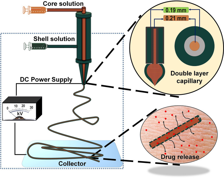
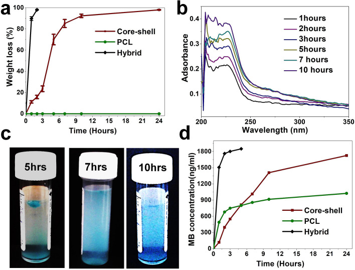
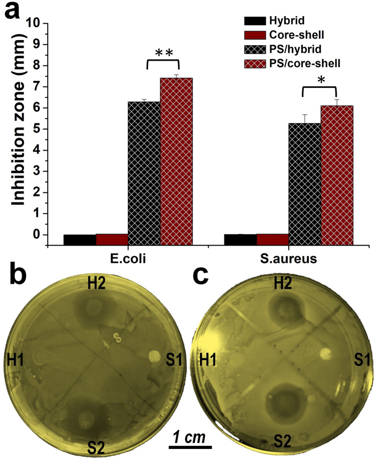
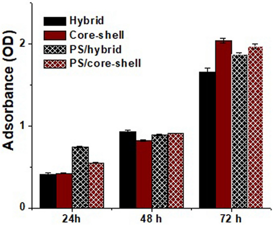
Non-healing wounds have placed an enormous stress on both patients and healthcare systems worldwide. Severe complications induced by these wounds can lead to limb amputation or even death and urgently require more effective treatments. Electrospun scaffolds have great potential for improving wound healing treatments by providing controlled drug delivery. Previously, we developed fibrous scaffolds from complex carbohydrate polymers [i.e. chitin-lignin (CL) gels]. However, their application was limited by solubility and undesirable burst drug release. Here, a coaxial electrospinning is applied to encapsulate the CL gels with polycaprolactone (PCL). Presence of a PCL shell layer thus provides longer shelf-life for the CL gels in a wet environment and sustainable drug release. Antibiotics loaded into core-shell fibrous platform effectively inhibit both gram-positive and -negative bacteria without inducting observable cytotoxicity. Therefore, PCL coated CL fibrous gel platforms appear to be good candidates for controlled drug release based wound dressing applications.


Conflict of interest statement

The authors declare no competing interests.

Figures

Figure 1
Figure 1
Systematic representation of chitin-lignin (CL) based hybrid fiber encapsulation by PCL using coaxial electrospinning technique and theoretical assumption for its drug release behavior. Here, a mixture of CL sol–gel solution and PGS solution in 9:1 volume ratio is used as a core solution, and PCL solution is used as a shell solution to produce the core–shell fiber. The schematic was prepared by first author Tuerdimaimaiti Abudula.
Figure 2
Figure 2
SEM micrographs and fiber size distribution of hybrid fiber (a,b), PCL fiber (c,d) and core–shell fiber (e,f). The result shows morphological and dimensional similarity of the core–shell fibers with the hybrid fibers alone.
Figure 3
Figure 3
TEM image of the core–shell fiber (a); FTIR spectra of the electrospun scafflds (b), and deconvolved XPS spectra of carbon in the scaffolds (c). Overall results suggests that the shell layer was much thinner compared to the core layer in the resulted core–shell fiber.
Figure 4
Figure 4
Stress–strain curve (a) and DSC curve (b) of the electrospun scaffolds. The result indicates insignificant effects of PCL encapsulation on thermal transition and mechanical behavior of the CL based hybrid fibrous scaffold.
Figure 5
Figure 5
Weight change of the electrospun fiber under PBS (pH 7.4) (a), UV spectra change and representative color change of the PBS solution after immersing the core–shell fibrous sheet (b,c), and methylene blue release profile from the electrospun scaffolds (d). The photo was taken by first author Tuerdimaimaiti Abudula. Overall results imply the role of PCL shell layer coating on the CL based hybrid scaffold in preventing burst release and providing prolonged drug release profile.
Figure 6
Figure 6
Inhibition zone of the PS loaded fibrous scaffolds against E.coly and S.arous. (a) Diameter of the inhibition zone, calculated according to triple experiment. (b) Representative inhibition zone of the scaffolds against E.coly. (c) Representative inhibition zone of the scaffolds against S.arous. Where H1 and S1 represents hybrid and core–shell fibrous scaffold, H2 and S2 represents PS loaded hybrid and core–shell fibrous scaffold. PS loaded core–shell fibrous scaffolds showed better antibacterial performance than that of hybrid scaffolds due to controlled release of the antibiotics. The photo was taken by first author Tuerdimaimaiti Abudula.
Figure 7
Figure 7
MTT assay following culture of bone marrow mesenchymal stem cells (BM-MSCs) for 24, 48 and 72 h. Individual fibers show increases in cell proliferation with time. The values are expressed as mean ± SD from three independent experiments. All the scaffolds showed a good biocompatibility, and the incorporation of PS as an antibiotic within PCL coated hybrid scaffolds did not show any significant negative effects on the cell proliferation.

References

    1. Saghazadeh S, et al. Drug delivery systems and materials for wound healing applications. Adv. Drug Del. Rev. 2018;127:138–166. - PMC - PubMed
    1. Tamayol A, et al. Biodegradable elastic nanofibrous platforms with integrated flexible heaters for on-demand drug delivery. Sci. Rep. 2017;7:1–10. - PMC - PubMed
    1. Ardeshirzadeh B, Anaraki NA, Irani M, Rad LR, Shamshiri S. Controlled release of doxorubicin from electrospun PEO/chitosan/graphene oxide nanocomposite nanofibrous scaffolds. Mater. Sci. Eng. C. 2015;48:384–390. - PubMed
    1. Kenawy E-R, Worley SD, Broughton R. The chemistry and applications of antimicrobial polymers: A state-of-the-art review. Biomacromol. 2007;8:1359–1384. - PubMed
    1. Singer AJ, Clark RA. Cutaneous wound healing. N. Engl. J. Med. 1999;341:738–746. - PubMed

Publication types

MeSH terms