

The Appropriateness of Digital Diabetic Retinopathy Screening Images for a Computer-Aided Glaucoma Screening System
- PMID: 33235429
- PMCID: PMC7678698
- DOI: 10.2147/OPTH.S273659
The Appropriateness of Digital Diabetic Retinopathy Screening Images for a Computer-Aided Glaucoma Screening System
Abstract
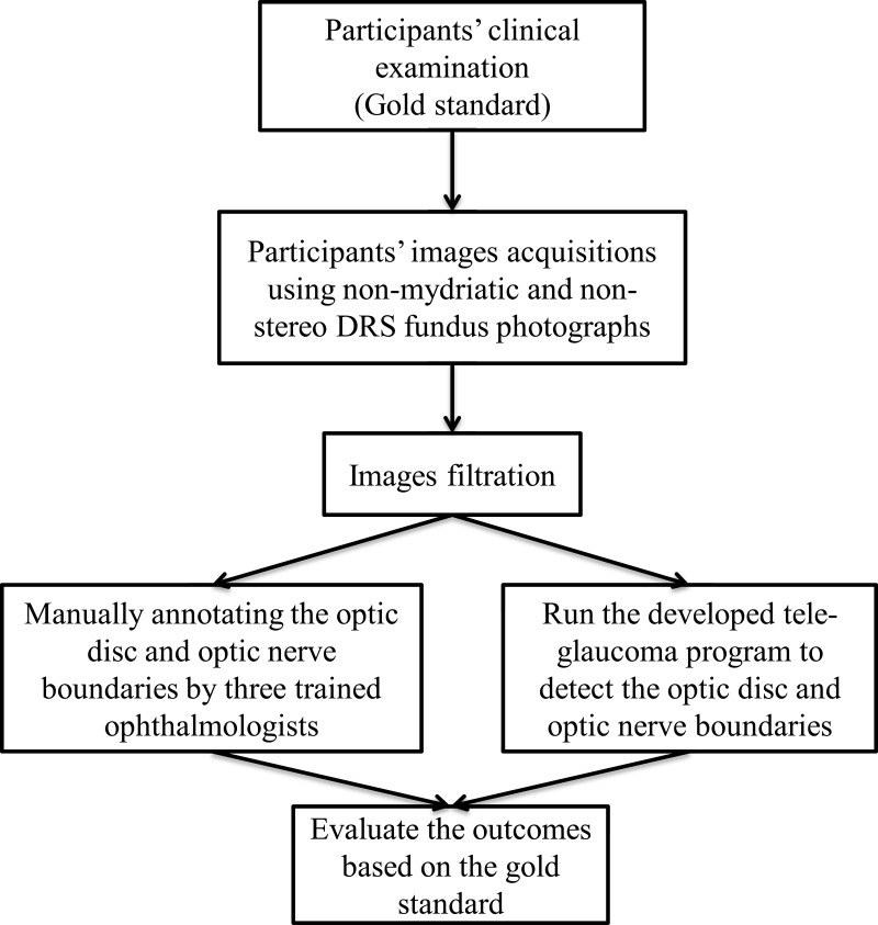
Purpose: The purpose of this study was to evaluate the ability to screen for glaucoma using a Food Drug Administration (FDA) Class II diagnostic digital fundus photography system used for diabetic retinopathy screening (DRS).
Methods: All research participants underwent a comprehensive eye examination as well as non-mydriatic 45°single photograph retinal imaging centered on the macula. Optic nerve images within the 45° non-mydriatic and non-stereo DRS image were evaluated by two methods: 1) grading by three glaucoma specialists, and 2) a computer-aided automated segmentation system to determine the vertical cup-to-disc ratio (VCDR). Using VCDR from clinical assessment as gold standard, VCDR results from two methods were compared to that from clinical assessment. Inter-grader agreement was assessed by computing intraclass correlation coefficient (ICC). In addition, sensitivity and specificity were calculated.
Results: Among 245 fundus photos, 166 images met quality specifications for analysis. Fifty images were not processed by the automated system due to the poor quality of the optic disc, and 29 images did not include the optic nerve head due to the patient movement during the photo acquisition. The ICC value for the VCDR between the gold standard clinical exam and the automated system was 0.41, indicating fair agreement. The ICC value between the three ophthalmologists and the gold standard was 0.51, 0.56, and 0.69, respectively, indicating fair to moderate agreement.
Discussion: Assessing the VCDR on non-mydriatic and non-stereo DRS fundus photographs by either the computer-aided automated segmentation system or by glaucoma specialists showed similar fair to moderate agreement. In summary, optic nerve assessment for glaucoma from these 45° non-mydriatic and non-stereo DRS images is not yet suitable for tele-glaucoma screening.
Keywords: automated screening system; diabetic retinopathy screening; glaucoma screening; tele-glaucoma; telemedicine.
© 2020 Almazroa et al.
Conflict of interest statement
Maria A Woodward reports grants from National Eye Institute, during the conduct of the study. Paula Anne Newman-Casey reports grants from the National Eye Institute, Career Development Award from Research to Prevent Blindness, during the conduct of the study. Dr Manjool M Shah reports personal fees from Allergan, nothing from Glaukos, nothing from Katena, outside the submitted work. Sayoko E Moroi reports grants from University of Michigan, during the conduct of the study; grants from NIH, nothing from Allergan, Royalties unrelated to this manuscript from Wolters Kluwer, outside the submitted work. The authors declare that they have no other actual or potential conflict of interests relevant to the content of this project.
Figures


References
-
- FDA permits marketing of artificial intelligence-based device to detect certain diabetes-related eye problems. Food and drug administration. 2018. Available from: https://www.fda.gov/newsevents/newsroom/pressannouncements/ucm604357.htm. Accessed February26, 2019.
-
- Open-angle Glaucoma defined. National eye institute. 2018. Available from: https://nei.nih.gov/eyedata/glaucoma#5. Accessed February26, 2019.
LinkOut - more resources
Full Text Sources
Miscellaneous
