. 2020 Nov 17:13:11769-11779.
doi: 10.2147/OTT.S260671. eCollection 2020.

Simultaneous Treatment with Soluble Forms of GAS1 and PTEN Reduces Invasiveness and Induces Death of Pancreatic Cancer Cells

Affiliations

Simultaneous Treatment with Soluble Forms of GAS1 and PTEN Reduces Invasiveness and Induces Death of Pancreatic Cancer Cells

Lizbeth Daniel-García et al. Onco Targets Ther. .

Abstract

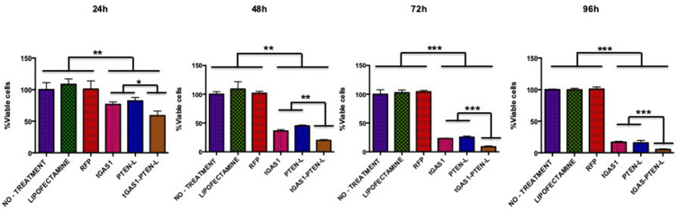
Introduction: Pancreatic carcinoma cells exhibit a pronounced tendency to invade along and through intra and extrapancreatic nerves, even during the early stages of the disease, a phenomenon called perineural invasion (PNI). Thus, we sought to determine the effects of the simultaneous expression of soluble forms of GAS1 and PTEN (tGAS1 and PTEN-L) inhibiting tumor growth and invasiveness.

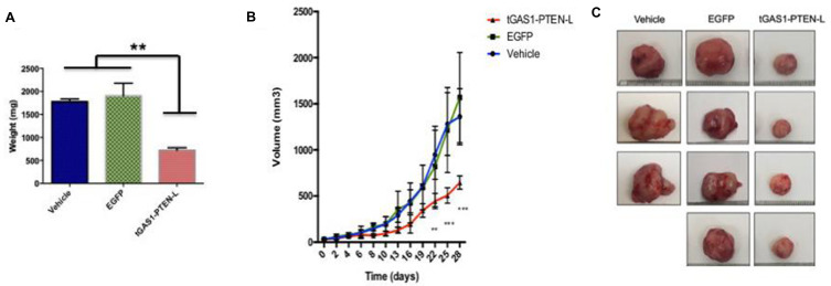
Materials and methods: We employed a lentiviral system to simultaneously express tGAS1 and PTEN-L; in order to determine the effects of the treatments, cell viability and apoptosis as well as the expression of the transgenes by ELISA and intracellular signaling as ascertained by the activation of AKT and ERK1/2 were measured; cell invasiveness was determined using a Boyden chamber assay; and the effects of the treatment were measured in vivo in a mouse model.

Results: In the present work, we show that the combined treatment with tGAS1 and PTEN-L inhibits the growth of pancreatic cancer cells, by reducing the activities of both AKT and ERK 1/2, decreases cell invasiveness, and restrains tumor growth in a mouse model.

Conclusion: The combined administration of tGAS1 and PTEN-L could be a valuable adjunct therapy for the treatment of pancreatic cancer.

Keywords: GFRα3; PTEN-L; artemin; growth arrest specific 1; pancreatic cancer; perineural invasion.


Conflict of interest statement

The authors declare that they have no conflict of interest.

Figures

Figure 1
Figure 1
Characterization of HPAF-II pancreatic cancer cells. (A) HPAF-II cells do not express gas1, gfrα1, ret, or gdnf but express both artemin and its receptor gfrα3. SHSY5Y and U87-MG cells were used as positive controls. (B) HPAF-II cells do not express PTEN, as assessed by Western blot analysis; SH-SY5Y cells were used as positive control; β-actin is the loading control.
Figure 2
Figure 2
Comparison of the effect of soluble forms of GAS1 and PTEN on cell viability. The graphs show the percentage of viable HPAF-II cells, as determined by Trypan blue exclusion at different times after transfection with GAS1, PTEN, or both simultaneously transfected (24, 48, 72 and 96 h). Controls were non-treated HPAF-II cells, cells treated with Lipofectamine or transfected with the Red Fluorescent Protein plasmid (RFP). tGAS1 and PTEN-L were more effective together than when individually transfected. One-way ANOVA followed by Duncan´s test. *p< 0.05, **p<0.01, ***p<0.001; n= 3.
Figure 3
Figure 3
Effects of soluble forms of GAS1 and PTEN on intracellular signaling pathways and apoptosis. (A) Effects of the soluble forms of GAS1 and PTEN, and when transfected simultaneously on the activity of AKT (pAKT is the phosphorylated molecule and tAKT is the total); (B) effects of the soluble forms of GAS1 and PTEN, and transfected together on the activity of ERK1/2 (pERK1/2 is the phosphorylated molecule and tERK1/2 is the total); left panels show representative experiments and right panels, the statistical analysis of three independent experiments; (C) apoptosis demonstrated by the activation of caspase-3, β-Actin is the loading control. One-way ANOVA followed by Tukey’s multiple comparisons test. *p< 0.05, **p<0.01, ***p<0.001; n=3.
Figure 4
Figure 4
Effect of soluble forms of GAS1 and PTEN, and when simultaneously applied on pancreatic cancer cell invasiveness. (A) Levels of the soluble forms of GAS1 and PTEN in conditioned media from HPAF-II producer cells (left transfected with tGAS1; middle with PTEN-L; right with tGAS1 and PTEN-L); (B) number of DRG viable cells in the presence of the different media; (C) Immunofluorescence of DRG cultures against β-III Tubulin (left panel), nuclei revealed by DAPI (middle panel) and merge of the two channels (right panel). (D) Percentage of HPAF-II cancer cells that crossed the membrane towards DRG cells incubated in the presence of the different media; right panel shows a representative experiment. One-way ANOVA followed by Tukey’s multiple comparisons test. **p<0.01, ***p<0.001; n=3.
Figure 5
Figure 5
Effect of the expression of tGAS1-PTEN-L on tumor growth. HPAF-II cells were subcutaneously inoculated into the flanks of nu/nu mice, and when tumors reached approximately 30 mm3, they received the same volume of vehicle (culture medium without serum), EGFP-expressing virus, or tGAS1-PTEN-L-expressing virus. (A) Weight of tumors after the different treatments (B) Volume of tumors treated with tGAS1-PTEN-L compared with controls through time. (C) Size of the tumors after the treatments (ruler is in cm). ANOVA followed by Tukey´s (A); and Duncan´s post-hoc test (B). **p<0.01, ***p<0.001; n=3 for vehicle; n=4 for EGFP and n=4 for tGAS1-PTEN-L treatments.
Figure 6
Figure 6
Effects of tGAS1-PTEN-L on tumor cells intracellular pathways and apoptosis. (A and B) Levels of Gas1 and PTEN in tumors; (C) activity of AKT in tumors, left representative experiment, right statistical analysis; (D) activity of ERK1/2 in tumors, left representative experiment, right statistical analysis; (E) apoptosis of tumor cells as shown by caspase-3 activity. One-way ANOVA followed by Tukey’s multiple comparisons test. **p<0.01, ***p<0.001; n=3–4 (AD).

References

    1. Haeberle L, Esposito I. Pathology of pancreatic cancer. Transl Gastroenterol Hepatol. 2019;4:1–12. doi:10.21037/tgh.2019.06.02 - DOI - PMC - PubMed
    1. Amit M, Na’Ara S, Gil Z. Mechanisms of cancer dissemination along nerves. Nat Rev Cancer. 2016;16(6):399–408. doi:10.1038/nrc.2016.38 - DOI - PubMed
    1. Alrawashdeh W, Jones R, Dumartin L, et al. Perineural invasion in pancreatic cancer: proteomic analysis and in vitro modelling. Mol Oncol. 2019;13(5):1075–1091. doi:10.1002/1878-0261.12463 - DOI - PMC - PubMed
    1. Liang D, Shi S, Xu J, et al. New insights into perineural invasion of pancreatic cancer: more than pain. Biochim Biophys Acta Rev Cancer. 2016;1865(2):111–122. doi:10.1016/j.bbcan.2016.01.002 - DOI - PubMed
    1. Kuang AG, Nickel JC, Andriole GL, Castro-Santamaria R, Freedland SJ, Moreira DM. Both acute and chronic inflammation are associated with less perineural invasion in men with prostate cancer on repeat biopsy. BJU Int. 2019;123(1):91–97. doi:10.1111/bju.14428 - DOI - PubMed