. 2020 Nov 17:12:11723-11733.
doi: 10.2147/CMAR.S271710. eCollection 2020.

Up-Regulation of miR-26a-5p Inhibits E2F7 to Regulate the Progression of Renal Carcinoma Cells

Affiliations

Up-Regulation of miR-26a-5p Inhibits E2F7 to Regulate the Progression of Renal Carcinoma Cells

Chuanyu Cheng et al. Cancer Manag Res. .

Abstract

Background: Metastasis is the main cause of renal cell carcinoma (RCC) tumor death, and effective inhibition of RCC metastasis is an essential means to meliorate the prognosis of RCC patients. MicroRNAs (miRs) have been proved to be stable and important biomarkers for several malignancies. This study is therefore set out to explore the metastasis-related miR and its mechanism in RCC.

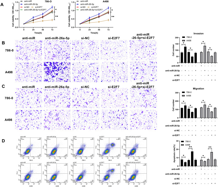
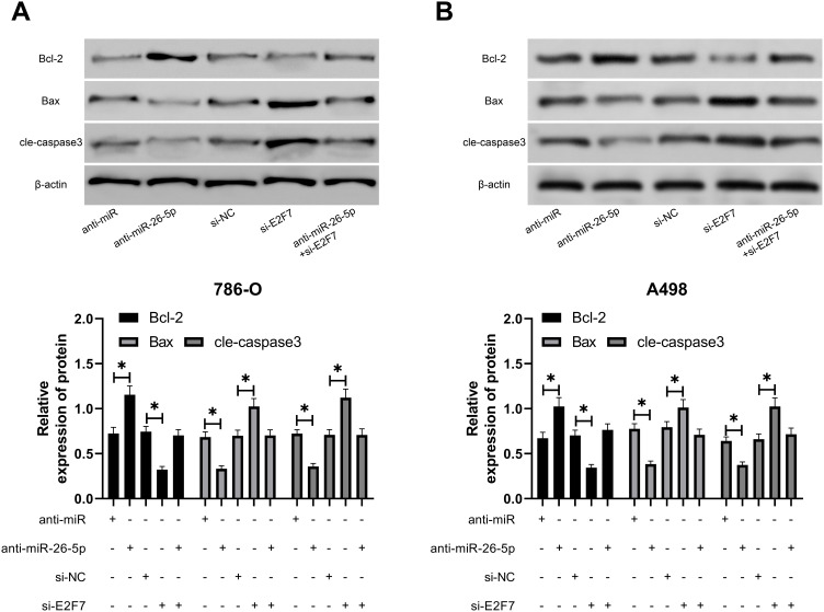
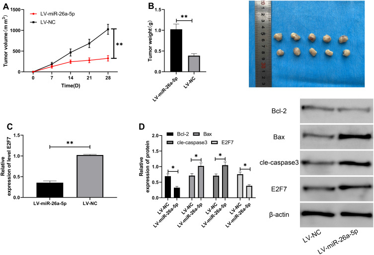
Methods: The expression of miR- 26a -5p in RCC was analyzed using the expression profile in the Cancer Genome Atlas (TCGA). MiR-26a-5p and E2F transcription factor 7 (E2F7) in RCC patients were detected by qRT-PCR. Cell Counting Kit-8 (CCK-8) was adopted to assess cell proliferation, Transwell was utilized to evaluate migration and invasion, and flow cytometry (FC) was used to determine apoptosis. Mouse cell-derived and patient-derived xenotransplantation models were established to evaluate the effect of miR-26a-5p on tumor growth and metastasis in vivo. The molecular mechanism of miR-26a-5p was analyzed by dual-luciferase reporter (DLR) gene analysis, qRT-PCR, and Western blot (WB) both in vivo and in vitro.

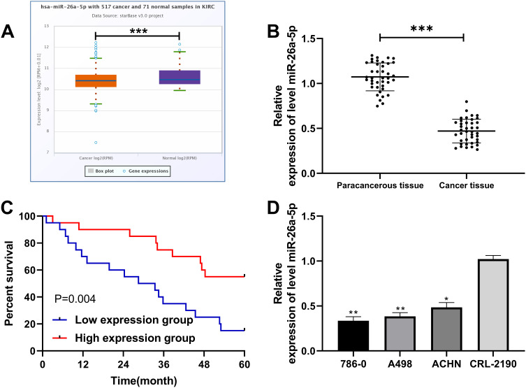
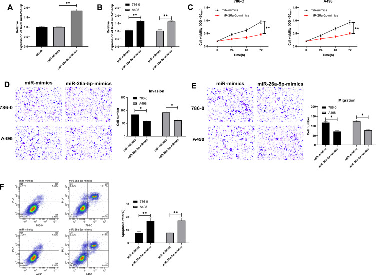
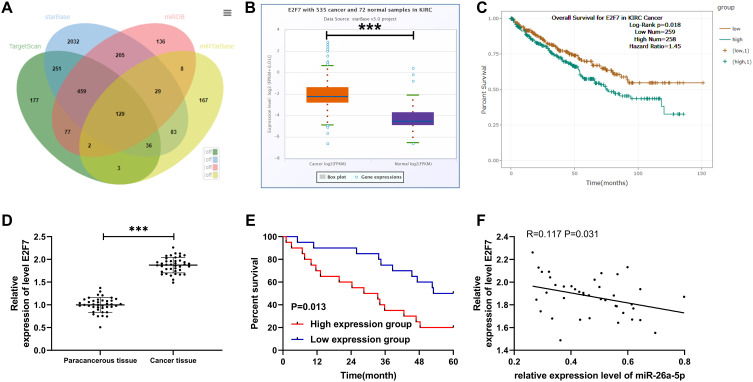
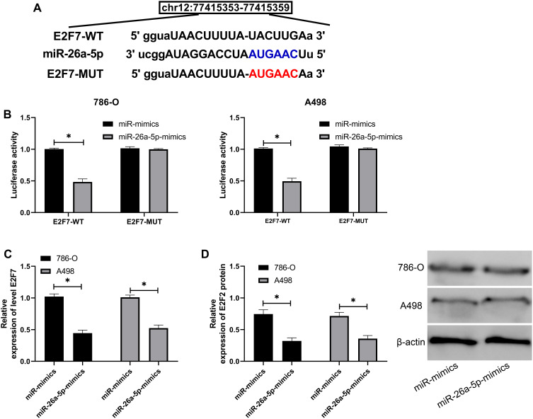
Results: MiR-26a-5p was reduced in renal carcinoma cells and may serve as a biomarker for renal cancer metastasis and prognosis. MiR-26a-5p up-regulation inhibited migration and invasion in renal cell lines and tumor metastasis in vivo. Bioinformatics target prediction and RNA-seq results showed that E2F7 was among the targets of miR-26a-5p and was significantly inhibited by miR-26a-5p in vivo and in vitro.

Conclusion: MiR-26a-5p presents low expression in RCC and promotes RCC cell apoptosis and prevents cells from proliferating and invading by targeting E2F7, which is a promising therapeutic target for RCC.

Keywords: E2F7; invasion; miR-26a-5p; proliferation; renal cell carcinoma.


Conflict of interest statement

The authors report no conflicts of interest for this work.

Figures

Figure 1
Figure 1
MiR-26a-5p expression and survival in RCC patients. (A) StarBase online website analysis of the expression of miR-26a-5pin RCC patients in TCGA database. ***Indicates P<0.001. (B) qRT-PCR detection of the relative expression of miR-26a-5p in tumor tissue of RCC patients. ***Indicates P<0.001. (C) K-M survival analysis of 5-year survival of patients with high and low expression of miR-26a-5p. (D) qRT-PCR detection of the relative expression of miR-26a-5p in RCC cells. *Indicates P<0.05 vs CRL-2190, and **Indicates P<0.01 vs CRL-2190.
Figure 2
Figure 2
Effects of up-regulating miR-26a-5p on the growth of RCC cells. (A) qRT-PCR detection of the relative expression of miR-26a-5p after transfection with miR-26a-5p-mimics. **Indicates P<0.01. (B) qRT-PCR detection of the relative expression of miR-26a-5p in RCC cells after transfection with miR-26a-5p-mimics. **Indicates P<0.01. (C) CCK-8 assay of the proliferation of RCC cells after transfection with miR-26a-5p-mimics. **Indicates P<0.01. (DE) Transwell detection of the changes of invasion and migration of RCC cells after transfection with miR-26a-5p-mimics. *Indicates P<0.05. (F) Flow cytometry of the change of RCC cell apoptosis rate after transfection with miR-26a-5p-mimics. **Indicates P<0.01.
Figure 3
Figure 3
E2F7was highly expressed in RCC and was negatively correlated with miR-26a-5p. (A) Venn diagram was used to map the online tools of TargetScan, starBase, miRDB and miRTarBase to predict the common target gene of miR-26a-5p. (BC) StarBase online site analysis of the relative expression of E2F7and the survival of RCC patients. (D) qRT-PCR detection of the relative expression of E2F7in RCC patients. ***Indicates P<0.001. (E) K-M test analysis of the survival of RCC patients with high and low E2F7expression. (F) The relationship between miR-26a-5p and E2F7tested by Pearson.
Figure 4
Figure 4
MiR-26a-5p can target E2F7. (A) The specific binding site of miR-26a-5p and E2F7. (B) Dual-luciferase reporter detected that miR-26a-5ptargeted E2F7. *Indicates P<0.05. (C and D) qRT-PCR and Western blot detection of the relative mRNA and protein expression of E2F7in cells transfected with miR-26a-5p-mimics. *Indicates P<0.05.
Figure 5
Figure 5
MiR-26a-5p regulated E2F7to participate in the occurrence of RCC. (A) CCK-8 assay of the changes of proliferation ability of RCC cells transfected with anti-miR-26a-5p and si-E2F7. *Indicates P<0.05, and **Indicates P<0.01. (B and C) Transwell assay showed that the number of invasion and migration of RCC cells increased after transfection of miR-26a-5p-mimics. *Indicates P<0.05. (D) Flow cytometry of the change of RCC cell apoptosis rate after transfection with miR-26a-5p-mimics. *Indicates P<0.05, and **Indicates P<0.01. *Indicates P<0.05.
Figure 6
Figure 6
Effect of MiR-26a-5p regulated E2F7on apoptotic protein of RCC cells. (A and B) Western blot of the changes of apoptosis-related proteins in RCC cells transfected with anti-miR-26a-5p and si-E2F7. *Indicates P<0.05.
Figure 7
Figure 7
Up-regulation of miR-26a-5p inhibited the growth of tumor volume in nude mice. (A) Changes of tumor volume in nude mice. **Indicates P<0.01. (B) Changes of tumor weight in nude mice within 28 days. **Indicates P<0.01. (C) qRT-PCR detection of the expression of E2F7in tumor tissues of nude mice. **Indicates P<0.01. (D) Western blot detection of the relative expression of apoptosis-related proteins and E2F7in tumor tissues of nude mice. *Indicates P<0.05.

Similar articles

Cited by

References

    1. arcinoma: standards and controversies. World J Urol. 2018;36:1889–1890. [PMID: 30209553]. doi:10.1007/s00345-018-2490-5 - DOI - PubMed
    1. Molina AMD. A multidisciplinary approach for the management of earlier stage renal cell carcinoma. Urol Oncol. 2018;36:15–16. [PMID: 29198585]. doi:10.1016/j.urolonc.2017.09.020 - DOI - PubMed
    1. Siegel RL, Miller KD, Jemal A. Cancer statistics, 2019. CA Cancer J Clin. 2019;69:7–34. [PMID: 30620402]. doi:10.3322/caac.21551 - DOI - PubMed
    1. De Meerleer G, Khoo V, Escudier B, et al. Radiotherapy for renal-cell carcinoma. Lancet Oncol. 2014;15:e170–e177. [PMID: 24694640]. doi:10.1016/S1470-2045(13)70569-2 - DOI - PubMed
    1. Roos FC, Rubben H, Stief C, Stockle M, Thuroff JW. [Surgical treatment for renal cell carcinoma]. Aktuelle Urol. 2010;41(4):252–256. German. doi:10.1055/s-0030-1247480 - DOI - PubMed