

Impact of Magnetic Resonance Imaging (MRI) Findings on Management of Symptomatic Patients Following Radiofrequency Ablation (RFA) of Osteoid Osteoma (OO)
- PMID: 33235978
- PMCID: PMC7681937
- DOI: 10.1007/s42399-020-00514-7
Impact of Magnetic Resonance Imaging (MRI) Findings on Management of Symptomatic Patients Following Radiofrequency Ablation (RFA) of Osteoid Osteoma (OO)
Abstract
Object: To assess the impact of MRI findings on management of symptomatic patients following RFA of OO.
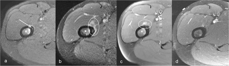
Materials & methods: Retrospective review of 43 patients with RFA for OO between June 2010 and June 2017 was performed. Patient, nidus and ablation data were reviewed. Pre- and 6-8 weeks post-procedural MRI (n=32) were compared for coverage of nidus by ablation zone, bone marrow edema, nidus hyperintensity and other findings. Baseline pain levels and analgesic use were compared with post-procedural follow-up visit at 6-8 weeks. Three groups of clinical and MRI outcomes of complete (CR), partial (PR) and no response (NR) were defined. A weighted-kappa statistic was used to assess for agreement.
Results: Clinical responses were CR in 34/43 (79.1%, 95%CI: 64.0-90.0%), PR in 8/43 (18.6%) and NR in 1/43 (2.3%) patients. All 19/32 patients with MRI CR experienced clinical CR. One patient with MRI NR had clinical NR. All 7/32 patients with clinical PR had MRI PR. All 4/43 complications were in MRI PR or NR groups. Substantial agreement was observed between MRI and clinical outcomes (kappa:0.69, 95%CI:0.45-0.95). MRI helped determine etiologies in all symptomatic patients and their management (n=8).
Conclusions: MRI is recommended for symptomatic patients after ablation.
Keywords: Clinical Outcomes; MRI; Osteoid Osteoma; Radiofrequency Ablation.
Conflict of interest statement
Conflict of Interest: None.
Figures


Similar articles
-
Intraarticular Osteoid Osteoma: MRI Characteristics and Clinical Presentation Before and After Radiofrequency Ablation Compared to Extraarticular Osteoid Osteoma.Rofo. 2020 Dec;192(12):1190-1199. doi: 10.1055/a-1181-9041. Epub 2020 Jul 8. Rofo. 2020. PMID: 32643768 English, German.
-
A comparative study assessing the efficacy and safety of radiofrequency ablation versus surgical treatment for osteoid osteoma: retrospective analysis in a single institution.Insights Imaging. 2024 Mar 22;15(1):82. doi: 10.1186/s13244-024-01656-1. Insights Imaging. 2024. PMID: 38517657 Free PMC article.
-
Correlation of 3-T MRI and CT findings with patient symptoms and treatment outcome in radiofrequency ablation of osteoid osteoma.Acta Orthop Traumatol Turc. 2019 Jul;53(4):239-247. doi: 10.1016/j.aott.2019.04.015. Epub 2019 May 16. Acta Orthop Traumatol Turc. 2019. PMID: 31104885 Free PMC article.
-
Diffusion weighted MRI of osteoid osteomas: Higher ADC values after radiofrequency ablation.Eur J Radiol. 2016 Jul;85(7):1284-8. doi: 10.1016/j.ejrad.2016.03.028. Epub 2016 Apr 9. Eur J Radiol. 2016. PMID: 27235875
-
CT-guided radiofrequency ablation of osteoid osteoma: established concepts and new ideas.Br J Radiol. 2020 Oct 1;93(1114):20200266. doi: 10.1259/bjr.20200266. Epub 2020 Jun 24. Br J Radiol. 2020. PMID: 32520586 Free PMC article. Review.
Cited by
-
Spinal osteoid osteoma in the pediatric population: A management algorithm and systematic review.J Child Orthop. 2023 Aug 30;17(5):428-441. doi: 10.1177/18632521231192477. eCollection 2023 Oct. J Child Orthop. 2023. PMID: 37799321 Free PMC article. Review.
-
Interventional Radiology's Osteoid Osteoma Management: Percutaneous Thermal Ablation.J Clin Med. 2022 Jan 29;11(3):723. doi: 10.3390/jcm11030723. J Clin Med. 2022. PMID: 35160184 Free PMC article. Review.
-
Osteoid osteoma: which is the best mininvasive treatment option?Eur J Orthop Surg Traumatol. 2021 Dec;31(8):1611-1624. doi: 10.1007/s00590-021-02946-w. Epub 2021 Apr 11. Eur J Orthop Surg Traumatol. 2021. PMID: 33839926 Free PMC article. Review.
References
-
- Boscainos PJ, Cousins GR, Kulshreshtha R, Oliver TB, Papagelopoulos PJ. Osteoid osteoma. Orthopedics 2013;36:792–800. - PubMed
-
- Chai JW, Hong SH, Choi JY, Koh YH, Lee JW, Choi JA, Kang HS. Radiologic diagnosis of osteoid osteoma: from simple to challenging findings. Radiographics 2010;30:737–749. - PubMed
-
- Napoli A, Bazzocchi A, Scipione R, Anzidei M, Saba L, Ghanouni P, Cozzi DA, et al. Noninvasive Therapy for Osteoid Osteoma: A Prospective Developmental Study with MR Imaging-guided High-Intensity Focused Ultrasound. Radiology 2017;285:186–196. - PubMed
-
- Vanderschueren GM, Taminiau AH, Obermann WR, van den Berg-Huysmans AA, Bloem JL, van Erkel AR. The healing pattern of osteoid osteomas on computed tomography and magnetic resonance imaging after thermocoagulation. Skeletal Radiol 2007;36:813–821. - PubMed
-
- Rehnitz C, Sprengel SD, Lehner B, Ludwig K, Omlor G, Merle C, Kauczor HU, et al. CT-guided radiofrequency ablation of osteoid osteoma: correlation of clinical outcome and imaging features. Diagn Interv Radiol 2013;19:330–339. - PubMed
Grants and funding
LinkOut - more resources
Full Text Sources
Research Materials