. 2021 Feb;8(1):447-460.
doi: 10.1002/ehf2.13095. Epub 2020 Nov 24.

Crosstalk between the activated Slit2-Robo1 pathway and TGF-β1 signalling promotes cardiac fibrosis

Affiliations

Crosstalk between the activated Slit2-Robo1 pathway and TGF-β1 signalling promotes cardiac fibrosis

Yunqi Liu et al. ESC Heart Fail. 2021 Feb.

Abstract

Aims: Previous reports indicated that the Slit2-Robo signalling pathway is involved in embryonic heart development and fibrosis in other solid organs, but its function in adult cardiac fibrosis has not been investigated. Here, we investigate the role of the Slit2-Robo1 signalling pathway in cardiac fibrosis.

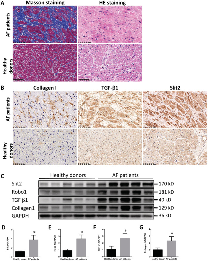
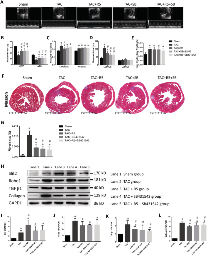
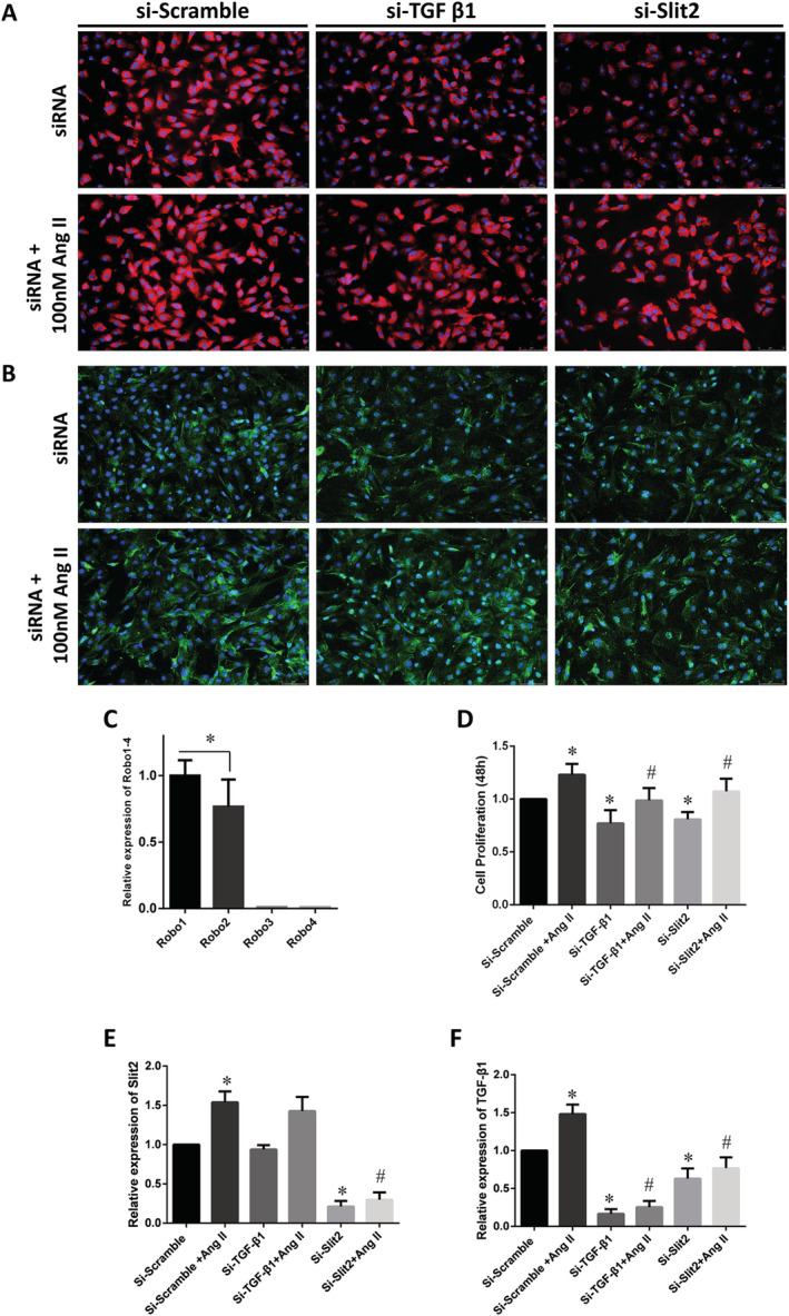
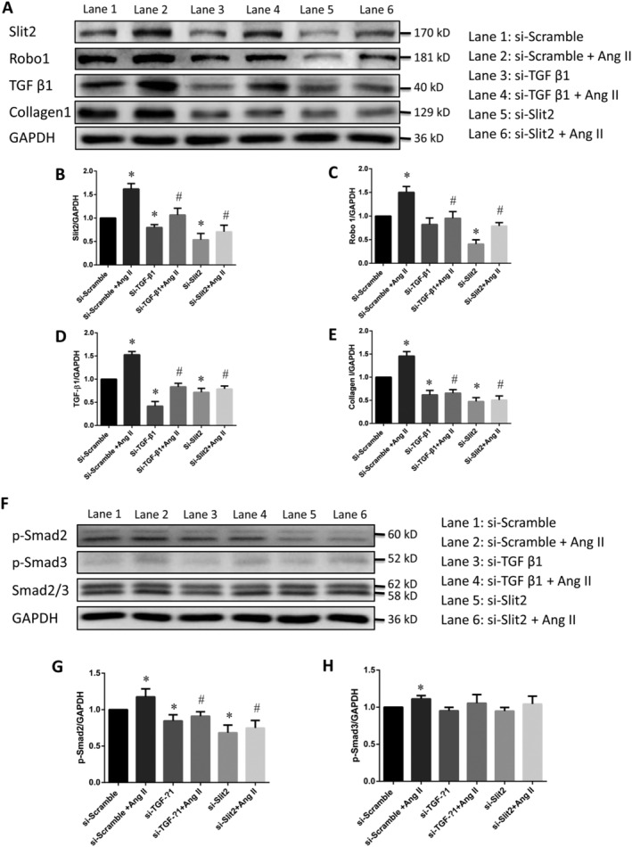
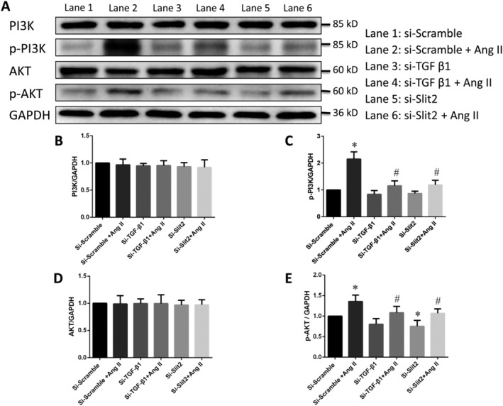
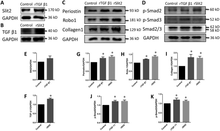
Methods and results: The right atrial tissue samples were obtained from patients with valvular heart disease complicated by atrial fibrillation during heart valve surgery and from healthy heart donors. The fibrotic animal model is created by performing transverse aortic constriction (TAC) surgery. The Robo1, Slit2, TGF-β1, and collagen I expression levels in human and animal samples were evaluated by immunohistochemistry and western blot analysis. Echocardiography measured the changes in heart size and cardiac functions of animals. Angiotensin II (Ang II), Slit2-siRNA, TGF-β1-siRNA, recombinant Slit2, and recombinant TGF-β1 were transfected to cardiac fibroblasts (CFs) respectively to observe their effects on collagen I expression level. The right atrial appendage of patients with valvular heart disease complicated by atrial fibrillation found significantly up-regulated Slit2, Robo1, TGF-β1, and collagen I expression levels. TAC surgery leads to heart enlargement, cardiac fibrosis, and up-regulation of Slit2, Robo1, TGF-β1, and collagen I expression levels in animal model. Robo1 antagonist R5 and TGF-β1 antagonist SB431542 suppressed cardiac fibrosis in TAC mice. Treatment with 100 nM Ang II in CFs caused significantly increased Slit2, Robo1, Smad2/3, TGF-β1, collagen I, PI3K, and Akt expression levels. Transfecting Slit2-siRNA and TGF-β1-siRNA, respectively, into rat CFs significantly down-regulated Smad2/3 and collagen I expression, inhibiting the effects of Ang II. Recombinant Slit2 activated the TGF-β1/Smad signalling pathway in CFs and up-regulated Periostin, Robo1, and collagen I expression.

Conclusions: The Slit2-Robo1 signalling pathway interfered with the TGF-β1/Smad pathway and promoted cardiac fibrosis. Blockade of Slit2-Robo1 might be a new treatment for cardiac fibrosis.

Keywords: Cardiac fibrosis; Crosstalk; Signalling pathway; Slit2; TGF-β1.


Conflict of interest statement

The authors declare that they have no conflict of interest.

Figures

Figure 1
Figure 1
Slit2–Robo1 signalling is activated in patients with valvular heart disease complicated by atrial fibrillation (AF). (A) Masson and haematoxylin–eosin (HE) staining of the right atrial myocardium. In the Masson‐stained sections, collagen fibres are stained blue, muscle fibres and erythrocytes red, and nuclei bluish‐brown. Substantial collagen deposition was identified in the right atrial tissue of patients, and no significant collagen deposition was observed in that of healthy donors. HE staining indicated disorder and significant hypertrophy of myocardial cells. (B) Immunocytochemistry of collagen I, TGF‐β1, and Slit2 in the right atrial myocardium. The expression levels of collagen I, TGF‐β1, and Slit2 were significantly higher in cardiac fibroblasts of patients with valvular heart disease complicated by AF than in those of healthy donors. (C) Western blot analysis of Slit2, Robo1, TGF‐β1, and collagen I expression levels. (D–G) Bar chart of the expression ratios from western blotting normalized to GAPDH. Data are presented as the means ± standard deviations, n = 5. *P < 0.05 vs. the healthy donor group.
Figure 2
Figure 2
Cardiac remodelling and function in the mouse model of transverse aortic constriction (TAC). (A) Echocardiographic short‐axis images and cardiac function measurements of all groups. (B–E) Quantification of cardiac function measurements and heart weight/body weight in all mouse groups. LVPW, left ventricular posterior wall thickness; LVSD, left ventricular end‐systolic diameter; LVDV, left ventricular end‐diastolic volume; IVSD, interventricular septal thickness at end‐diastole; LVSV, left ventricular stroke volume; LVFS, left ventricular fractional shortening; LVEF, left ventricular ejection fraction. (F) Masson staining of mouse left ventricles. Collagen fibres are stained blue, muscle fibres and erythrocytes red, and nuclei bluish‐brown (×200). (G) Masson staining showed that the collagen content in the muscle bundles of myocardial tissue from the TAC and the drug‐treated groups was significantly increased compared with that in the sham group. (H) Representative western blot of left ventricle tissue. (I–L) Quantitative analysis of Slit2, Robo1, TGF‐β1, Smad2/3, and collagen I expressions. GAPDH was used as the internal control. Data are presented as the means ± standard deviations, n = 6. *P < 0.05 vs. the sham control group; #P < 0.05 vs. the TAC control group.
Figure 3
Figure 3
The effect of siRNA and angiotensin II (Ang II) on cardiac fibroblasts (CFs). CFs were transfected with si‐TGFβ1 and si‐Slit2 and then treated with or without Ang II for 24 h in serum‐free medium. (A) Immunofluorescence staining of collagen I; (B) immunofluorescence staining of Vimentin. Collagen I is stained red. Vimentin is stained green. Scale bar = 75 μm (images acquired at ×200). (C) Expression levels of the four Robo receptors, as determined by quantitative PCR. The Robo1 expression level was significantly higher than that of Robo2, and those of Robo3 and Robo4 were negligible. (D) Cell Counting Kit‐8 cell proliferation assay. Compared with the si‐Scramble group, the si‐TGF‐β1 and si‐Slit2 groups exhibited significantly inhibited CF proliferation; Ang II only partially restored CF proliferation. (E–F) Bar chart showing the ratio of the TGF‐β1 and Slit2 mRNA expression levels in CFs as determined by quantitative PCR and normalized to GAPDH. Data are presented as the means ± standard deviations, n = 5. *P < 0.05 vs. the si‐Scramble group without Ang II; #P < 0.05 vs. the si‐Scramble + Ang II group.
Figure 4
Figure 4
The Slit2–Robo1 signalling pathway is involved in cardiac fibrosis. (A) si‐Slit2 siRNA and si‐TGF‐β1 suppressed collagen I expression in cardiac fibroblasts. Cardiac fibroblasts were transfected with si‐TGF‐β1 or si‐Slit2 and then treated with or without Ang II for 24 h in serum‐free medium. (B–E) Bar chart showing the ratio of total Slit2, Robo1, TGF‐β1, and collagen I protein expression levels normalized to GAPDH. (F) Western blot analysis of p‐Smad2, p‐Smad3, and Smad2/3 expressions after treatment with siRNAs or siRNAs + Ang II. (G–H) Bar charts showing the expression ratios of related proteins normalized to GAPDH. Data are presented as the means ± standard deviations, n = 5. *P < 0.05 vs. the corresponding control group without Ang II; #P < 0.05 vs. the si‐Scramble + Ang II group.
Figure 5
Figure 5
The PI3K/Akt pathway is involved in the regulation of Slit2–Robo1 signalling in cardiac fibroblasts. (A) Western blot results for PI3K, p‐PI3K, Akt, and p‐Akt. (B–E) Bar charts of the expression ratios of related proteins normalized to GAPDH for each group. Data are presented as the means ± standard deviations, n = 5. *P < 0.05 vs. the corresponding control group without Ang II; #P < 0.05 vs. the si‐Scramble + Ang II group.
Figure 6
Figure 6
Recombinant Slit2 (rSlit2) promotes Periostin expression and activates Smad signalling. (A) Recombinant TGF‐β1 (rTGF‐β1) promoted Slit2 expression. (B) rSlit2 significantly promoted TGF‐β1 expression. (C–D) Recombinant proteins promoted Periostin, Robo1, and collagen I expressions and Smad2/3 phosphorylation. (E–K) Bar charts of the expression ratios of related proteins normalized to GAPDH for each group. Data are presented as the means ± standard deviations, n = 5. *P < 0.05 vs. the corresponding control group.

Similar articles

Cited by

References

    1. Tomasoni D, Adamo M, Lombardi CM, Metra M. Highlights in heart failure. ESC Heart Fail 2019; 6: 1105–1127. - PMC - PubMed
    1. Guo Y, Gupte M, Umbarkar P, Singh AP, Sui JY, Force T, Lal H. Entanglement of GSK‐3β, β‐catenin and TGF‐β1 signaling network to regulate myocardial fibrosis. J Mol Cell Cardiol 2017; 110: 109–120. - PMC - PubMed
    1. Meagher P, Adam M, Connelly K. It's not all about the cardiomyocyte: fibroblasts, empagliflozin, and cardiac remodelling. Can J Cardiol 2020; 36: 464–466. - PubMed
    1. Souders CA, Bowers SL, Baudino TA. Cardiac fibroblast: the renaissance cell. Circ Res 2009; 105: 1164–1176. - PMC - PubMed
    1. Gourdie RG, Dimmeler S, Kohl P. Novel therapeutic strategies targeting fibroblasts and fibrosis in heart disease. Nat Rev Drug Discov 2016; 15: 620–638. - PMC - PubMed

Publication types

Substances

LinkOut - more resources