

Role of Contrast-Enhanced Ultrasound as a Second-Line Diagnostic Modality in Noninvasive Diagnostic Algorithms for Hepatocellular Carcinoma
- PMID: 33236540
- PMCID: PMC7909851
- DOI: 10.3348/kjr.2020.0973
Role of Contrast-Enhanced Ultrasound as a Second-Line Diagnostic Modality in Noninvasive Diagnostic Algorithms for Hepatocellular Carcinoma
Abstract
Objective: To investigate the diagnostic performance of contrast-enhanced ultrasound (CEUS) and its role as a second-line imaging modality after gadoxetate-enhanced MRI (Gd-EOB-MRI) in the diagnosis of hepatocellular carcinoma (HCC) among at risk observations.
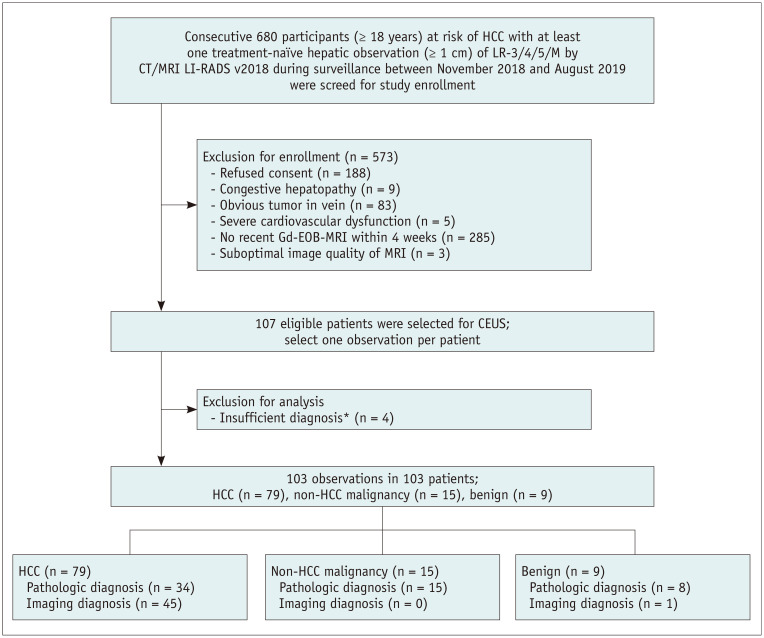
Materials and methods: We prospectively enrolled participants at risk of HCC with treatment-naïve solid hepatic observations (≥ 1 cm) of Liver Imaging Reporting and Data System (LR)-3/4/5/M during surveillance and performed Gd-EOB-MRI. A total of one hundred and three participants with 103 hepatic observations (mean size, 28.2 ± 24.5 mm; HCCs [n = 79], non-HCC malignancies [n = 15], benign [n = 9]; diagnosed by pathology [n = 57], or noninvasive method [n = 46]) were included in this study. The participants underwent CEUS with sulfur hexafluoride. Arterial phase hyperenhancement (APHE) and washout on Gd-EOB-MRI and CEUS were evaluated. The distinctive washout in CEUS was defined as mild washout 60 seconds after contrast injection. The diagnostic ability of Gd-EOB-MRI and of CEUS as a second-line modality for HCC were determined according to the European Association for the Study of the Liver (EASL) and the Korean Liver Cancer Association and National Cancer Center (KLCA-NCC) guidelines. The diagnostic abilities of both imaging modalities were compared using the McNemar's test.
Results: The sensitivity of CEUS (60.8%) was lower than that of Gd-EOB-MRI (72.2%, p = 0.06 by EASL; 86.1%, p < 0.01 by KLCA-NCC); however, the specificity was 100%. By performing CEUS on the inconclusive observations in Gd-EOB-MRI, HCCs without APHE (n = 10) or washout (n = 12) on Gd-EOB-MRI further presented APHE (80.0%, 8/10) or distinctive washout (66.7%, 8/12) on CEUS, and more HCCs were diagnosed than with Gd-EOB-MRI alone (sensitivity: 72.2% vs. 83.5% by EASL, p < 0.01; 86.1% vs. 91.1% by KCLA-NCC, p = 0.04). There were no false-positive cases for HCC on CEUS.
Conclusion: The addition of CEUS to Gd-EOB-MRI as a second-line diagnostic modality increases the frequency of HCC diagnosis without changing the specificities.
Keywords: Contrast enhanced ultrasound; Hepatocellular carcinoma; Liver; Magnetic resonance imaging.
Copyright © 2021 The Korean Society of Radiology.
Conflict of interest statement
The authors have no potential conflicts of interest to disclose.
Figures

References
-
- Marrero JA, Kulik LM, Sirlin CB, Zhu AX, Finn RS, Abecassis MM, et al. Diagnosis, staging, and management of hepatocellular carcinoma: 2018 Practice Guidance by the American Association for the Study of Liver Diseases. Hepatology. 2018;68:723–750. - PubMed
-
- Cruite I, Tang A, Sirlin CB. Imaging-based diagnostic systems for hepatocellular carcinoma. AJR Am J Roentgenol. 2013;201:41–55. - PubMed
-
- Tang A, Cruite I, Sirlin CB. Toward a standardized system for hepatocellular carcinoma diagnosis using computed tomography and MRI. Expert Rev Gastroenterol Hepatol. 2013;7:269–279. - PubMed
-
- Tang A, Cruite I, Mitchell DG, Sirlin CB. Hepatocellular carcinoma imaging systems: why they exist, how they have evolved, and how they differ. Abdom Radiol (NY) 2018;43:3–12. - PubMed
-
- European Association for the Study of the Liver. EASL Clinical Practice Guidelines: management of hepatocellular carcinoma. J Hepatol. 2018;69:182–236. - PubMed
Publication types
MeSH terms
Substances
Grants and funding
LinkOut - more resources
Full Text Sources
Other Literature Sources
Medical
