Review
. 2021 Apr;22(4):547-558.
doi: 10.3348/kjr.2019.0774. Epub 2020 Nov 19.

MR Imaging of the Perihepatic Space

Affiliations
Review

MR Imaging of the Perihepatic Space

Angèle Bonnin et al. Korean J Radiol. 2021 Apr.

Abstract

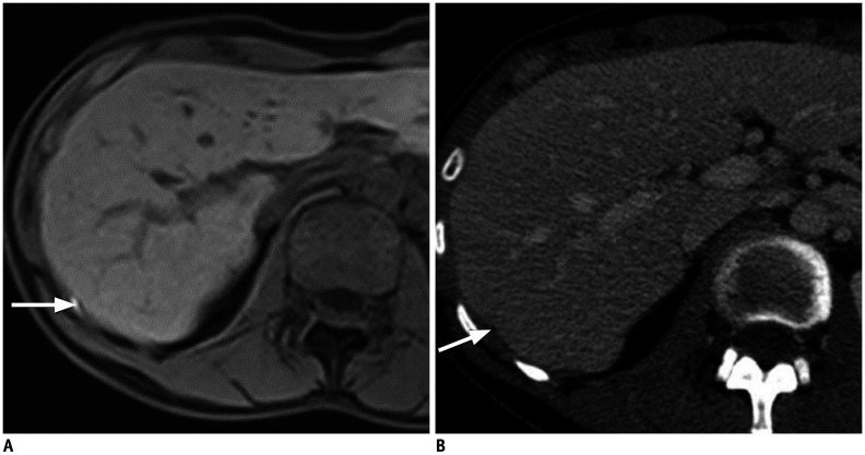
The perihepatic space is frequently involved in a spectrum of diseases, including intrahepatic lesions extending to the liver capsule and disease conditions involving adjacent organs extending to the perihepatic space or spreading thanks to the communication from intraperitoneal or extraperitoneal sites through the hepatic ligaments. Lesions resulting from the dissemination of peritoneal processes may also affect the perihepatic space. Here we discuss how to assess the perihepatic origin of a lesion and describe the magnetic resonance imaging (MRI) features of normal structures and fluids that may be abnormally located in the perihepatic space. We then review and illustrate the MRI findings present in cases of perihepatic infectious, tumor-related, and miscellaneous conditions. Finally, we highlight the value of MRI over computed tomography.

Keywords: Computed tomography; Liver; Magnetic resonance imaging; Peritoneum.


Conflict of interest statement

The authors have no potential conflicts of interest to disclose.

Figures

Fig. 1
Fig. 1. Anatomy of the perihepatic space.
Falciform ligament (yellow arrows, A) separates the left and right hepatic lobes, and the right from the left subphrenic space (dashed arrows). On axial T2-weighted MR images (B) shows the spread of hepatobiliary disease to the perihepatic space and peritoneum through the falciform ligament (yellow arrow). Pancreatic and gastric disease spread to the porta hepatis and liver through the lesser omentum (red arrow). MR = magnetic resonance
Fig. 2
Fig. 2. Biliary leakage post-cholecystectomy.
A 69-year-old female ten days after a cholecystectomy complicated by fluid collection drained under radiological guidance. Axial T1-weighted sequence one and a half hours after intravenous administration of gadobenate dimeglumine shows high-signal intensity fluid seen in the hepatic hilum (arrow), confirmed the bilious nature of the leak.
Fig. 3
Fig. 3. Abscess related to dropped gallstone.
A 76-year-old male, heavy smoker, was referred to the hospital two months after a subtotal cholecystectomy performed for cholecystitis. He presented with fatigue and marked recent weight loss. A. Axial portal venous phase CT image shows a perihepatic, nodular, hypoattenuating lesion, associated with perihepatic, focal, peritoneal thickening, suggestive of peritoneal metastasis (arrow). B. Axial fat-suppressed T2-weighted MR image reveals a small perihepatic abscess (arrowheads) with foci of signal loss indicating tiny gallstones that were not seen on CT. CT = computed tomography
Fig. 4
Fig. 4. Alveolar echinococcosis ruptured in the peritoneum.
A 61-year-old male patient presenting with diarrhea for six months without any weight loss and mild abdominal pain. He had been eating lettuce from his garden in the countryside. A. Axial contrast-enhanced CT slices show partially calcified cystic lesions in the liver (arrow) and peritoneal fluid (arrowhead). B, C. Axial HASTE T2-weighted MR images confirm the abnormalities seen on CT and show microvesicules floating in the peritoneal fluid (arrows) around the liver and thickened peritoneum (arrowhead). These findings are related to the intraperitoneal rupture of a microcystic alveolar echinococcosis lesion in the right liver (dashed arrow) with subsequent peritonitis.
Fig. 5
Fig. 5. Peritoneal metastases of a low-grade serous adenocarcinoma.
A 63-year-old female treated for low-grade serous adenocarcinoma with peritoneal metastases. Axial high b value diffusion-weighted MR image (A) optimally depicts the metastatic peritoneal implants of the perihepatic space, of the subphrenic space (arrowhead), and the implants located on both sides of the falciform ligament (arrows). These lesions are less easily detected and seen only in retrospect on axial gadolinium-enhanced MR (B) images (arrows and arrowhead). Intraoperative photograph (C) shows the lesions in the subphrenic space (arrows)
Fig. 6
Fig. 6. Peritoneal metastases of an ovarian serous adenocarcinoma.
Preoperative evaluation in a 66-year-old female with peritoneal recurrence of a moderately differentiated ovarian serous adenocarcinoma. A. Axial fat-suppressed gadolinium-enhanced T1-weighted MR image optimally shows the enhancement and thickening of both the visceral and parietal layers of the perihepatic peritoneum along with the diaphragmatic cupola, Glisson's sheath, the lesser omentum, and the ductus venosus (arrows). These lesions were confirmed during laparotomy, thus jeopardizing curative resection. B. The peritoneal thickening is barely visible on the corresponding contrast-enhanced axial CT image, although CT was performed on the same day as the MRI (arrows). MRI = magnetic resonance imaging
Fig. 7
Fig. 7. Mucinous perihepatic implants of pseudomyxoma peritonei of appendicular origin.
A 35-year-old female patient with low-grade pseudomyxoma peritonei of an appendicular origin. MR images (axial T2 (A) and fat-suppressed gadolinium-enhanced T1-weighted (B)) better depict the mucinous lesions than CT (C) (arrows, A, B, and C). Moreover, MRI allows the characterization of the mucinous implants, which enhance (dotted arrow, B) and display intermediate signal intensity (dotted arrow, A) versus amorphous mucinous ascites, which do not enhance and display a fluid-like signal (arrows, A, B).
Fig. 8
Fig. 8. Better depiction of small mucinous implants using MRI than with CT.
A 65-year-old male patient with high-grade pseudomyxoma peritonei of appendicular origin. Detection and localization of the mucinous peritoneal lesions are best performed on axial T2-weighted MRI (A) rather than axial contrast-enhanced CT image (B). Of note are the small implants seen on the wall of the lesser curvature of the stomach (black arrowhead, A), behind the inferior vena cava (white arrowhead, A), along the left portal vein (arrow, A), and in the splenic hilum (dotted arrow, A).
Fig. 9
Fig. 9. Peritoneal fibromatosis in a 37-year-old female with abdominal pain.
A 37-year-old female patient with a history of peritoneal fibromatosis operated on at the age of five with complete cytoreduction complaining of abdominal pain. A. Axial contrast-enhanced CT image displays well-delineated peritoneal nodules containing calcifications, one of which is in the perihepatic space (arrows). B. Axial T2-weighted MR image reveals the fibrous nature of the nodules displaying low signal intensity (arrows).
Fig. 10
Fig. 10. MRI characteristics can make the diagnosis of splenosis.
A 53-year-old male, with a vehicle-accident history with subsequent surgical removal of the spleen some years ago. A liver lesion was fortuitously discovered. Axial (A) CT images obtained at a portal phase after contrast medium injection shows a rounded, well-defined, homogeneous, subhepatic lesion that seems to be extrahepatic, similar to liver parenchyma on this phase (arrow). Axial fat-suppressed T2-weighted (B), as well as fat-suppressed gadolinium-enhanced T1-weighted MR images at different phases (not shown)confirm the extrahepatic location of the lesion (arrows). The lesion displays mild hyperintensity to the liver on (B), slightly higher enhancement on portal phase, and lower enhancement on late phase. Red blood cell scintigraphy (C) confirms the diagnosis of perihepatic splenosis (arrow).
Fig. 11
Fig. 11. Fitz-Hugh-Curtis syndrome.
A 30-year-old female patient with abdominal pain and documented pelvic infection (Chlamydia trachomatis). Axial fat-suppressed gadolinium-enhanced T1-weighted MRI acquired at an arterial phase shows mild capsular perihepatic enhancement (arrow). Laparoscopy confirmed the presence of perihepatic adhesions typical of Fitz-Hugh-Curtis syndrome.
Fig. 12
Fig. 12. Endometriosis of the right subphrenic space.
A 28-year-old female patient followed-up for severe endometriosis and presenting with pain in the upper right quadrant and in the scapula during menstruation. Coronal T1-weighted MRI (arrow, A) optimally detects high-signal intensity, diffuse deposits on the right diaphragmatic cupola. However, on the corresponding coronal contrast-enhanced CT acquired at a portal phase (arrow, B), the diaphragmatic lesion is not visible. Furthermore, characterization of the lesions is not possible with CT based on their appearance.
Fig. 13
Fig. 13. MRI is the best imaging technique for the diagnosis of endometriosis.
A 32-year-old female was presenting with isolated unexplained lower thoracic pain which started seven years ago. Axial fat-suppressed abdominal T1-weighted MRI (A) detects a small posterolateral implant between the diaphragm and the liver, displaying a high signal intensity and suggesting endometriosis (arrow). The corresponding contrast-enhanced axial CT image (B) displays a non-specific hypoattenuating focus, only visible in retrospect (arrow). The radiologist interviewed the patient after the MRI, and she confirmed the catamenial nature of her thoracic pain. Three implants were seen during laparoscopy, and the diagnosis of perihepatic endometriosis was confirmed.
Fig. 14
Fig. 14. Pseudolipoma of Glisson's capsule.
Axial in (A) and out-of-phase (B) MRI sequences show a small, subcapsular, well-circumscribed lesion (arrows) displaying high signal intensity on the in-phase and signal drop on the out-of-phase sequence, suggestive of fatty content.

References

    1. Kim S, Kim TU, Lee JW, Lee TH, Lee SH, Jeon TY, et al. The perihepatic space: comprehensive anatomy and CT features of pathologic conditions. Radiographics. 2007;27:129–143. - PubMed
    1. Tirkes T, Sandrasegaran K, Patel AA, Hollar MA, Tejada JG, Tann M, et al. Peritoneal and retroperitoneal anatomy and its relevance for cross-sectional imaging. Radiographics. 2012;32:437–451. - PubMed
    1. DeMeo JH, Fulcher AS, Austin RF., Jr Anatomic CT demonstration of the peritoneal spaces, ligaments, and mesenteries: normal and pathologic processes. Radiographics. 1995;15:755–770. - PubMed
    1. Patel CM, Sahdev A, Reznek RH. CT, MRI and PET imaging in peritoneal malignancy. Cancer Imaging. 2011;11:123–139. - PMC - PubMed
    1. Oh SN, Rha SE, Byun JY, Kim JY, Song KY, Park CH. Chilaiditi syndrome caused by Fitz-Hugh-Curtis syndrome: multidetector CT findings. Abdom Imaging. 2006;31:45–47. - PubMed

MeSH terms

Supplementary concepts