Review
. 2021 Mar 1;320(3):G241-G257.
doi: 10.1152/ajpgi.00373.2020. Epub 2020 Nov 25.

Sarcopenia in chronic liver disease: mechanisms and countermeasures

Affiliations
Review

Sarcopenia in chronic liver disease: mechanisms and countermeasures

Sophie L Allen et al. Am J Physiol Gastrointest Liver Physiol. .

Abstract

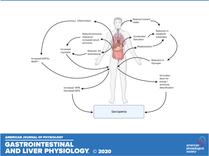
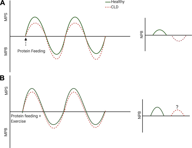
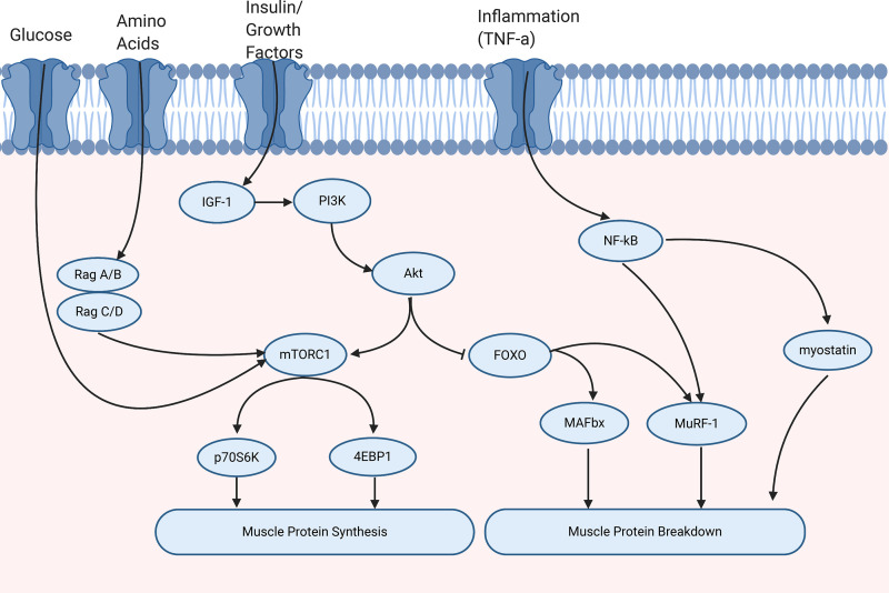
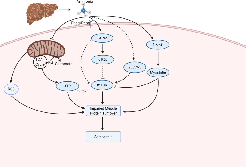
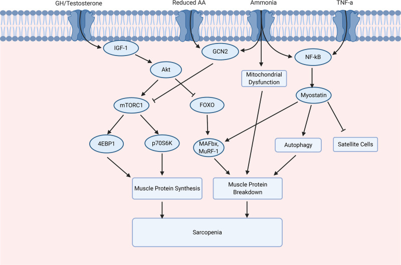
Sarcopenia, a condition of low muscle mass, quality, and strength, is commonly found in patients with cirrhosis and is associated with adverse clinical outcomes including reduction in quality of life, increased mortality, and posttransplant complications. In chronic liver disease (CLD), sarcopenia is most commonly defined through the measurement of the skeletal muscle index of the third lumbar spine. A major contributor to sarcopenia in CLD is the imbalance in muscle protein turnover, which likely occurs due to a decrease in muscle protein synthesis and an elevation in muscle protein breakdown. This imbalance is assumed to arise due to several factors including accelerated starvation, hyperammonemia, amino acid deprivation, chronic inflammation, excessive alcohol intake, and physical inactivity. In particular, hyperammonemia is a key mediator of the liver-gut axis and is known to contribute to mitochondrial dysfunction and an increase in myostatin expression. Currently, the use of nutritional interventions such as late-evening snacks, branched-chain amino acid supplementation, and physical activity have been proposed to help the management and treatment of sarcopenia. However, little evidence exists to comprehensively support their use in clinical settings. Several new pharmacological strategies, including myostatin inhibition and the nutraceutical Urolithin A, have recently been proposed to treat age-related sarcopenia and may also be of use in CLD. This review highlights the potential molecular mechanisms contributing to sarcopenia in CLD alongside a discussion of existing and potential new treatment strategies.

Keywords: chronic liver disease; exercise; muscle protein synthesis; nutrition; sarcopenia.


Conflict of interest statement

The views expressed are those of the authors and not necessarily those of the National Institute for Health Research or the Department of Health and Social Care. No conflicts of interest, financial or otherwise, are declared by the authors.

Figures

None
Graphical abstract
Figure 1.
Figure 1.
Schematic representation of muscle protein turnover in response to anabolic stimuli (protein feeding with/without exercise) in healthy and chronic liver disease (CLD). A: we hypothesize that the primary reason for muscle loss in patients with CLD is blunted muscle protein synthesis (MPS) in response to protein ingestion that coincides with an increase in muscle protein breakdown (MPB). This likely equates to a reduction in net protein balance (NPB) within patients with CLD, in response to proposed alterations in muscle protein turnover. B: exercise in combination with protein ingestion may however partially restore the MPS response within patients with CLD patients.
Figure 2.
Figure 2.
Molecular regulation of muscle protein turnover. Muscle protein synthesis is regulated by several signals including energy status, amino acids, and growth factors, for example, insulin growth factor-1 (IGF-1). This leads to the activation of phosphoinositide 3-kinase (PI3K). Protein kinase B (Akt) regulates muscle protein turnover through the activation of mammalian target of rapamycin complex 1 (mTORC1) and inhibition of Forkhead box O (FOXO). In turn, the activation of mTORC1 leads to the phosphorylation of translation initiation factor 4E-binding protein 1 (4EBP1) and ribosomal protein S6 kinase (p70S6K) that contribute to muscle protein synthesis. Muscle protein breakdown can be activated by inflammatory cytokines, for example, tumor necrosis factor-α (TNF-α), which subsequently leads to the activation of nuclear factor-κB (NF-κB), myostatin, and the ubiquitin ligase Muscle RING finger-1 (MuRF-1). Furthermore, activation of the ubiquitin proteasome pathway through FOXO can increase MuRF-1 and muscle atrophy-box (MAFbx).
Figure 3.
Figure 3.
The proposed molecular alterations that contribute to ammonia-induced changes in muscle protein turnover. Ammonia enters the skeletal muscle through the Rhcg/Rhbg receptors. Subsequently, ammonia contributes to mitochondrial dysfunction, an impaired integrated stress response, and activation of myostatin. These contribute to an impairment in protein turnover and sarcopenia. 4EBP1, translation initiation factor 4E-binding protein 1; eIF2α, eukaryotic initiation factor 2α; IGF-1, insulin growth factor 1; MAFbx, muscle atrophy-box; mTORC1, mammalian target of rapamycin complex 1; MuRF1, muscle RING finger-1; NF-κB, nuclear factor-κB; PI3K, phosphoinositide 3-kinase; p70S6K, ribosomal protein S6 kinase; Rhbg, Rh B glycoprotein; Rhcg, Rh C glycoprotein; ROS, reactive oxygen species; TNF-α, tumor necrosis factor-α; TCA cycle, tricarboxylic acid cycle; α-KG, α ketoglutarate.
Figure 4.
Figure 4.
The molecular regulation of muscle protein synthesis and muscle protein breakdown in chronic liver disease (CLD). The mammalian target of rapamycin complex 1 (mTORC1) can be activated in response to the activation of insulin growth factor-1 (IGF-1), which leads to the activation of protein kinase B (Akt), and inhibited through general control nondepressed 2 (GCN2), activated by a reduction in amino acids (AAs) and ammonia. mTORC1 activation leads to the activation of translation initiation factor 4E-binding protein 1 (4EBP1) and ribosomal protein S6 kinase (p70S6K). Akt regulates FOXO, and the subsequent activation of muscle atrophy-box (MAFbx) and Muscle Ring Finger-1 (MuRF-1). Inflammatory cytokines, for example, tumor necrosis factor-α (TNF-α) and nuclear factor-κB (NF-κB) activate, leading to the activation of myostatin. Myostatin results in an increase in autophagy and inhibition of satellite cells. FOXO, Forkhead box O.

References

    1. Cruz-Jentoft AJ, Bahat G, Bauer J, Boirie Y, Bruyère O, Cederholm T, Cooper C, Landi F, Rolland Y, Sayer AA, Schneider SM, Sieber CC, Topinkova E, Vandewoude M, Visser M, Zamboni M; Writing Group for the European Working Group on Sarcopenia in Older People 2 (EWGSOP2), and the Extended Group for EWGSOP2. Sarcopenia: revised European consensus on definition and diagnosis. Age Ageing 48: 16–31, 2019. doi:10.1093/ageing/afy169. - DOI - PMC - PubMed
    1. Periyalwar P, Dasarathy S. Malnutrition in cirrhosis: contribution and consequences of sarcopenia on metabolic and clinical responses. Clin Liver Dis 16: 95–131, 2012. doi:10.1016/j.cld.2011.12.009. - DOI - PMC - PubMed
    1. Rosenberg IH. Summary comments: epidemiological and methodological problems in determining nutritional status of older persons. Am J Clin Nutr 50: 1231–1233, 1989. doi:10.1093/ajcn/50.5.1231. - DOI
    1. Cruz-Jentoft AJ, Baeyens JP, Bauer JM, Boirie Y, Cederholm T, Landi F, Martin FC, Michel J-P, Rolland Y, Schneider SM, Topinkova E, Vandewoude M, Zamboni M. Sarcopenia: European consensus on definition and diagnosis: report of the European Working Group on Sarcopenia in Older People. Age Ageing 39: 412–423, 2010. doi:10.1093/ageing/afq034. - DOI - PMC - PubMed
    1. Evans WJ, Morley JE, Argiles J, Bales C, Baracos V, Guttridge D, Jatoi A, Kalantar-Zadeh K, Lochs H, Mantovani G, Marks D, Mitch WE, Muscaritoli M, Najand A, Ponikowski P, Rossi Fanelli F, Schambelan M, Schols A, Schuster M, Thomas D, Wolfe R, Anker SD. Cachexia: a new definition. Clin Nutr 27: 793–799, 2008. doi:10.1016/j.clnu.2008.06.013. - DOI - PubMed

Publication types