. 2020 Dec 16;48(22):12711-12726.
doi: 10.1093/nar/gkaa1090.

The deubiquitinase USP36 Regulates DNA replication stress and confers therapeutic resistance through PrimPol stabilization

Affiliations

The deubiquitinase USP36 Regulates DNA replication stress and confers therapeutic resistance through PrimPol stabilization

Yuanliang Yan et al. Nucleic Acids Res. .

Abstract

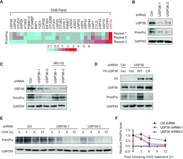
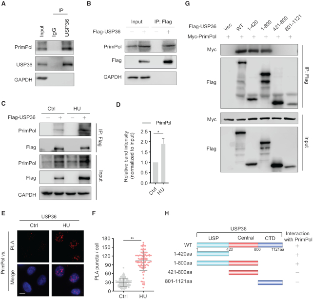
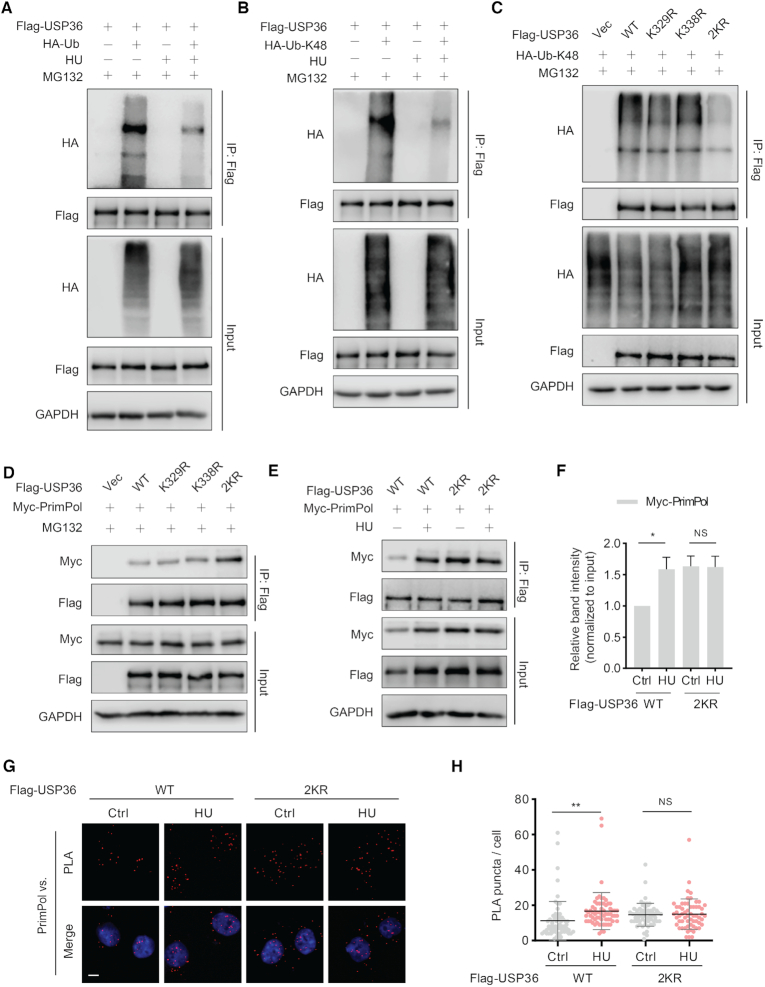
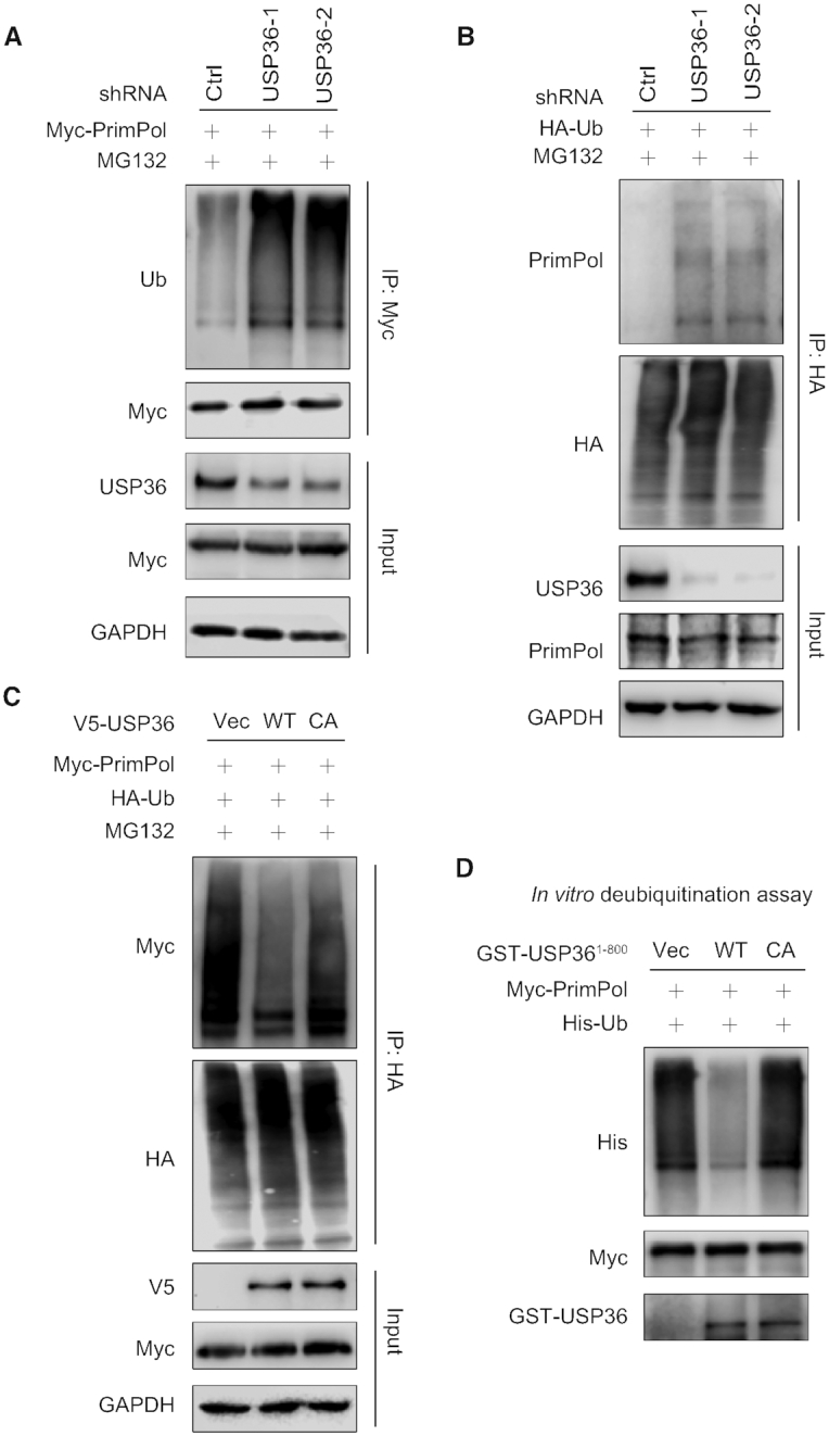
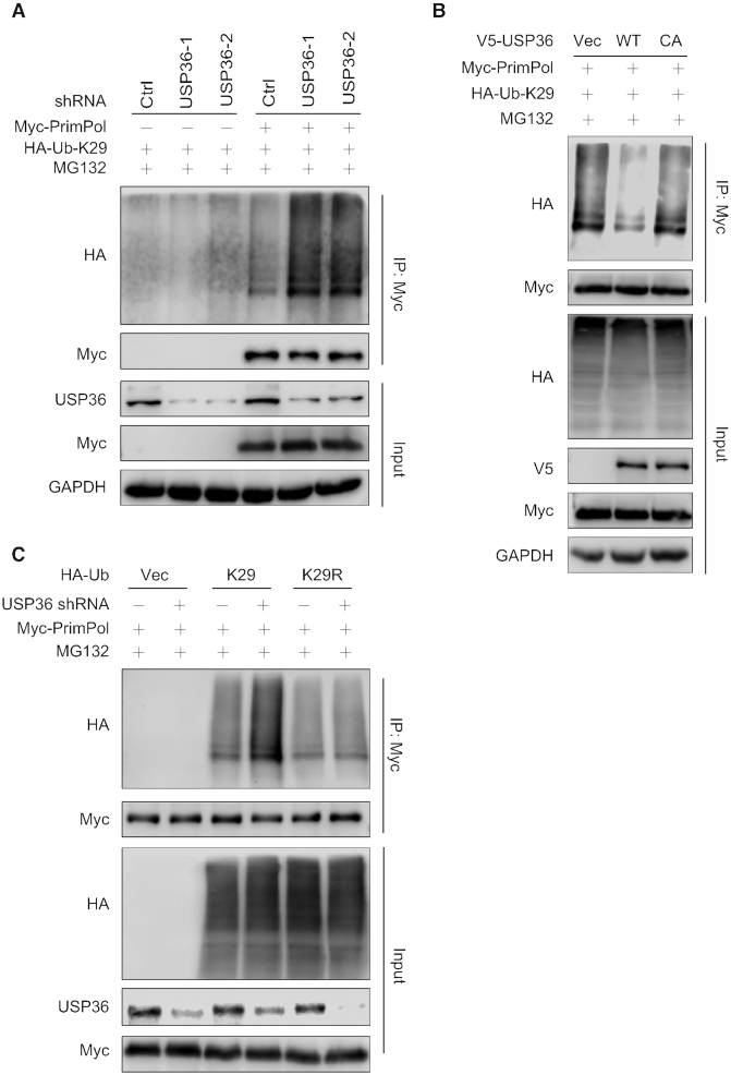
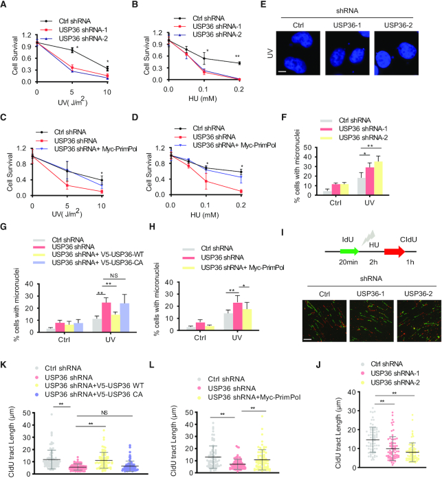
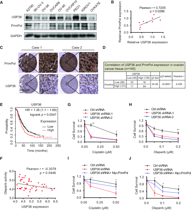
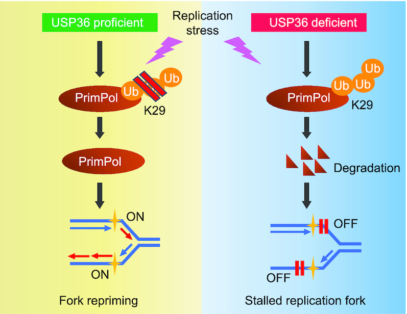
PrimPol has been recently identified as a DNA damage tolerant polymerase that plays an important role in replication stress response. However, the regulatory mechanisms of PrimPol are not well defined. In this study, we identify that the deubiquitinase USP36 interferes with degradation of PrimPol to regulate the replication stress response. Mechanistically, USP36 is deubiquitinated following DNA replication stress, which in turn facilitates its upregulation and interaction with PrimPol. USP36 deubiquitinates K29-linked polyubiquitination of PrimPol and increases its protein stability. Depletion of USP36 results in replication stress-related defects and elevates cell sensitivity to DNA-damage agents, such as cisplatin and olaparib. Moreover, USP36 expression positively correlates with the level of PrimPol protein and poor prognosis in patient samples. These findings indicate that the regulation of PrimPol K29-linked ubiquitination by USP36 plays a critical role in DNA replication stress and chemotherapy response.


Figures

Figure 1.
Figure 1.
USP36 stabilizes PrimPol protein. (A) A panel of deubiquitinases was expressed in HEK293T cells, and the protein levels of PrimPol were assayed using western blot. A heatmap representing the PrimPol changes after overexpression of a panel of deubiquitinases in three repeat experiments. Red and blue represent the increase and decrease in the PrimPol expression level, respectively. With three biological replicates, a total of three deubiquitinases (USP36, USP7 and VCPIP1) were found to upregulate PrimPol protein level (fold-change > 2.0). Among all of them, USP36 was identified to induce the most significant upregulation to PrimPol protein level. (B) USP36 depletion reduces PrimPol protein level. HEK293T cells were transduced with lentivirus encoding control (Ctrl) or USP36 shRNAs, and cell lysates were blotted with indicated antibodies. (C) HEK293T cells stably expressing Ctrl or USP36 shRNA were treated with vehicle or MG132 (50 μM) for 3 h. Cell lysates were then blotted with indicated antibodies. (D) HEK293T cells stably expressing Ctrl or USP36 shRNA-2 were transiently transfected with the wild-type (WT) and catalytically inactive C131A mutant (CA) of V5-tagged USP36. The cells were lysed and western blot was performed with the indicated antibodies. (E) HEK293T cells stably expressing Ctrl or USP36 shRNAs were treated with CHX (0.1 mg/ml), and harvested at the indicated times. Cells were lysed and cell lysates were then blotted with the indicated antibodies. (F) Quantification of the PrimPol protein levels relative to GAPDH. The graphs represent mean ± SD, two-tailed, Student's t-test. *P < 0.05.
Figure 2.
Figure 2.
USP36 is a PrimPol binding protein. (A) Interaction between endogenous USP36 and PrimPol. HEK293T cell were collected and subjected to immunoprecipitation using control IgG and anti-USP36 antibodies. Blots were then probed with the indicated antibodies. (B) Interaction between exogenous USP36 and endogenous PrimPol. Cell lysates from HEK293T cells transiently transfected with Flag-USP36 were subjected to immunoprecipitation with anti-Flag agarose beads, and then blotted with the indicated antibodies. (C) HEK293T cells were transiently transfected with Flag-USP36 for 48h, then left untreated or treated with HU (10 mM) for 8 h. The cell lysates were subjected to immunoprecipitation with anti-Flag agarose beads, and immunoprecipitated lysates were equalized for Flag-USP36. Then cell lysates were blotted with the indicated antibodies. (D) Quantification of the amount of PrimPol in the pull-down relative to the PrimPol input level. The graphs represent mean ± SD, two-tailed, Student's t-test. *P < 0.05. (E) In situ PLA between endogenous USP36 and PrimPol after treated with/without HU (10 mM) for 4 h in OVCAR8 cells. Representative images are shown with merged PLA and nuclei (DAPI) channels from PLA experiments. Scale bar in the bottom left is 10 μm. **P < 0.01. Each red dot represents the detection of USP36-PrimPol interaction complex, and the mean ± SD are shown in (F). (G) The N-terminal USP domain of USP36 is required for binding to PrimPol. HEK293T cells were transiently transfected with Myc-PrimPol together with Wild-type (WT) and truncated mutants (1-420, 1–800, 421–800 and 801–1121aa) of Flag-USP36. The protein interaction was assayed by immunoprecipitation with anti-Flag agarose beads, and then blotted with the indicated antibodies. Schematic representation of the Flag-USP36 and its deletion mutants are shown in (H). USP: Ub-specific protease domain. CTD: Carboxy-terminal domain.
Figure 3.
Figure 3.
USP36 interacts with PrimPol is dependent on its ubiquitination sites K329 and K338 upon DNA replication stress. (A) USP36 stably knocked-down HEK293T cells were transiently transfected with Flag-USP36 and HA-Ub for 48h. Cells were pretreated with HU (10 mM) for 5h, then cotreated with MG-132 (50 μM) for additional 3 h. Cell lysates were subjected to immunoprecipitation with anti-Flag agarose beads, and then blotted with the indicated antibodies. (B) USP36 stably knocked-down HEK293T cells were transiently transfected with Flag-USP36 and HA-tagged Ub-K48 for 48h. Cells were pretreated with HU (10 mM) for 5h, then cotreated with MG132 (50 μM) for additional 3 h. Cell lysates were subjected to immunoprecipitation with anti-Flag agarose beads, and then blotted with the indicated antibodies. (C) Identification of the ubiquitination sites of USP36 for its K48-specific polyubiquitination. USP36 stably knock-down HEK293T cells were transiently transfected with indicated constructs. After 48 h, cells were treated with MG-132 (50 μM) for 3 h before collecting. Cell lysates were subjected to immunoprecipitation with anti-Flag agarose beads, and then blotted with the indicated antibodies. (D) Flag-tagged USP36 expression constructs and Myc-PrimPol plasmid were transfected into USP36 stably knock-down HEK293T cells. After 48 h, cells were treated with MG-132 (50 μM) for 3 h before collecting. Cell lysates were then immunoprecipitated with anti-Flag beads and immunoblotted as indicated. (E) USP36 stably knocked-down HEK293T cells were transiently transfected with indicated constructs, then left untreated or treated with HU (10 mM) for 8 h. The cell lysates were subjected to immunoprecipitation with anti-Flag agarose beads, and immunoprecipitated lysates were equalized for Flag-USP36. Then cell lysates blotted with the indicated antibodies. (F) Quantification of the amount of Myc-PrimPol in the pull-down relative to the Myc-PrimPol input level. The graphs represent mean ± SD, two-tailed, Student's t-test. *P < 0.05; NS = not significant. (G) Representative images of merged PLA and nuclei (DAPI) channels from PLA experiments. USP36 stably knock-down OVCAR8 cells were transiently transfected with USP36-wild-type (WT) or the USP36-WT 2KR mutant, then left untreated or treated with HU (10 mM) for 4 h. In situ PLA was used to assess the interaction between USP36 and endogenous PrimPol. Scale bar in the bottom left is 10 μm. Each red dot represents the detection of USP36-PrimPol interaction complex, and the graphs represent mean ± SD are shown in (H). **P < 0.01; NS = not significant.
Figure 4.
Figure 4.
USP36 deubiquitinates PrimPol. (A) HEK293T cells stably expressing control (Ctrl) or USP36 shRNAs were transiently transfected with Myc-tagged PrimPol. After 48 h, cells were treated with MG132 (50 μM) for 3 h. Cell lysates were subjected to immunoprecipitation with anti-Myc agarose beads, and then blotted with the indicated antibodies. (B) HEK293T cells stably expressing Ctrl or USP36 shRNAs were transiently transfected with HA-tagged ubiquitin. After 48 h, cells were treated with MG132 (50 μM) for 3 h. Cell lysates were subjected to immunoprecipitation with anti-HA agarose beads, and then blotted with the indicated antibodies. (C) HEK293T cells with USP36 stably knocked-down were transiently transfected with indicated constructs. After 48 h, cells were treated with MG132 (50 μM) for 3 h before collecting. Cell lysates were subjected to immunoprecipitation with anti-HA agarose beads, and then blotted with the indicated antibodies. (D) Deubiquitination of PrimPol in vitro by USP36. Ubiquitinated PrimPol was incubated with purified USP361–800-WT or USP361–800-CA in vitro. Cell lysates were subjected to immunoprecipitation with anti-Myc agarose beads, and then blotted with the indicated antibodies.
Figure 5.
Figure 5.
USP36 regulates K29-linked polyubiquitination of PrimPol. (A) Knock-down of USP36 promotes K29-linked polyubiquitination of PrimPol. HEK293T cells stably expressing control (Ctrl) or USP36 shRNAs were transiently transfected with HA-tagged Ub-K29 and Myc-tagged PrimPol. After 48 h, cells were treated with MG132 (50 μM) for 3 h before collecting. Cell lysates were subjected to immunoprecipitation with anti-Myc agarose beads, and then blotted with the indicated antibodies. (B) USP36 stably knock-down HEK293T cells were transiently transfected with indicated constructs. After 48 h, cells were treated with MG132 (50 μM) for 3 h before collecting. Cell lysates were subjected to immunoprecipitation with anti-Myc agarose beads, and then blotted with the indicated antibodies. (C) Ctrl or USP36 stably knock-down HEK293T cells were transiently transfected with indicated HA-K29 lysine-specific mutant constructs and Myc-tagged PrimPol. After 48 h, cells were treated with MG132 (50 μM) for 3 h before collecting. Cell lysates were subjected to immunoprecipitation with anti-Myc agarose beads, and then blotted with the indicated antibodies.
Figure 6.
Figure 6.
USP36 participates in DNA replication stress through a PrimPol-dependent manner. (A, B) OVCAR8 cells from Supplementary Figure S7A were treated with indicated does of UV or HU. Cell survival was determined by colony formation assay. Error bars represent ± SD from three independent experiments. (C, D) OVCAR8 cells from Supplementary Figure S7B were treated with indicated does of UV or HU. Cell survival was determined by colony formation assay. Error bars represent ± SD from three independent experiments. (E) Representative images of micronuclei labeled with the DNA dye DAPI in OVCAR8 USP36-knockdown cells from Supplementary Figure S7A. Cells were left untreated or treated with UV (5 J/m2) for 48 h. Cells were fixed, permeabilized in 0.5% Triton X-100, stained with DAPI, then analyzed by Nikon eclipse 80i fluorescence microscope for micronuclei. The micronuclei assays were repeated for three times, and at least 200 cells were counted for each experiment. Scale bar in the bottom left is 10 μm. (F) Quantification of micronuclei in cells from E. (G) OVCAR8 USP36-knockdown cells reconstituted with V5-tagged USP36 (WT and C131A mutant) from Supplementary Figure S7C were used for measuring micronuclei. (H) OVCAR8 USP36-knockdown cells reconstituted with Myc-PrimPol from Supplementary Figure S7B were used for measuring micronuclei. (I) Representative images of DNA fibers in OVCAR8 USP36-knockdown cells from Supplementary Figure S7A. Cells were labeled with IdU for 20 minutes as a control to label the active replication, and followed by incubation with 4 mM HU for 2 h. Cells were then labeled with CIdU for 1h to evaluate the DNA replication upon genotoxic stress. DNA fibers were stretched on a microscope slide, stained with IdU and CIdU antibodies, imaged, and the lengths of fiber tracks measured. Scale bar in the bottom left is 10 μm. (J) Quantification of DNA fibers in cells from I. (K) OVCAR8 USP36-knockdown cells reconstituted with V5-tagged USP36 (WT and C131A mutant) from Supplementary Figure S7C were used to detect the restart efficiency of stalled replication forks. (L) OVCAR8 USP36-knockdown cells reconstituted with Myc-PrimPol from Supplementary Figure S7B were used to detect the restart efficiency of stalled replication forks. The graphs represent mean ± SD, two-tailed, Student's t-test. *P < 0.05; **P < 0.01; NS = not significant.
Figure 7.
Figure 7.
USP36 expression is correlated with PrimPol levels in ovarian cancer, and regulates the treatment response of cancer cells through PrimPol stabilization. (A) Cell lysates from several ovarian cancer cell lines were blotted with USP36 and PrimPol antibodies. (B) Correlation analysis of USP36 and PrimPol in ovarian cancer cells. Statistical analyses were performed with the χ2 test. The Pearson r indicates correlation coefficient. (C) Representative images of immunohistochemical staining of USP36 and PrimPol on tissue microarray of ovarian cancer specimens (n = 140). Scale bars are indicated. (D) USP36 expression correlates with PrimPol levels in tissue microarray of ovarian cancer samples. Protein levels of USP36 and PrimPol were quantified in ovarian cancer specimens. Statistical analyses were performed with the χ2 test. (E) Survival analysis of ovarian cancer patients by the Kaplan–Meier Plotter. (F) Negative correlation between olaparib activity and USP36 expression in 43 ovarian cancer cells using CellMinerCDB. (G-H) Control and OVCAR8 cells with USP36 stably knock-down from Supplementary Figure S7A were subjected to cisplatin or olaparib treatment. Survival of the cells was assessed by colony formation assay. (I, J) OVCAR8 USP36-knockdown cells reconstituted with Myc-PrimPol from Supplementary Figure S7B were subjected to cisplatin or olaparib treatment. Survival of the cells was assessed by colony formation assay. Error bars represent ± SD from three independent experiments. *P < 0.05; **P < 0.01.
Figure 8.
Figure 8.
A schematic for PrimPol stabilization by deubiquitinase USP36, which promotes replication fork progression following DNA replication stress. Overexpressed USP36 binds to PrimPol and interferes with its K29-linked polyubiquitination and degradation. The PrimPol protein is markedly stabilized by USP36 overexpression and promotes stalled fork restart in the response to DNA replication stress in cancer cells.

Similar articles

Cited by

References

    1. Martin S.K., Wood R.D.. DNA polymerase zeta in DNA replication and repair. Nucleic Acids Res. 2019; 47:8348–8361. - PMC - PubMed
    1. Jain R., Aggarwal A.K., Rechkoblit O.. Eukaryotic DNA polymerases. Curr. Opin. Struct. Biol. 2018; 53:77–87. - PubMed
    1. Garcia-Gomez S., Reyes A., Martinez-Jimenez M.I., Chocron E.S., Mouron S., Terrados G., Powell C., Salido E., Mendez J., Holt I.J. et al. .. PrimPol, an archaic primase/polymerase operating in human cells. Mol. Cell. 2013; 52:541–553. - PMC - PubMed
    1. Mouron S., Rodriguez-Acebes S., Martinez-Jimenez M.I., Garcia-Gomez S., Chocron S., Blanco L., Mendez J.. Repriming of DNA synthesis at stalled replication forks by human PrimPol. Nat. Struct. Mol. Biol. 2013; 20:1383–1389. - PubMed
    1. Bailey L.J., Bianchi J., Hegarat N., Hochegger H., Doherty A.J.. PrimPol-deficient cells exhibit a pronounced G2 checkpoint response following UV damage. Cell Cycle. 2016; 15:908–918. - PMC - PubMed

Publication types

MeSH terms