. 2021 Jan;30(1):92-101.
doi: 10.1111/exd.14244. Epub 2020 Dec 7.

Single-cell transcriptomic analysis of small and large wounds reveals the distinct spatial organization of regenerative fibroblasts

Affiliations

Single-cell transcriptomic analysis of small and large wounds reveals the distinct spatial organization of regenerative fibroblasts

Quan M Phan et al. Exp Dermatol. 2021 Jan.

Abstract

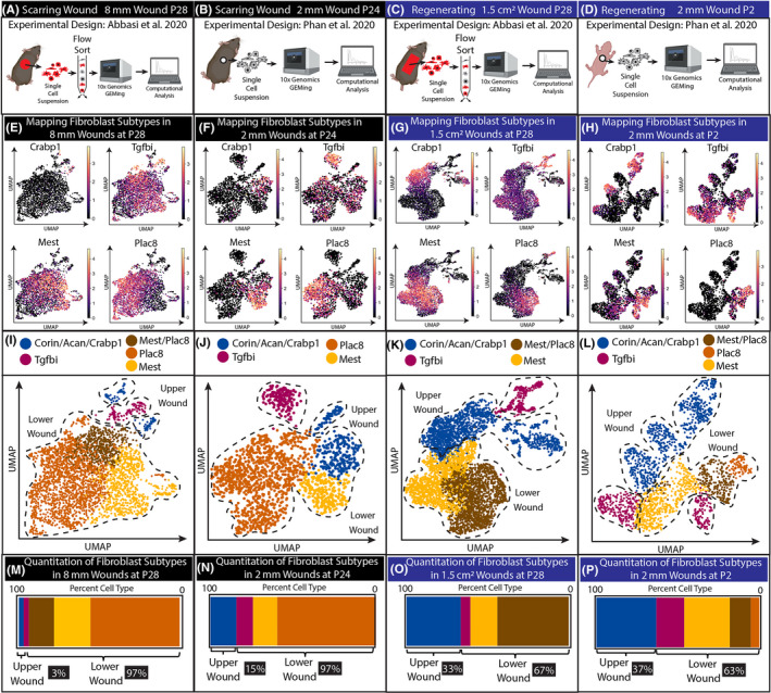
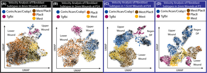
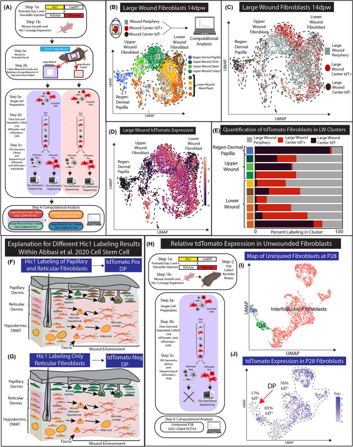
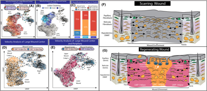
Wound-induced hair follicle neogenesis (WIHN) has been an important model to study hair follicle regeneration during wound repair. However, the cellular and molecular components of the dermis that make large wounds more regenerative are not fully understood. Here, we compare and contrast recently published scRNA-seq data of small scarring wounds to wounds that regenerate in hope to elucidate the role of fibroblasts lineages in WIHN. Our analysis revealed an over-representation of the newly identified upper wound fibroblasts in regenerative wound conditions, which express the retinoic acid binding protein Crabp1. This regenerative cell type shares a similar gene signature to the murine papillary fibroblast lineage, which are necessary to support hair follicle morphogenesis and homeostasis. RNA velocity analysis comparing scarring and regenerating wounds revealed the divergent trajectories towards upper and lower wound fibroblasts and that the upper populations were closely associated with the specialized dermal papilla. We also provide analyses and explanation reconciling the inconsistency between the histological lineage tracing and the scRNA-seq data from recent reports investigating large wounds. Finally, we performed a computational test to map the spatial location of upper wound fibroblasts in large wounds which revealed that upper peripheral fibroblasts might harbour equivalent regenerative competence as those in the centre. Overall, our scRNA-seq reanalysis combining multiple samples suggests that upper wound fibroblasts are required for hair follicle regeneration and that papillary fibroblasts may migrate from the wound periphery to the centre during wound re-epithelialization. Moreover, data from this publication are made available on our searchable web resource: https://skinregeneration.org/.

Keywords: dermal papilla; fibroblast heterogeneity; scRNA-seq; web resource.


Conflict of interest statement

The authors have no conflict of interest.

Figures

Figure 1
Figure 1
Regenerating wounds contain a higher proportion of upper wound fibroblasts compared with scarring wounds. (A‐D) Experimental design to generate scRNA data from Phan et al., 2020 and Abbasi et al., 2020. (E‐H) Identifying the clusters in UMAPs utilizing upper wound marker Crabp1, and lower wound markers Mest and Plac8. Tgfbi cluster is a newly identified fibroblast cluster in wounds. (I‐L) UMAP of upper, lower and Tgfbi clusters in scarring and regenerative wounds. (M‐P) Quantification of cells within upper, lower and Tgfbi clusters in scarring and regenerative wounds
Figure 2
Figure 2
RNA velocity reveals distinct trajectories for upper and lower wound fibroblasts in both scarring and regenerative wounds. (A‐D) Stochastic model of RNA velocity analysis from scarring and regenerative wounds
Figure 3
Figure 3
Analysis of Hic1CreERt2‐labelled tdTomato expression in scRNA‐seq data of large wound fibroblast populations. (A) Schematic of experimental design. (B) Recomputed UMAP analysis of all large wound fibroblasts aligned with a genome containing tdTomato. Both wound periphery files (LWP 14dpw Pos and Neg) are coloured grey. (C) UMAP projection colour coded by 10× Genomics libraries of large wounds. (D) UMAP of large wounds with tdTomato expression. (E) Quantification of the number of cells within the Leiden cluster by 10× Genomic library. (F‐G) A proposed explanation for inconsistent results between Hic1CreERT2 histological lineage tracing (F) and scRNA‐seq data (G). (H‐J) An alternative explanation for the lack of tdTomato‐positive cell scRNA‐seq lineage tracing. UMAP projection of fibroblasts subclustered using original Louvain algorithm and grouped as ‘interfollicular fibroblasts’, ‘DP’ or ‘DS’ based on canonical markers (I) (Figure S3). tdTomato expression overlayed on uninjured fibroblast UMAP (J)
Figure 4
Figure 4
Computational test to identify the identify regeneration‐competent fibroblasts in large wounds. (A) Overlaying RNA velocity analysis in the context of four 10× genomics libraries generated for large wound periphery 14dpw tdTomato positive (LWP 14dpw Pos), large wound periphery 14dpw tdTomato negative (LWP 14dpw Neg), large wound centre 14dpw tdTomato Pos (LWC 14dpw Pos) and large wound centre 14dpw tdTomato negative (LWC 14dpw Neg). (B) Leiden clustering reveals three distinct clusters of Crabp1 upper wound fibroblasts. (C) Quantification of the number of cells from 10× genomic libraries represented in Leiden clusters. (D‐E) Computational test to determine the origin of the regenerative fibroblast and dermal papilla. (D) Velocity analysis of LWC 14dpw Pos and LWC 14dpw Neg libraries. (E) Velocity analysis of LWC 14dpw Neg and LWP 14dpw Pos libraries. (F) Proposed model for fibroblast heterogeneity in small scarring wounds. Papillary fibroblasts are green, while reticular/hypodermal/DWAT fibroblasts are yellow. (G) Proposed model for the contribution of different fibroblasts subtypes in large wounds

References

    1. Griffin MF, desJardins‐Park HE, Mascharak S, Borrelli MR, Longaker MT. Understanding the impact of fibroblast heterogeneity on skin fibrosis. Dis Model Mech. 2020;1310.1242/dmm.044164 - DOI - PMC - PubMed
    1. Driskell RR, Watt FM. Understanding fibroblast heterogeneity in the skin. Trends Cell Biol. 2015;25:92–99. - PubMed
    1. Driskell RR, Lichtenberger BM, Hoste E,, et al. Distinct fibroblast lineages determine dermal architecture in skin development and repair. Nature. 2013;504:277–281. - PMC - PubMed
    1. Sorrell JM, Caplan AI. Fibroblast heterogeneity: more than skin deep. J Cell Sci. 2004;117:667–675. - PubMed
    1. Mine S, Fortunel NO, Pageon H, Asselineau D. Aging alters functionally human dermal papillary fibroblasts but not reticular fibroblasts: a new view of skin morphogenesis and aging. PLoS One. 2008;3:e4066. - PMC - PubMed

Publication types

MeSH terms

Substances