. 2020 Dec 23;120(24):13480-13544.
doi: 10.1021/acs.chemrev.0c00672. Epub 2020 Nov 25.

Design and Applications of Water-Soluble Coordination Cages

Affiliations

Design and Applications of Water-Soluble Coordination Cages

Edmundo G Percástegui et al. Chem Rev. .

Abstract

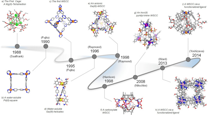
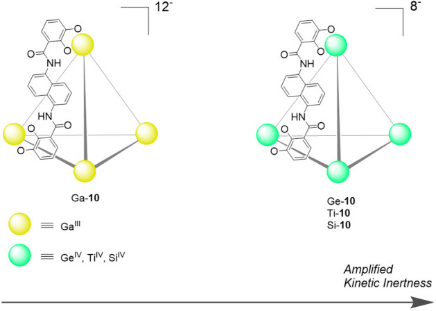
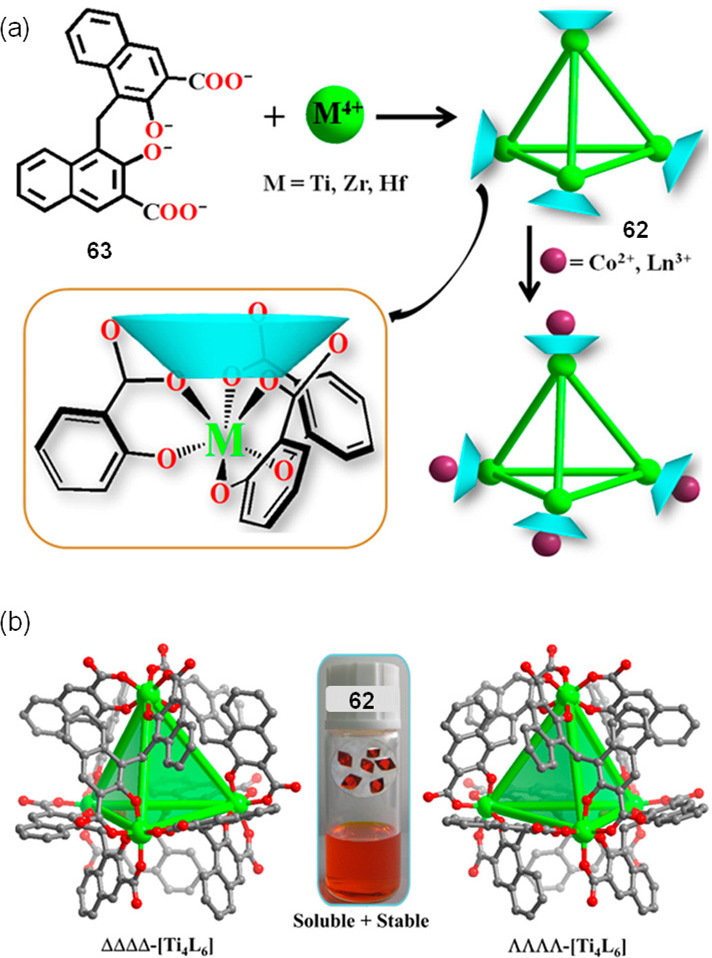
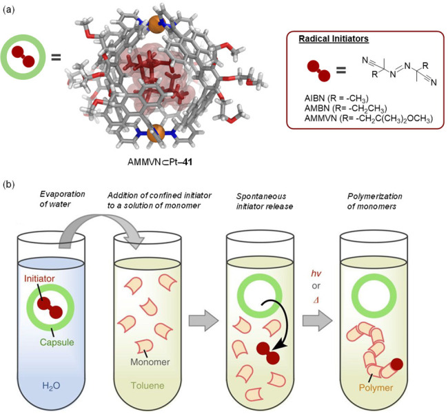
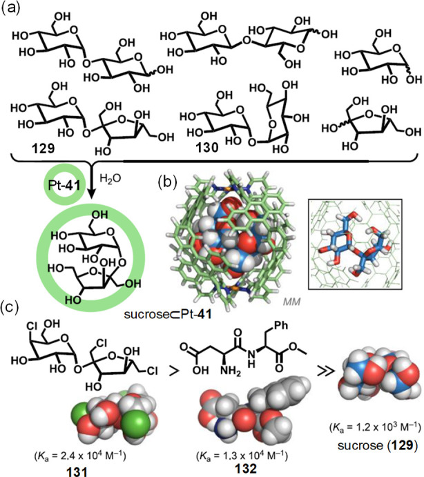
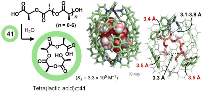
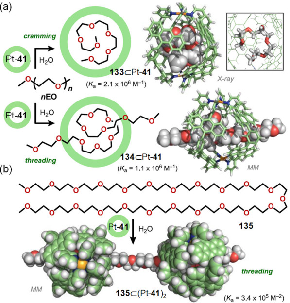
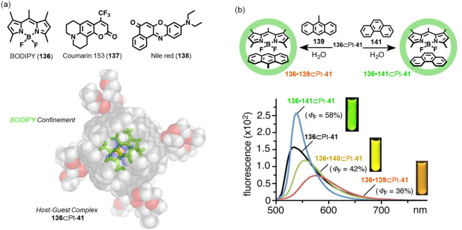
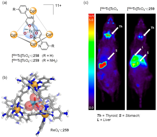
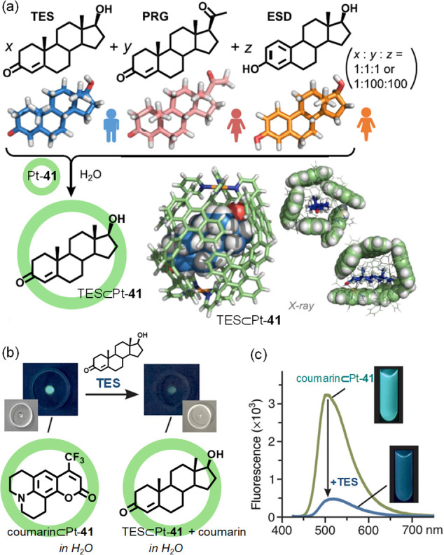
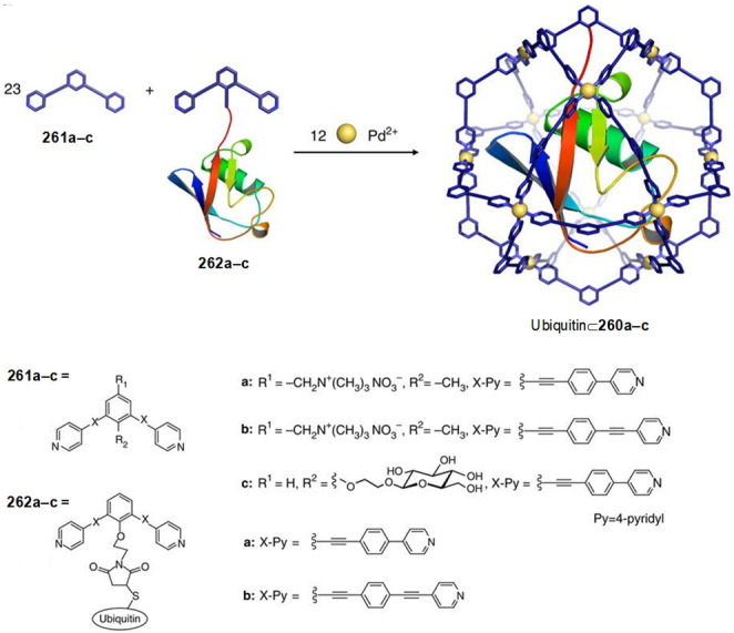
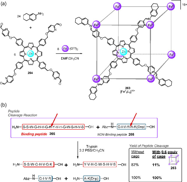
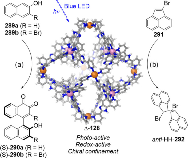
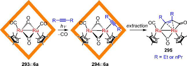
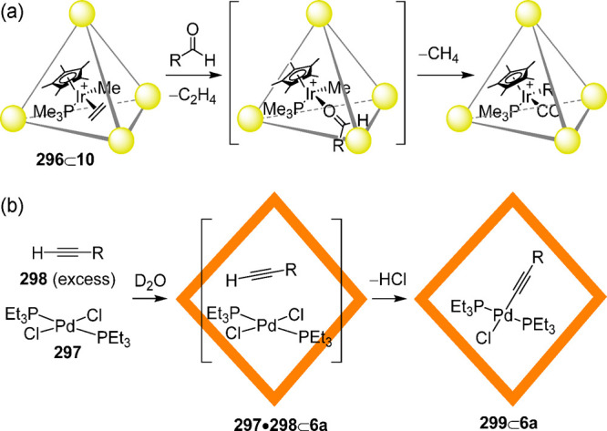
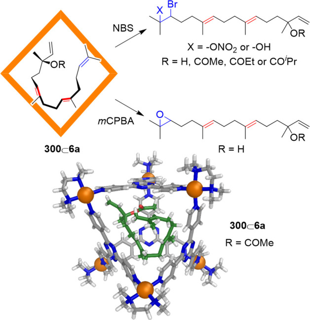
Compartmentalization of the aqueous space within a cell is necessary for life. In similar fashion to the nanometer-scale compartments in living systems, synthetic water-soluble coordination cages (WSCCs) can isolate guest molecules and host chemical transformations. Such cages thus show promise in biological, medical, environmental, and industrial domains. This review highlights examples of three-dimensional synthetic WSCCs, offering perspectives so as to enhance their design and applications. Strategies are presented that address key challenges for the preparation of coordination cages that are soluble and stable in water. The peculiarities of guest binding in aqueous media are examined, highlighting amplified binding in water, changing guest properties, and the recognition of specific molecular targets. The properties of WSCC hosts associated with biomedical applications, and their use as vessels to carry out chemical reactions in water, are also presented. These examples sketch a blueprint for the preparation of new metal-organic containers for use in aqueous solution, as well as guidelines for the engineering of new applications in water.


Conflict of interest statement

The authors declare no competing financial interest.

Figures

Figure 1
Figure 1
Timeline showing major advances in supramolecular coordination chemistry that have led from (a) the first cage complex to (b–f) early water-soluble complexes in the 1990s, and to (g–i) recent examples of WSCCs synthesized from ligands functionalized with solubilizing functional groups. The name of the group for each work is shown for reference.
Figure 2
Figure 2
Synthesis of the MgII-based cage (1) obtained by Saalfrank. The X-ray crystal structure of anionic [Mg4L6]4– (1) is shown.
Figure 3
Figure 3
Synthesis of Fujita’s water-soluble square (4) and the cationic portion of its X-ray crystal structure.
Figure 4
Figure 4
(a) Self-assembly of water-soluble octahedron 6 reported by Fujita. Analogous octahedra can be prepared using different cis-capped metallic corners in place of the [(en)Pd]2+ units. (b) The X-ray crystal structure of 6.
Figure 5
Figure 5
(a) The C2-symmetric catechol ligand 7 combined with trivalent metal ions to yield dinuclear helicates of type 8; the anionic part of the X-ray crystal structure of a gallium(III) helicate (8a) obtained from a modified version of ligand 7 is shown. (b) The similar ligand 9, based on a naphthalene spacer, selectively self-assembles with GaIII or FeIII to afford water-soluble tetrahedron 10; the anionic part of the X-ray structure of complex K5(Et4N)7[Fe4L6] containing an encapsulated Et4N+ is shown.
Figure 6
Figure 6
(a) Synthesis of capsule 12 built from two 11 ligands and CoII ions. (b) X-ray structure of 12 showing the four cobalt(II) ions around the periphery.
Figure 7
Figure 7
Reactions of [(en)Pd(NO3)2] (2) with rigid oligo(pyridine/pyrimidine) “panels” enable the formation of WSCCs. Examples of these ligands and the X-ray crystal structures of the corresponding cages are shown.
Figure 8
Figure 8
Aqueous subcomponent self-assembly reaction to prepare the tetramethylammonium salt of cage 27; the anionic portion of its X-ray crystal structure is depicted.
Figure 9
Figure 9
(a) Enantioselective formation of ΔΔΔΔ-29 from (S,S)-28; the universal force field (UFF) model of ΔΔΔΔ-29 is shown. (b) Previously reported 4,4′′-diamino-p-terphenyls 3033 led to water-insoluble cages.
Figure 10
Figure 10
(a) Self-assembly of CoII ions with 34 led to water-insoluble cube 35. The analogous reaction using 36, equipped with hydroxymethyl groups yielded water-soluble cage 37. (b) View of the X-ray structure of WSCC 37, emphasizing the windows leading to the central cavity, and the decoration of the external surface with hydroxyl groups.
Figure 11
Figure 11
(a) Reaction of PdII with ligands 38 and 40, containing a different number of solubilizing chains, led to organic-soluble cage 39(76) and its water-soluble analogue 41, respectively. (b) X-ray structure of the water-soluble cage 41 showing its 12 pendant solubilizing chains.
Figure 12
Figure 12
(a) Self-assembly of PdII ions with W-shaped ligands 42 and 43 (bearing four and six solubilizing chains, respectively) yielded 44 and the more water-soluble 45 dual cavity cages. (b) X-ray structure of the water-insoluble cation 44.
Figure 13
Figure 13
(a) The self-assembly of PdII ions with 46 afforded water-insoluble capsule 47. (b) Water-soluble analogues were obtained using functionalized ligands 48(80) or 49(81) instead of 46.
Figure 14
Figure 14
(a) Cationic ligand 50 selectively self-assembles with PdII ions under different conditions to yield water-soluble capsule 51 or tube 52. (b) The combination of the elongated cationic ligand 53 with PdII affords expanded water-soluble capsule 54. The X-ray structures of the products are depicted.
Figure 15
Figure 15
Examples of WSCCs based on palladium(II) ions and pyridinium ligands. (a) Synthesis of Mukherjee’s “molecular dice” (56) and (b) Sun’s nanocapsule 58. X-ray structures of the cages are depicted.
Figure 16
Figure 16
Preparation of water-insoluble cube 60 and reversible anion exchange to obtain water-soluble cube 61, the direct synthesis of which is not possible. Adapted with permission from ref (88). Copyright 2017 Wiley-VCH.
Figure 17
Figure 17
[Ga4L6]12– tetrahedron Ga-10 and its less-labile congeners [M4L6]8– (M = GeIV, TiIV, SiIV), built using tetravalent metal ions.,
Figure 18
Figure 18
(a) Assembly of tetrahedral Ti4L6 cage 62, the vertices of which bound CoII or LnIII ions. (b) X-ray structures of the ΔΔΔΔ and ΛΛΛΛ enantiomers of 62. Inset: photo of cage crystals in DMF/H2O solution. Adapted with permission from ref (97). Copyright 2017 American Chemical Society.
Figure 19
Figure 19
(a) Crystal structures of zirconium tetrahedra 6467. Color code: Zr, turquoise; O, red; N, blue; C, black. Yellow balls represent the cage inner void. (b) Scheme for the postassembly modification (PAM) of cage 65 containing up to six functionalized sites per cage. Adapted with permission from ref (101). Copyright 2018 American Chemical Society.
Figure 20
Figure 20
(a) Self-assembly of subcomponents 6872 with metal(II)-ions (M = CoII, NiII, ZnII, and CdII) yielded cages M-7377. Kinetically stable WSCC assemblies were obtained in the cases noted.
Figure 21
Figure 21
(a) Self-assembly of water-soluble ZnII-helicates and CdII-tetrahedra from tritopic amines TREN (78) and TRPN (79) and C2-symmetric subcomponents 72 and 8082. Cationic portions of the X-ray structures of (b) helicate Zn-84, (c) tetrahedron Cd-88, (d) helicate Zn-85, and (e) tetrahedron Cd-90. Adapted with permission from ref (89). Copyright 2019 Royal Society of Chemistry.
Figure 22
Figure 22
Stability of cubes M-73 in water noting their decomposition conditions. (a) Half-lives (t1/2), (b) ionic radii (IR, Å), and (c) ligand-exchange rates for water (kH2O, s–1) for the different metal ions are shown for comparison. Fe-76 is low-spin, but kH2O for high-spin FeII is given as reference. Adapted with permission from ref (89). Copyright 2019 Royal Society of Chemistry.
Figure 23
Figure 23
Reaction of 5 with PtII-complex 91 yielded a kinetic mixture of oligomers containing the target cage Pt-6. Addition and subsequent removal of a template led to the clean formation of the kinetically robust product Pt-6.
Figure 24
Figure 24
(a) Ligand 92 assembles with CoII to afford cobalt(II) tetrahedron 93, which is transformed into the more robust cobalt(III) tetrahedron 94 through oxidation. (b) X-ray crystal structure of a cobalt(III) cage (94a) obtained from a derivative of ligand 92; peripheral groups are omitted.
Figure 25
Figure 25
(a) Conditions for the selective preparation of tetrahedron 96 and prism 97 and their interconversion. The cationic parts of the crystal structures of (b) 96 and (c) 97.
Figure 26
Figure 26
(a) Schematic representation of the template-controlled syntheses of WSCCs 98 and 99. (b) X-ray structure of tetrahedron 99 with CBr4 in its cavity. (a) Adapted with permission from ref (121). Copyright 2000 American Chemical Society.
Figure 27
Figure 27
(a) Template-induced self-assembly of monoend-capped tube 103 and dimeric open tube 104. (b) Crystal structure of 104 with two equivalents of biphenylcarboxylate included. (a) Reproduced with permission from ref (122). Copyright 2003 Wiley-VCH.
Figure 28
Figure 28
Equilibrium between catenane 105 and monomeric ring 106 shifts to either side depending on the polarity of the solvent employed.
Figure 29
Figure 29
Self-assembly of ligand 107 with CoII ions results in an equilibrium between three assemblies 118, 109 and 110 in D2O. The X-ray crystal structures of each assembly are shown.
Figure 30
Figure 30
Crystal structures of coordination cages containing water clusters in the central cavity. (a) Fujita’s PdII6L4 octahedron 6c(149) and the (b) CoII8L12 cube 35 made by Ward.
Figure 31
Figure 31
Initial WSCCs as examples of binding of hydrophobic neutral organic molecules. (a) Fujita octahedra can bind up to four identical guests or two different ones. Cages reported by (b) Harrison, and (c) Raymond also bind hydrophobic molecules in water.
Figure 32
Figure 32
Contrasting binding ability of two isostructural FeII4L6 tetrahedra. (a) Cage 111 binds only small inorganic anions in acetonitrile but not larger anions or neutral organic molecules. (b) Analogous cage 27 binds a wide variety of neutral organic molecules, some gases, and P4 in water. (c–g) X-ray crystal structures of selected host–guest complexes of each cage are depicted.
Figure 33
Figure 33
(a) Subcomponent self-assembly of cage 113 and its crystal structure. Guest molecules for 113. In D2O (b) all molecules shown were encapsulated (outer blue box) in 113, whereas in CD3CN (c) only a subset (inner red box) were bound.
Figure 34
Figure 34
Binding of biologically relevant guests was only observed in aqueous solution using the water-soluble cube 61 obtained upon anion exchange from the MeCN-soluble cube 60.
Figure 35
Figure 35
Guests used to compare binding between hosts 35 and 37 in acetonitrile and water, respectively. The highest binding association was observed between host 37 and 2-hydroxyquinoline (amide tautomer) in water.
Figure 36
Figure 36
(a) Ketone guests for 37 in water. The plot of binding free energy vs number of C atoms for guest series A, and the X-ray crystal structure of the cycloundecanone⊂37 complex are shown. (b) The three guests used in the pH-swing experiment and their binding constants within cage 37 as a function of protonation state, together with a diagram showing which host–guest species predominates as a function of pH.
Figure 37
Figure 37
Cage 27 as a protective environment for guests. (a) Crystal structure of P427. (b) Extraction of P4 from 27 by n-heptane was not possible, whereas replacing P4 with benzene results in phosphorus release into the organic phase. (b) Reproduced with permission from ref (165). Copyright 2009 American Association for the Advancement of Science. (c) Schematic representation of a cage-controlled Diels–Alder reaction: encapsulation of furan by 27 prevented its reaction with maleimide. Addition of benzene released furan, triggering reaction.
Figure 38
Figure 38
Stabilization of otherwise unfavorable guest configurations by cage 6 in aqueous media. (a) Ruthenium(II) complex 117 exists in equilibrium among four isomers; cage 6 stabilizes the 117a form upon encapsulation. (b) Chemical structures of the lactone (118a) and the quinone (118b) forms of phenolphthalein, along with a cartoon representation of the cavity-directed stabilization of the ordinarily unstable lactone dianion. (b) Adapted with permission from ref (179). Copyright 2015 American Chemical Society.
Figure 39
Figure 39
(a) Self-assembly of barrel 120 from ligand 119 and cis-[(tmeda)Pd(NO3)2]; the X-ray structure of 120 is shown. (b) Encapsulation of 121-MC and 122-MC into the cavity of 120 led to stable green and pink aqueous solutions of the host–guest complexes. (b) Adapted with permission from ref (183). Copyright 2018 American Chemical Society.
Figure 40
Figure 40
(a) Self-assembly of barrel 124 and the unusual stabilization of the open forms of DASA molecules 125 and 126; the X-ray crystal structure of 124 is depicted. (b) Photoswitching behavior of DASA molecules in organic solvents or water in the presence and absence of 124.
Figure 41
Figure 41
(a) Self-assembly of heterometallic cage 128 from ruthenium(II)-ligand 127 and palladium(II). (b) The X-ray structure of 128, and (c) the aromatic and photosensitive guests bound by 128 are shown. (d) Preresolved ligands Δ/Λ-127 assemble with PdII to yield WSCCs Δ/Λ-128, which crystallized in the presence of S- or R-BINOL. The crystal structures of Δ/Λ-128 showing S-/R-BINOL guests residing in the window pockets. (e) Chiral guests tested for enantioselective resolution.
Figure 42
Figure 42
(a) X-ray crystal structure of AMMVN⊂Pt-41 and diagram of encapsulated radical initiators. (b) Addition of the confined initiator (red) to an organic solution of monomers induces the spontaneous release of the initiator from the capsule and the polymerization initiates by light or thermal stimuli. (b) Adapted with permission from ref (77). Copyright 2014 Springer Nature.
Figure 43
Figure 43
(a) Selective sucrose recognition by Pt-41 in water and (b) the X-ray crystal structure of sucrose⊂Pt-41. (c) Hierarchy of binding affinity of artificial and natural sweeteners by Pt-41. Reproduced with permission from ref (136). Copyright 2019 American Chemical Society.
Figure 44
Figure 44
Preferential binding of lactic acid tetramer by 41. The X-ray crystal structure and specific host–guest interactions are shown. Reproduced with permission from ref (136). Copyright 2019 American Chemical Society.
Figure 45
Figure 45
(a) Coiling of 133 and threading of 134 into Pt-41 in water, showing the X-ray and computationally optimized structures of 133⊂Pt-41 and 134⊂Pt-41 complexes, respectively. (b) Formation of pseudorotaxane complex 135⊂(Pt-41)2. Reproduced with permission from ref (136). Copyright 2019 American Chemical Society.
Figure 46
Figure 46
(a) Fluorescent guests 136138 for Pt-41 and the X-ray crystal structure of 136⊂Pt-41 with the cage represented as transparent spheres and the included BODIPY in green. (b) Schematic representation of the formation of 136•139⊂Pt-41 and 136•141⊂Pt-41 along with the fluorescence spectra (H2O, λex = 495 nm, rt) of 136•139⊂Pt-41, 136•140⊂Pt-41, 136•141⊂Pt-41, and 136⊂Pt-41. (b) Adapted with permission from ref (194). Copyright 2015 American Chemical Society.
Figure 47
Figure 47
Water-soluble triangular prisms 142147 prepared through the use of different bipyridyl pillars.
Figure 48
Figure 48
(a) Prism 142 encapsulates tetraazaporphyrin 148, preserving its red emission. (b) Electrostatic interactions in discrete stack 144; the D–A heterocomplex (149150)⊂144 is shown along with its X-ray crystal structure. (c) Formation of triple-decker cluster (151•AgI151•AgI151)⊂146; the X-ray crystal structure is shown. Structural formulas of pillared cages shown in (a, b, and c) are reproduced with permission from refs (196), (197), and (198), respectively. Copyrights 2009 and 2010, American Chemical Society, and 2012 Wiley-VCH.
Figure 49
Figure 49
Crystal structures of (a) the (152153)⊂142 inclusion complex, highlighting H-bonds between nucleotides, and (b) the (154)2143 duplex formed in a taller cage.
Figure 50
Figure 50
Cation binding within cage 10. (a) Cage 10 exhibits a preference for the inclusion of Et4N+ over the smaller Me4N+ and larger Pr4N+. (b) Stabilization of iminium ions generated in water from pyrrolidine and various ketones. (c) Examples of amines and phosphines that bind within 10 upon protonation.
Figure 51
Figure 51
(a) The organometallic intermediates 186189 are stabilized in water within 10. (b) Structural formula of RuII complexes 190193 that are partially encapsulated.
Figure 52
Figure 52
Binding of guanidinium to the faces of 27. A space-filling representation is shown of the crystal structure of host 27, as viewed perpendicular to a face capped by one guanidinium ion (194, depicted in blue).
Figure 53
Figure 53
Self-assembly of the urea-functionalized anion hosts constructed from (a) nickel(II) and (b) zinc(II). Representative X-ray structures of the host–guest complexes are depicted.
Figure 54
Figure 54
Subcomponent self-assembly of cages 200 and 201. The X-ray crystal structure of 200 is shown along with the hydrogen bonding between the central SO42– and the urea functions.
Figure 55
Figure 55
Schematic representation of the encapsulation of trifluoroborates by capsule 51 in water.
Figure 56
Figure 56
(a) Schematic representation of the cage isomers XT-205 and XC3-205 obtained by aqueous self-assembly in the presence of template anions. Adapted with permission from ref (218). Copyright 2017 American Chemical Society. (b) X-ray structure of the OTfT-205 complex. (c) X-ray crystal structure of ReO4205. (d) Selective extraction of ReO4 from water into an organic phase in the presence of competing anions. (e) Liquid–liquid extraction of ReO4 from an organic phase into water. Adapted with permission from ref (219). Copyright 2018 Wiley-VCH.
Figure 57
Figure 57
(a) Self-assembly reaction to form fluorescent tetrahedron 208; an MM3-optimized molecular model of the cage is depicted. (b) While neutral molecules and nucleobases were not encapsulated, their anionic congeners, including nucleotides, were bound in fast exchange; affinity constants were measured from NMR titration experiments.
Figure 58
Figure 58
Helicate Ga-210 transforms into tetrahedron Ga-211 following the addition of Me4N+.
Figure 59
Figure 59
Guest-induced Pd18L6–Pd24L8 cage–cage conversion. X-ray structures of 212 and (215)4213 complexes as well as structural formulas of the ligand and guests are shown.
Figure 60
Figure 60
Cage–Bowl conversion driven by encapsulation of hydrolyzed guest 222. X-ray structures of 218 and 222219 are shown.
Figure 61
Figure 61
(a) Synthesis of hexaruthenium prism 223 for transporting M(acac)2 (M = PdII, PtII) complexes into cells. Reproduced with permission from ref (240). Copyright 2008 Wiley-VCH. (b) Crystal structure of the platinum containing adduct 226223. (c) A pyrenyl-floxuridine derivative encapsulated within 223.
Figure 62
Figure 62
Structural formula of hexa- (223) and octa-ruthenium (229) cages encapsulating porphyrin 228. Fluorescence microscopy of HeLa cells incubated with [228229]8+: (A) white light and (B) fluorescence. Reproduced with permission from ref (242). Copyright 2012 American Chemical Society.
Figure 63
Figure 63
(a) Scheme of click reaction to obtain 232. (b) Chemical structure of 5-fluorouracil (231) and the release of 231 from control (square) and cage 232. Adapted with permission from ref (243). Copyright 2011 Wiley-VCH.
Figure 64
Figure 64
Representation of the release of cisplatin upon reduction of (233)4⊂Pt-6 by ascorbic acid.
Figure 65
Figure 65
(a) Synthesis of WSCC 238 and encapsulation of coronene; the X-ray structure of the (coronene)2238 complex is shown. (b) Micrographs reveal that perylene does not enter the cell on its own (no fluorescence) but that blue emission corresponding to perylene was observed from the cytoplasm of HeLa cells after the incubation with an aqueous solution of perylene⊂238. (b) Adapted with permission from ref (247). Copyright 2015 American Chemical Society.
Figure 66
Figure 66
(a) Synthesis of barrel 239. The crystal structure of 239 and structural formula of cucurmin guest 241 are shown. (b) Plot showing the decrease in cancer cell growth when treated with 241239 as compared to 239 alone. (b) Reproduced with permission from ref (248). Copyright 2017 American Chemical Society.
Figure 67
Figure 67
Cage 242, assembled from 243 and PdII ions, forms the heteroternary complex 245 with CB[8] and prodrug 244 for the delivery of doxorubicin. Adapted with permission from ref (249). Copyright 2016 American Chemical Society.
Figure 68
Figure 68
Self-assembly of cages 246 and 251 and schematic representation of the stimuli-responsive release of guests from the hydrophobic cavity of 251 by 252. Chemical structures of compounds used are shown. Adapted with permission from ref (250). Copyright 2017 American Chemical Society.
Figure 69
Figure 69
(a) Self-assembly of zirconium(IV) cages 254 and 255 from carboxylate molecular rotors 256 and 257. (b) Cell imaging of 254 (top) and 255 (bottom) in HeLa cells. Adapted with permission from ref (252). Copyright 2020 Wiley-VCH.
Figure 70
Figure 70
(a) Chemical structure of 99mTcO4258/259 complexes. (b) X-ray crystal structure of ReO4259. (c) Comparison of 99mTcO4 uptake in mice (left) vs 99mTcO4259 (right). (c) Reproduced with permission from ref (253). Copyright 2018 American Chemical Society.
Figure 71
Figure 71
(a) Selective testosterone binding by Pt-41 and X-ray structure of TES⊂Pt-41. (b) Schematic representation of nanogram-scale fluorescent detection of TES with one drop of 137⊂Pt-41 solution on a Petri dish. (c) Fluorescence spectra and pictures of a H2O solution of 137⊂Pt-41 before and after addition of TES. Adapted with permission from refs (136) and (254). Copyright 2019 American Chemical Society. Copyright 2019 American Association for the Advancement of Science.
Figure 72
Figure 72
Aqueous self-assembly of ubiquitin-containing spheres 260ac. Reproduced with permission from ref (256). Copyright 2012 Springer Nature.
Figure 73
Figure 73
(a) Self-assembly of 263 from porphyrin 264. (b) Trypsin treatment of peptides 265 and 266 in the presence and absence of 263. Red arrows indicate the cleavage positions of peptides: Abz, 2-aminobenzoic acid; Dnp, 2,4-dinitrophenyl.
Figure 74
Figure 74
Self-assembly of cages 267ad with (b) the crystal structure of complex 267a shown. (c) Illustration of the end-stacking binding of cage 267 to two G-quadruplexes. (c) Reproduced with permission from ref (265). Copyright 2016 Royal Society of Chemistry.
Figure 75
Figure 75
(a) Schematic representation of the fluorescence quenching of DNA structures upon binding with cage 269. (b) Molecular structure of cage 269. (c) Quenching efficiency based on the ratio of 269 to the DNA. Adapted with permission from ref (267). Copyright 2019 American Chemical Society.
Figure 76
Figure 76
Examples of pericyclic reactions mediated by Fujita’s PdII and PtII octahedral cages. (a) [2 + 2] Photodimerization of acenaphthylenes. (b) [2 + 2] Cross-photoaddition of acenaphthylene and 5-ethoxy-1,4-naphthoquinone. (c) Asymmetric [2 + 2] cross-photoaddition of fluoranthenes and N-cyclohexylmaleimide. Diels–Alder reactions of N-cyclohexylmaleimide with (d) 9-hydroxymethylanthracene to give a 1,4-adduct and with normally unreactive (e) triphenylene and (f) 2,3-diethylnaphthalene. The X-ray structure of Diels–Alder adduct 2806a is shown.
Figure 77
Figure 77
Regio- and stereoselective light-mediated 1,4-radical addition within 6. The crystal structure of 2862856c is shown.
Figure 78
Figure 78
(a) Schematic representation showing WSCCs as photosensitizers for reactions. (b) Example showing demethylenation of 288via photoinduced guest-to-host electron transfer in cage 6a under UV radiation.
Figure 79
Figure 79
Regio- and stereoselective reactions mediated by Δ-128. (a) Photoinduced biaryl coupling of 289 to give 290. (b) Photodimerization of 291.
Figure 80
Figure 80
Organometallic transformation of dinuclear ruthenium complex 293 encapsulated in 6a. The complex undergoes photosubstitution of a CO ligand by an alkyne, followed by rearrangement upon extraction from the cage.
Figure 81
Figure 81
Cavity-promoted C−H activation by WSCCs. (a) C−H activation of aldehydes, where R is a short alkyl group, by an encapsulated IrIII host−guest complex. (b) C−H activation of a terminal alkyne, where R = nBu or iBu, by an encapsulated PdII complex. In both, the cavities of the WSCCs exhibited shape selectivity for their substrates.
Figure 82
Figure 82
Site-selective electrophilic addition reactions of diterpenoids within 6a. The crystal structure of 3006a is shown.
Figure 83
Figure 83
(a) Hydrolysis of twisted amides in the cavity of 6a. (b) X-ray structure of (301)26c with cutout of the dimer composed of two inequivalent amides, which are twisted by 34° and 30° around their C–N–C(O)–C bonds.
Figure 84
Figure 84
Catalytic acceleration of the aza-Cope rearrangement of allyl enammonium ions, promoted by constrictive binding effects within 10.,
Figure 85
Figure 85
Examples of catalysis via enhancement of substrate basicity through encapsulation within 10. (a) Hydrolysis of orthoformates in basic solution., (b) Nazarov cyclization, which also benefits from conformational restriction.
Figure 86
Figure 86
(a) Cyclization of monoterpene (±)-citronellal (307) catalyzed by 10 gives alkene 308a as the major product, whereas diol 309 is obtained for the acid catalyzed reaction in bulk solution. (b) Structure of water-soluble enantiopure tetrahedron 310 used as an enantioselective catalyst, showing the X-ray structure of ΔΔΔΔ-310.
Figure 87
Figure 87
(a) Aza-Prins reaction catalyzed by 10 to give a different product to the one observed in bulk solution as a result of stabilization of a more spherical transition state in the cage cavity. (b) Three-component Aza-Darzens reaction catalyzed by 10.
Figure 88
Figure 88
(a) Stereoretentive solvolysis reaction catalyzed by cages 10 and 310. (b) Proposed intermediate showing a transient carbocation interacting with one of the six naphthalene walls through cation−π interactions (X = leaving group).
Figure 89
Figure 89
Knoevenagel condensation of an aromatic aldehyde with Meldrum’s acid in cage 6 showing the structure of the cage-stabilized anionic intermediates formed during the reaction, which then undergo dehydration in the hydrophobic cavity.
Figure 90
Figure 90
(a) Self-assembly of tetrahedron 320 from ligand 321 and cis-[(cyhex)Pt(NO3)2]; the X-ray structure of 320 is shown. (b) Michael addition of aromatic nitroalkene guests with indole promoted by cage 320.
Figure 91
Figure 91
(a) Catalytic reaction cycle for the Kemp elimination catalyzed by cage 37. (b) Autocatalysis of this reaction occurs in cage 35 in the presence of chloride. (c) X-ray structure of 32537. (d) Structure of phosphoester guests bound by the chloride salt of cage 35 and X-ray structure of the BF4 salt crystallized with dichlorvos 327, showing both the internally and externally interacting guests. In both structural representations the guests are shown in space-filling mode, and the tetrafluoroborate anions that occupy the cage windows are shown with the frontmost anion omitted for clarity in both cases.
Figure 92
Figure 92
Selective monohydrogenation of 330 using cage 332 and an encapsulated rhodium catalyst.
Figure 93
Figure 93
(a) Structure of porphyrin-based cage 334 and encapsulated porphyrin catalysts. (b) Size-selective cyclopropanation catalyzed by 333334 controlled by the pore size of the cage. Adapted with permission from ref (362). Copyright 2014 Wiley-VCH.
Figure 94
Figure 94
One-pot multicatalytic relay system, in which all steps take place in water at room temperature at pH 4.0.
Figure 95
Figure 95
Tandem enzymatic and transition-metal catalysis sequences mediated by 10. (a) Esterase- or lipase-mediated acetate hydrolysis followed by Me3PAu+10-catalyzed hydroalkoxylation. (b) RuII-mediated olefin isomerization of allyl alcohol to give propanal followed by reduction to propanol via ADH.

Similar articles

Cited by

References

    1. Szostak J. D.; Bartel D. P.; Luisi P. L. Synthesizing Life. Nature 2001, 409, 387–390. 10.1038/35053176. - DOI - PubMed
    1. Radzicka A.; Wolfenden R. A Proficient Enzyme. Science 1995, 267, 90–93. 10.1126/science.7809611. - DOI - PubMed
    1. Appleby T. C.; Kinsland C.; Begley T. P.; Ealick S. E. The Crystal Structure and Mechanism of Orotidine 5′-Monophosphate Decarboxylase. Proc. Natl. Acad. Sci. U. S. A. 2000, 97, 2005–2010. 10.1073/pnas.259441296. - DOI - PMC - PubMed
    1. Rosenbaum D. M.; Rasmussen S. G. F.; Kobilka B. K. The Structure and Function of G-Protein-Coupled Receptors. Nature 2009, 459, 356–363. 10.1038/nature08144. - DOI - PMC - PubMed
    1. Stank A.; Kokh D. B.; Fuller J. C.; Wade R. C. Protein Binding Pocket Dynamics. Acc. Chem. Res. 2016, 49, 809–815. 10.1021/acs.accounts.5b00516. - DOI - PubMed

Publication types