. 2020 Nov 24;33(8):108408.
doi: 10.1016/j.celrep.2020.108408.

Transiently "Undead" Enterocytes Mediate Homeostatic Tissue Turnover in the Adult Drosophila Midgut

Affiliations

Transiently "Undead" Enterocytes Mediate Homeostatic Tissue Turnover in the Adult Drosophila Midgut

Alla Amcheslavsky et al. Cell Rep. .

Abstract

We reveal surprising similarities between homeostatic cell turnover in adult Drosophila midguts and "undead" apoptosis-induced compensatory proliferation (AiP) in imaginal discs. During undead AiP, immortalized cells signal for AiP, allowing its analysis. Critical for undead AiP is the Myo1D-dependent localization of the initiator caspase Dronc to the plasma membrane. Here, we show that Myo1D functions in mature enterocytes (ECs) to control mitotic activity of intestinal stem cells (ISCs). In Myo1D mutant midguts, many signaling events involved in AiP (ROS generation, hemocyte recruitment, and JNK signaling) are affected. Importantly, similar to AiP, Myo1D is required for membrane localization of Dronc in ECs. We propose that ECs destined to die transiently enter an undead-like state through Myo1D-dependent membrane localization of Dronc, which enables them to generate signals for ISC activity and their replacement. The concept of transiently "undead" cells may be relevant for other stem cell models in flies and mammals.

Keywords: Dronc; Drosophila melanogaster; Duox; JNK; Myo1D; apoptosis-induced proliferation; enterocyte; hemocyte; posterior midgut; undead state.


Conflict of interest statement

Declaration of Interests The authors declare no competing interests.

Figures
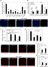
Figure 1.
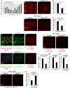
Figure 1.. Myo1D Is Non-Cell-Autonomously Required for Mitotic Activity of ISCs
(A) Myo1D is partially required for mitotic activity of ISCs. Various allelic combinations of Myo1D mutants were tested. Mitotic activity was determined by PH3 labelings. Even heterozygous Myo1D mutants display reduced mitotic activity. Myo1DEY is Myo1DEY08859. NP1-Gal4 is a Gal4 insertion in the Myo1D gene and a partial loss-of-function mutant. PH3 counts were analyzed by ordinary one-way ANOVA for multiple comparisons. Plotted is relative mitotic activity ± SEM. ***p < 0.001; **p < 0.01; *p < 0.05. p values are relative to w1118. (B) Myo1D controls mitotic activity of ISCs in ECs. esg-Gal4 and Su(H)-Gal4 are expressed in progenitor cells; NP1-Gal4 and 5966::GS are expressed in ECs. Controls are the Gal4 drivers over +. In this and the following figures, the ts annotation indicates the tub-Gal80ts transgene for temporal control of Gal4 activity. Conditional expression from the 5966::GS driver was induced by feeding RU486 to the animals. PH3 counts were analyzed by ordinary one-way ANOVA for multiple comparisons. Plotted is relative mitotic activity ± SEM. **p < 0.01; ***p < 0.001. ns, not significant. (C–E) Representative examples of PH3 (green) labelings in posterior midguts with either normal (C), reduced (D), or increased (E) levels of Myo1D. DAPI (blue) labels nuclei. (F) Expression of a wild-type UAS-Myo1D transgene in ECs using NP1-Gal4 can rescue the ISC mitotic phenotype of the indicated Myo1D mutants. Please note that NP1-Gal4 itself is a loss-of-function allele of Myo1D. Therefore, the Myo1D mutants are trans-heterozygous with NP1-Gal4. Control (ctrl) is NP1-Gal4/+. PH3 counts were analyzed by ordinary one-way ANOVA for multiple comparisons. Plotted is relative mitotic activity ± SEM. ns, not significant. (G) Several upstream activating sequence (UAS)-based transgenes encoding mutant forms of Myo1D (Myo1DΔIQ, Myo1DΔtail, Myo1DΔAbs) are dominant-negative alleles. Control (ctrl) is NP1-Gal4/+. PH3 counts were analyzed by ordinary one-way ANOVA for multiple comparisons. Plotted is relative mitotic activity ± SEM. *p < 0.05; **p < 0.01. p values are relative to ctrl. (H–K) Heterozygosity of the amorphic Myo1DL152 allele results in partial loss of precursor cells, labeled by esg-lacZ for ISCs (H and I) and Su(H)-lacZ for EBs (J and K). (L) Quantification of (H)–(K). β-Gal positive cells were analyzed by unpaired t test, two tailored, and plotted ± SEM. *p < 0.05; **p < 0.01. (M–P) Depletion of Myo1D by RNAi results in partial loss of precursor cells, labeled by esg-lacZ for ISCs (M and N) and Su(H)-lacZ for EBs (O and P). (Q) Quantification of (M)–(P). β-Gal positive cells were analyzed by unpaired t test, two tailored, and plotted ± SEM. ****p < 0.0001.
Figure 2.
Figure 2.. Myo1D Is Required for Various Signaling Events between ECs and Stem Cells/Muscle Cells
(A) qRT-PCR analysis of the expression of select signaling genes in the midgut as a function of EC-specific Myo1D RNAi. Primer sequences are listed in Table S1. The values for pairwise comparisons (control versus Myo1D RNAi) were analyzed by unpaired t test, two tailored. Plotted is mean signal ± SEM. **p < 0.01; ***p < 0.001; ****p < 0.0001. ns, not significant. (B and C) The amorphic Myo1DL152 allele dominantly affects upd3-lacz expression. (D) Quantification of (B) and (C). β-Gal signal intensities were determined and analyzed by unpaired t test, two-tailored. Plotted is mean intensity ± SE. **p < 0.01. (E and F) Depletion of Myo1D by RNAi partially impairs upd3-lacZ expression. Control is NP1-Gal4/+. (G) Quantification of (E) and (F). β-Gal signal intensities were determined and analyzed by unpaired t test, two-tailored. Plotted is mean intensity ± SEM. **p < 0.01. (H–I′′) ISC-specific STAT activity (STAT-GFP) and muscle-specific vein (vn-lacZ) expression is partially dependent on Myo1D. Quantified in (L) (STAT-GFP) and (M) (vn-lacZ). (J and K) EC-specific knockdown of Myo1D affects vein expression (vn-lacZ) in the visceral muscle. Quantified in (N). (L–N) Quantification of the data shown in (H)–(K). All three datasets were analyzed by unpaired t test, two tailored. In the case of STAT-GFP (H′ and I′), mean signal intensities were determined and plotted ± SEM (L). In the case of vn-lacZ (H′′, I′′, J, and K), mean β-Gal signal intensities per nucleus were determined and plotted ± SEM. *p < 0.05; ****p < 0.0001. (O and P) Muscle-specific vn-lacZ expression is strongly induced by EC-specific overexpression of Myo1D. (Q) Quantification of (O) and (P). β-Gal signal intensities per nucleus were determined and analyzed by unpaired t test, two-tailored. Plotted is mean intensity ± SEM. ****p < 0.0001. See also Table S1.
Figure 3.
Figure 3.. Myo1D Is Required for Dronc Localization to the Basal Side of the Plasma Membrane in ECs
(A–D) Analysis of the subcellular localization of Dronc protein in the posterior midgut. The schemes above the panels indicate the focal plane across the midgut (apical [A and C] or basal [B and D]), which was applied when the midgut labelings were imaged (black line). White arrows in (B) point to ECs with plasma membrane localization of Dronc; yellow arrowheads point to ECs with cytosolic Dronc. Blue arrowheads in (B) and (D) point to the smaller precursor cells. (C and D) EC-specific knockdown of Myo1D by NP1-Gal4ts. The membrane localization of Dronc in ECs is lost. (E) ECs with high levels of cytosolic Dronc are apoptotic as judged by the CC3 antibody. White arrows indicate examples. See also Figures S1 and S2.
Figure 4.
Figure 4.. Myo1D Is Required for ROS Generation and Hemocyte Recruitment to the Midgut
(A and B) The amorphic Myo1DL152 allele dominantly reduces the redox-reporter GstD-GFP. (C) Quantification of (A) and (B). GFP signal intensities were determined and analyzed by unpaired t test, two-tailored. Plotted is mean intensity ± SEM. ****p < 0.0001. (D–H) Analysis of the redox-reporter GstD-GFP in response to knockdown (E) or overexpression of Myo1D (F) and the dominant-negative Myo1DΔIQ (G) and Myo1DΔtail (H) transgenes. Control is NP1-Gal4/+. (I) Quantification of (D)–(H). GFP signal intensities were determined in defined areas and analyzed by ordinary one-way ANOVA. Plotted is mean intensity ± SEM. ****p < 0.0001. (J) Myo1D-induced mitotic activity of ISCs is dependent on extracellular ROS. Duox RNAi and overexpression of extracellular catalases hCatS (human secreted catalase) and IRC. Control (ctrl) is NP1-Gal4/+. PH3 counts were analyzed by ordinary one-way ANOVA for multiple comparisons. Plotted is relative mitotic activity ± SEM. **p < 0.01; ***p < 0.001. (K) The reduction of the mitotic activity caused by Myo1D RNAi can be partially rescued by overexpression of Duox. PH3 counts in the two datasets were analyzed by unpaired t test, two tailored. Plotted is relative mitotic activity ± SEM. **p < 0.01. ns – not significant. (L–O) Recruitment of hemocytes to the midgut is dependent on Myo1D and ROS. Depletion of Myo1D (M) and Duox (N) by RNAi as well as reduction of extracellular ROS by overexpression of hCatS partially reduces the recruitment of hemocytes to the midgut. A hmlΔ-DsRed transgene was used to label hemocytes. Control: NP1-Gal4/+. (P) Quantification of (L)–(O). Hemocyte counts were analyzed by ordinary one-way ANOVA for multiple comparisons. Plotted is number of hemocytes per gut ± SEM. **p < 0.01; ***p < 0.001. See also Figure S3.
Figure 5.
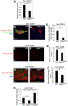
Figure 5.. JNK Activity Is Induced in Both Hemocytes and ECs
hmlΔ-Gal4 drives expression of UAS-based transgenes specifically in hemocytes. (A) Hemocyte-specific knockdown of Ask1 partially reduces ISC mitotic activity. PH3 counts were analyzed by unpaired t test with Welch’s correction, two tailored, and plotted as relative mitotic activity ± SEM. *p < 0.05. (B and C) JNK activity is induced in hemocytes. Shown are confocal images focusing on the surface of the midgut where hemocytes are attached. The NimC antibody labels hemocytes (red); β-Gal labeling (green) detects the JNK marker puc-lacZ. Normal hemocytes are β-Gal positive, suggesting that they contain active JNK (B). Hemocyte-specific depletion of Ask1 results in loss of JNK activity (C). (D) Quantification of (B) and (C). β-Gal intensity was determined in hemocyte clusters and analyzed by unpaired t test with Welch’s correction, two tailored. Plotted is mean intensity per cluster ± SEM. *p < 0.05. (E and F) Shown are confocal images focusing on ECs in the midgut. puc-lacZ is a marker of JNK activity. Hemocyte-specific depletion of Ask1 results in partial loss of JNK activity in ECs. (G) Quantification of (E) and (F). β-Gal intensity in ECs was analyzed by unpaired t test, two tailored. Plotted is mean intensity in ECs ± SEM. *p < 0.05. (H and I) Hemocyte-specific knockdown of eiger results in partial loss of JNK activity in ECs. JNK activity was detected using tetradecanoylphorbol acetate response element-red fluorescent protein (TRE-RFP). (J) Quantification of (H) and (I). RFP intensity was determined in defined areas and analyzed by unpaired t test, two tailored. Plotted is mean intensity per area ± SEM. *p < 0.05. (K) Hemocyte-specific loss (by RNAi) or increase (by UAS-eiger) of eiger reduces or increases the mitotic activity of ISCs, respectively. PH3 counts were analyzed by ordinary one-way ANOVA and plotted as relative mitotic activity ± SEM. *p < 0.05; **p < 0.01. See also Figure S4.
Figure 6.
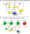
Figure 6.. Comparison of Undead AiP in Imaginal Discs and the Role of Transiently Undead ECs for Homeostatic Turnover in the Adult Posterior Midgut
(A) The hid,p35-expressing undead AiP model in larval imaginal discs. Myo1D has a key role by localizing Dronc to the basal side of the plasma membrane in close proximity to the NADPH oxidase Duox. ROS generated by Duox attract hemocytes, which release Eiger and trigger JNK signaling in undead cells. This leads to the production of the mitogens Wg, Dpp, and EGF (Spi) for AiP. Please note the apical-basal polarity of these cells. (B) Transiently undead ECs in the adult posterior midgut. Relevant parts are presented in red. Please note the similarities to the undead AiP model in imaginal discs (A). When ECs are getting ready for turnover, Dronc (red) is localized to the basal side of the plasma membrane in a Myo1D-dependent manner, rendering ECs transiently undead. Dronc stimulates Duox activity for ROS generation, hemocyte recruitment, and JNK activation in hemocytes and undead ECs. That releases Upd3 for stimulation of mitotic activity of ISCs. After that, undead ECs release Dronc from the membrane and become apoptotic. Please note that ISCs and visceral muscle cells are in direct contact with the basal side of ECs. See also Figure S5.

References

    1. Amcheslavsky A, Jiang J, and Ip YT (2009). Tissue damage-induced intestinal stem cell division in Drosophila. Cell Stem Cell 4, 49–61. - PMC - PubMed
    1. Amcheslavsky A, Wang S, Fogarty CE, Lindblad JL, Fan Y, and Bergmann A (2018). Plasma Membrane Localization of Apoptotic Caspases for Non-apoptotic Functions. Dev. Cell 45, 450–464.e453. - PMC - PubMed
    1. Ayyaz A, Li H, and Jasper H (2015). Haemocytes control stem cell activity in the Drosophila intestine. Nat. Cell Biol 17, 736–748. - PMC - PubMed
    1. Bach EA, Ekas LA, Ayala-Camargo A, Flaherty MS, Lee H, Perrimon N, and Baeg GH (2007). GFP reporters detect the activation of the Drosophila JAK/STAT pathway in vivo. Gene Expr. Patterns 7, 323–331. - PubMed
    1. Barylko B, Binns DD, and Albanesi JP (2000). Regulation of the enzymatic and motor activities of myosin I. Biochim. Biophys. Acta 1496, 23–35. - PubMed

Publication types

LinkOut - more resources